Note: Descriptions are shown in the official language in which they were submitted.
CA 02526276 2010-01-22
DESCRIPTION
Stent Supplying Device
Technical Field
This invention relates to a stent delivery system used for delivering a stent
fora vessel, implanted in a vessel of a living body, such as blood vessel,
trachea,
bile duct or urethra, for providing a support for the lumen of the vessel from
the
inside, to a target site for implantation in the vessel.
Background Art
If stenosis has occurred in a vessel of a living body, such as blood vessel, a
balloon forming portion provided in the vicinity of the distal end of a
balloon
catheter is inserted into this stenosis portion. This balloon forming portion
is dilated
to expand the stenosis portion of the blood vessel to improve the blood flow.
This
operation, known as percutaneous angioplasty (PTA), has so far been in
widespread
use.
However, after application of PTA to the site of occurrence of stenosis in a
blood vessel, acute occlusion, attributable to the dissection of the intima,
or
re-narrowing of the same site as that where narrowing in the blood vessel
(stenosis)
has occurred, that is, re-stenosis, tends to produced in a well-known manner
at a
CA 02526276 2005-11-17
2
high probability.
For preventing such acute occlusion or re-stenosis, the technique of
implanting a tubular stent at the target site where PTA has been applied, has
so far
been used. A stent, used for this purpose, is implanted into the blood vessel,
as it is
contracted in diameter, and subsequently enlarged in diameter so as to be
implanted
in the blood vessel to support the blood vessel wall from its inside.
For a stent implanted in the blood vessel, a stent made of metal has so far
been used. The metal stent is classified into a balloon expanding stent and a
self-expanding stent.
The balloon expanding stent is inserted into a targeted site for implantation
in
the blood vessel, in a state contracted in diameter, and subsequently enlarged
in
diameter with expansion of the balloon. Among the stents of this type, there
are a
stent comprised of a small-diameter tube of stainless steel provided with
numerous
incisions formed by e.g. a laser cutter to permit the tube to be enlarged in
diameter,
and a stent formed by braiding a fine metal filament into a tube, as disclosed
in US
Patent 4,950,227.
The self-expanded stent is contracted in diameter under application of an
external pressure and inserted in this contracted state in the target site for
implantation in the blood vessel. After removal of the external pressure, the
stent is
self-expanded in diameter to support the blood vessel from its inner wall
surface.
As typical of this type of the self-expanded stent, there is known such a one
CA 02526276 2005-11-17
3
obtained by spirally winding a fine metal wire to form a tube, as disclosed in
the
Japanese Laid-Open Patent Publication Hei-2-68052.
For implanting the above-described stent for a vessel in the target site in
the
blood vessel of a living body, a stent delivery system is used. The stent
delivery
system is of variable configurations, depending on the type of the stent to be
delivered, that is, on whether the stent delivered is the balloon expanding
stent or
the self-expanded stent.
The stent delivery system for delivering the balloon expanding stent within
the blood vessel includes a catheter inserted into the blood vessel, and a
balloon is
provided, as it is contracted in diameter, to the distal end of the catheter.
On this
balloon is mounted a stent as it is contracted in diameter. The stent, mounted
on the
balloon, is pressed from its outer peripheral side and retained against
detachment
from the balloon. The stent, thus mounted on the balloon, is delivered as far
as the
targeted site for implantation in the blood vessel, along with the balloon, by
progressively inserting the catheter into the blood vessel. The stent, thus
delivered
to the target site for implantation in the blood vessel, is expanded in
diameter on
plastic deformation caused by balloon expansion to support the blood vessel
wall
from its inner side.
For the stent delivery system, used for implanting the balloon expanding
stent in the blood vessel, it is basically only sufficient to include a means
for
mounting a stent, contracted in diameter, on the balloon provided to the
catheter.
CA 02526276 2005-11-17
4
As the stent delivery system for delivery of the balloon expanding stent,
there has been proposed such a system including a sheath covering up the stent
mounted on the balloon. The sheath used is provided for preventing the stent,
mounted on the balloon, from becoming detached from the balloon.
On the other hand, the stent delivery system for delivery of the balloon
expanding stent into the blood vessel is constructed so that a catheter
mounting a
stent contracted in diameter is inserted into a protective sheath. The stent,
mounted
in the state contracted in diameter in the catheter, is covered up by the
protective
sheath and thereby maintained in the state contracted in diameter. For
implanting
the stent in the target site for implantation, using the above-described stent
delivery
system, the catheter, mounting the stent, is inserted up to the target site
for
implantation in the blood vessel, along with the protective sheath. At this
time, the
catheter is fixed and only the protective sheath is retreated in the blood
vessel,
whereby the stent, mounted to the distal end of the catheter, is freed from
the sheath.
The stent, thus freed from the protective sheath, is self-expanded by
elasticity
proper to the stent itself, and is dilated in diameter to a size capable of
providing a
support for the inner wall of the blood vessel.
The stent delivery system, used for implanting the self-expanding stent in the
blood vessel, includes a catheter on which is mounted a stent, contracted in
diameter, and a protective sheath in which is housed the catheter, the stent
has been
mounted to, there being no necessity to provide a balloon for expanding the
stent.
CA 02526276 2005-11-17
Currently, there has not been established a method for treatment for an
instance where re-stenosis has occurred on a site where angioplasty has been
applied and a metal stent has been implanted.
Moreover, if metal, inherently a foreign substance for the living body, is
caused to remain for a prolonged time in the living body, there is a cause
that the
blood vessel may thereby be affected, such as by excessive intimal hyperplasia
occurring in the stent implant portion.
With a view to obviating the problems inherent in the conventional metal
stent, the present Assignee has already proposed a stent formed using a
biodegradable polymer (see US Patent specification No.6045568, Patent
No.2842943 and W000/13737).
The stent formed of the biodegradable polymer may be absorbed in the tissue
of the blood vessel after a preset time, has passed after it is implanted in
the blood
vessel, for example, after lapse of 6 to 12 months, such that the function of
providing a support for the blood vessel from the inner side thereof is no
longer
needed. Since the stent of this type may be absorbed in vivo, it becomes
possible to
suppress adverse effects which might be produced as a result of the stent, as
a
foreign material for the living body, being left over for a prolonged time.
In particular, the present Assignee has already proposed a stent for a vessel,
formed by braiding a yarn of a biodegradable polymer into a tube (US Patent
specification 6045568), a stent for a vessel prepared by forming a yarn of a
CA 02526276 2005-11-17
6
biodegradable polymer in a non-woven non-braided state (Patent 2842943) and a
stent for a vessel prepared by bending a yarn of biodegradable polymer in a
zigzag
design to form concatenated vee shapes, and by winding the resulting zig-zag
shaped yarn into a tube, with the stent for a vessel being expanded or
contracted in
diameter with vee shaped portions of the yarn as portions subjected to
displacements (WO00/13737). These stents were actually implanted in living
bodies.
The stent formed of the biodegradable polymer is formed into a tube and
subsequently heat-set, by way of heat treatment, for shape retention to a
desired
outer diameter. This heat-setting is carried out at a temperature not lower
than the
glass transition temperature and not higher than the melting point of the
biodegradable polymer making up the stent. The stent which is to be implanted
in
the blood vessel, and which has its shape retained to a desired outer
diameter, is
contracted in diameter for insertion into the blood vessel. This contraction
of the
stent is carried out under application of an external pressure with or without
heat
setting. The heat-setting here is carried out at a temperature lower than the
temperature for heat setting carried out for retention of the expanded state.
The stent made of the biodegradable polymer is expanded by a balloon
expansion method employing a balloon. This method is carried out for promptly
expanding the stent, inserted in a state contracted in diameter as far as the
site for
implantation in the blood vessel, to a size capable of reliably supporting the
inner
CA 02526276 2005-11-17
7
wall of the blood vessel.
Meanwhile, the stent, formed using the biodegradable polymer, may be
warmed and thereby given the self-expanding properties, that is, the
properties of
shape memory. When mounted on the catheter and inserted in this state into the
blood vessel of the living body, the stent, formed of the biodegradable
polymer, is
self-expanded, as it is warmed by body temperature of the living body. Since
the
stent has the self-expanding properties, it is tightly contacted with the
inner wall of
the blood vessel to maintain the force of dilating the blood vessel from its
inside,
and hence is able to distend the blood vessel from its inner wall over a
preset time
period until the time of biodegradation.
Thus, the stent formed of the biodegradable polymer has the self-expanding
properties, even though it necessitates expansion by the balloon. For
inserting this
sort of the stent into the blood vessel of the living body for implantation
therein,
there is needed, along with the balloon for expanding the stent, an expansion
inhibiting member for inhibiting self-expansion of the stent which is
otherwise
caused when the stent is warmed up by body temperature on insertion thereof
into
the blood vessel. That is, for preventing the occurrence of an accident in
which the
stent contracted in diameter is self-expanded on being inserted into the blood
vessel
and is disengaged from the balloon, it becomes necessary to provide a
protective
sheath to control the self-expansion of the stent mounted to the balloon.
There is also the possibility that the stent of the biodegradable polymer,
CA 02526276 2005-11-17
8
exhibiting the self-expanding properties, is jumped up from the protective
sheath by
its force of expansion and becomes disengaged from the catheter, when the
stent,
delivered to the targeted site for implantation in the blood vessel of the
living body,
is subsequently gradually freed of the support from the protective sheath,
such that
a given portion of the stent is protruded from the protective sheath. The
result is that
not only the stent cannot be implanted in the targeted site for implantation
in the
blood vessel but also the stent becomes unable to be expanded by the balloon.
Disclosure of the Invention
It is an object of the present invention to provide a stent delivery system
whereby a stent for a vessel, formed of a biodegradable polymer and afforded
with
the self-expanding properties, may correctly be implanted in a target site in
the
vessel.
It is another object of the present invention to provide a stent delivery
system
whereby a stent for a vessel, which is formed of the biodegradable polymer and
given the self-expanding properties, but which is furthermore in need of
expansion
by a balloon, may be enlarged in diameter such as to provide a reliable
support for
the inner wall of the vessel.
It is a further object of the present invention to provide a stent delivery
system in which the location of insertion of the stent relative to the vessel
may be
detected from outside the living body.
It is a further object of the present invention to provide a stent delivery
CA 02526276 2005-11-17
9
system whereby a stent for a vessel, mounted on the balloon, provided to the
catheter, may readily be delivered to a targeted site for implantation in the
vessel.
It is yet another object of the present invention to provide a stent delivery
system whereby a stent for a vessel may be delivered without falling off
within a
small vessel which may be bent, sinuous or hardened.
For accomplishing the above object, a stent delivery system, proposed by the
present invention, comprises a protective sheath inserted into a vessel of a
living
body, a catheter inserted into the protective sheath for performing a back-and-
forth
movement therein, a balloon arranged on an outer peripheral surface towards
the
distal end of the catheter protruded from the distal end of the protective
sheath, and
a stent for a vessel, formed of a biodegradable polymer. The balloon may be
expanded with a fluid supplied to the catheter. The stent for a vessel is
mounted in a
state contracted in diameter on the balloon and is moved back and forth along
with
the balloon relative to the protective sheath. At least one end of the stent
is retained
by a temporary holding member.
The temporary holding member temporarily holds one end side, located
towards the proximal side of the protective sheath, of the stent for a vessel,
housed
within the protective sheath. That is, the one end side, located towards the
proximal
side of the protective sheath, of the stent for a vessel, opposite to the side
of the
stent for a vessel protruded from the distal end of the protective sheath, is
retained
by the temporary holding member.
CA 02526276 2005-11-17
When freed from the retention by the protective sheath, the stent for a
vessel,
mounted on the balloon, has its one end side towards the proximal side of the
protective sheath retained by the temporary holding member, and hence is not
jumped up precipitously under the force of expansion.
Preferably, the temporary holding member, retaining the stent for a vessel, is
formed as a tube of an inner diameter smaller than the outer diameter of the
catheter.
The temporary holding member may hold only a part of the outer periphery
of the stent for a vessel.
Preferably, the temporary holding member is formed of an elastic material to
a tubular shape and retains the outer peripheral surface of the catheter as
far as the
one end of the stent for a vessel. At this time, the temporary holding member
preferably has at least a portion thereof towards the catheter side bonded to
the
catheter.
The stent for a vessel, employed in the present invention, is formed of a
biodegradable polymer to a tubular shape. This stent for a vessel is provided
with
the self-expanding function.
According to the present invention, a stent for a vessel, formed from a yarn
of a biodegradable polymer to a tubular structure, and provided with the
self-expanding function, is used. The stent for a vessel, formed of a
biodegradable
polymer, wound to a tube as the yarn is bent in a zigzag design, and expanded
or
CA 02526276 2005-11-17
11
contracted in diameter with the bends of the yarn as displacing portions, is
used.
The protective sheath preferably is formed of a material prohibited from
extension/ contraction along its longitudinal direction in order to render the
protective sheath scarcely extensible along the longitudinal direction.
The protective sheath has its distal end side towards a side for insertion
into a
vessel of a living body curved to conform to the shape of the vessel.
The protective sheath includes on its distal end side for insertion into the
vessel of the living body a flexible tubular section superior in flexibility
to its
proximal end side.
The protective sheath includes on the distal end side for insertion into the
vessel of the living body an insertion protecting part formed of a material
exhibiting
superior flexibility.
The protective sheath includes a radiopaque section, containing a radiopaque
material, on its distal end side from which is protruded the stent for a
vessel
mounted on the catheter.
The catheter inserted into the protective sheath includes a radiopaque section
indicating the mounting position of the stent for a vessel.
Since the radiopaque section containing a radiopaque material is provided on
the distal end side of the protective sheath, and the catheter inserted into
the
protective sheath includes a radiopaque section indicating the mounting
position of
the stent for a vessel, the relative positions of the radiopaque section on
the
CA 02526276 2005-11-17
12
protective sheath side and the radiopaque section on the catheter may be
confirmed,
when the protective sheath is moved relative to the stent for a vessel,
whereby it is
possible to determine the positions of the catheter and the stent for a vessel
relative
to the protective sheath.
Other objects and specified advantages of the present invention will become
more apparent from the following explanation of present embodiments of the
invention which will now be made hereinbelow by referring to the drawings.
Brief Description of the Drawings
Fig.l is a perspective view showing a system for delivery of a stent for a
vessel according to the present invention.
Fig.2 is a partial cross-sectional view showing the state in which a catheter
carrying the stent for a vessel has been inserted into a protective sheath.
Fig.3 is a partial perspective view showing the structure of a protective
sheath.
Fig.4 is a partial perspective view showing a distal end part of the
protective
sheath.
Fig.5 is a cross-sectional view showing the state in which the stent for a
vessel has been mounted on a balloon provided to the catheter.
Fig.6 is a cross-sectional view taken along line IV-IV of Fig.5.
Fig.7 is a perspective view showing an instance of the stent for a vessel used
in the present invention.
CA 02526276 2005-11-17
13
Fig.8 is a cross-sectional side view showing the state in which the stent for
a
vessel, mounted on the catheter, is held by a temporary holding member.
Fig.9 is a perspective view showing the state in which the stent for a vessel,
mounted on the catheter, is protruded from the protective sheath.
Fig. 10 is a side view showing the state in which the balloon is expanded to
enlarge the diameter of the stent for a vessel.
Fig. 1 l is a side view showing another instance of the state in which the
stent
for a vessel, mounted on the catheter, is retained.
Fig.12 is a cross-sectional view showing the proximal side end of the
catheter provided with a bend control part for preventing the bending of the
catheter.
Fig.13 is a cross-sectional view showing a second connection fixture
provided with a fixation unit for fixing the catheter to a protective sheath.
Fig.14 is a cross-sectional view showing the state in which the stent for a
vessel is inserted into the blood vessel of a living body and the balloon is
expanded
to dilate the stent in diameter.
Fig. 15 is a cross-sectional view showing the state in which the stent for a
vessel is inserted into the blood vessel of the living body and expanded in
diameter
and subsequently the balloon is contracted.
Fig. 16 is a perspective view showing a temporary holding member used for
the stent delivery system according to another embodiment of the present
invention.
CA 02526276 2005-11-17
14
Fig.17 is a cross-sectional view showing essential portions of the stent
supplying device according to a further embodiment of the present invention.
Fig. 18 is a perspective view showing a state of balloon expansion in a stent
delivery system according to the present invention.
Best Mode for Carrying out the Invention
Referring to the drawings, present embodiments of the present invention will
be explained in detail.
The stent delivery system according to the present invention is used for
delivering a stent, implanted in a vessel, such as blood vessel, trachea, bile
duct or
urethra of a living body, for supporting the lumen of the vessel from its
inner side
for maintaining a patency state of the lumen, to a target site for
implantation in the
vessel.
In the embodiment, as now explained, the present invention is applied to a
system for delivery in the blood vessel of a stent for a vascular vessel to be
implanted within the blood vessel of the living body.
The stent delivery system, embodying the present invention, includes a
protective sheath 1, inserted into the vessel, such as a blood vessel, of a
living body,
and a catheter 2, inserted into the protective sheath 1 and inserted along
with the
protective sheath 1 into the blood vessel, as shown in Fig.l.
The protective sheath 1 is formed as a long flexible tube so that it may be
smoothly inserted to conform to the shape e.g. of the blood vessel of the
living body.
CA 02526276 2005-11-17
The protective sheath 1 is of an outer diameter R1 of approximately 2 mm to 3
mm
and of a length approximately 100 cm, as shown in Fig.2.
In an inner bore opening 3 formed of the protective sheath 1, the catheter 2
is
inserted so as to be reciprocated therein as shown in Fig.2. The distal end of
the
catheter 2 carries a balloon 4 on which is mounted a vascular stent 5.
Meanwhile, the protective sheath 1, used for the stent delivery system,
according to the present invention, is movable relative to the catheter 2, so
as to
assume a state in which the vascular stent 5, mounted to the distal end of the
catheter 2, is housed within the protective sheath 1, and a state in which the
stent is
protruded outwards from the distal end. For reliably varying the position of
the
vascular stent 5 relative to the protective sheath 1, the length of the
protective
sheath 1, pulled out from the catheter 2, is accurately coincident with the
length of
the protective sheath 1 moved relative to the catheter 2. In addition, the
protective
sheath 1 is inserted into the inside of the blood vessel as the sheath is in
contact
with the inner wall of the blood vessel.
Hence, the protective sheath 1 is preferably formed of a material the
extension and contraction of which along the long axis of the sheath are
controlled
to render the sheath difficultly extensible along the long axis. It is also
desirable
that the protective sheath 1 may be inserted smoothly as it is deformed after
the
shape of the blood vessel of the living body and, in addition, the catheter 2
inserted
into the inner bore opening 3 may smoothly be reciprocated without any
significant
CA 02526276 2005-11-17
16
frictional resistance with respect to the protective sheath.
Thus, the protective sheath 1, used in the present invention, is constructed
and arranged as shown for example in Fig.3. This protective sheath 1 is formed
by a
readily flexible tubular member 6 formed by braiding fine wires of e.g.
stainless
steel in a meshed pattern, and outer and inner coating layers 7, 8 of
synthetic
polymer applied to the outer and inner peripheral surfaces of the tubular
member 6,
thereby suppressing the sheath from performing extension or contraction along
its
long axis. That is, the meshed tubular member 6, coated by the outer and inner
coating layers 7, 8 of synthetic polymer, is prevented from performing
extension or
contraction along its longitudinal direction.
The outer coating layer 7 of the protective sheath 1, contacted with the inner
wall of the blood vessel when the protective sheath is inserted into the
inside of the
blood vessel, is preferably formed of a highly anti-hygroscopic material, for
example, a polyamide-based synthetic material. On the other hand, the inner
coating
layer 8, having a sliding contact with the catheter 2, is preferably formed of
a
synthetic polymer material, low in friction and hence superior in lubricity,
for
example, polytetrafluoroethylene.
Moreover, the distal end of the protective sheath 1 of the stent delivery
system of the present invention, operating as an inserting end into the blood
vessel
of the living body, is formed as a flexible tubular part 9 exhibiting higher
flexibility
than its proximal side end, as shown in Fig.4. The outer coating layer 7 of
the
CA 02526276 2005-11-17
17
flexible tubular part 9 is formed of a flexible and flaccid material. The
protective
sheath 1, the distal end of which is formed by the flexible tubular part 9,
exhibiting
flexibility superior to that of the main sheath body part, may readily be
inserted into
the blood vessel to conform to its curved profile.
Referring to Fig.4, the flexible tubular part 9 is curved or sinuous to
conform
to the shape of the blood vessel, for example, artery, into which the
protective
sheath 1 is inserted along with the catheter 2. Since the flexible tubular
part 9 at the
distal end side of the protective sheath is sinuous in this manner, the
protective
sheath may be smoothly inserted without excessively loading the artery curved
strongly to e.g. a U-shape.
At the distal end of the protective sheath 1, there is provided an insertion
protecting part 10, formed of a material having superior flexibility, such as
silicone
rubber, as shown in Fig.4. Since the distal end of the protective sheath 1,
along the
direction of insertion of the sheath into the blood vessel, is provided with
the
insertion protecting part 10, described above, it becomes possible to insert
the
protective sheath into the blood vessel with the least risk of damage to the
vessel
wall.
The insertion protecting part 10, provided to the distal end towards the
inserting side of the protective sheath 1 into the blood vessel, is provided
with a
radiopaque section 11 containing a radiopaque material. By providing the
radiopaque section 11 to the distal end towards the inserting side of the
protective
CA 02526276 2005-11-17
18
sheath 1 into the blood vessel of the living body, the position of insertion
of the
protective sheath into the blood vessel may be visually determined by
illumination
of X-rays when the protective sheath is inserted into the blood vessel, while
it may
also be checked whether or not the stent as well as the catheter has been
protruded
accurately to outside the protective sheath at the time of implanting the
stent.
The radiopaque section 11 may also be provided to the distal end of the
protective sheath 1 per se.
The catheter 2, the vascular stent 5 is mounted to, and which is inserted into
the protective sheath 1, is formed of a synthetic polymer material, such as
polyethylene, for imparting flexibility to the catheter. Since the catheter 2
is used
for causing reciprocating movement of the vascular stent 5, mounted on the
balloon
4, provided towards the distal end of the catheter, between the position in
which the
stent is housed within the protective sheath 1 and the position in which the
vascular
stent 5 is protruded from the protective sheath 1, the catheter 2 has an
overall length
larger than the protective sheath 1 by a length at least corresponding to the
length of
reciprocation of the vascular stent 5 plus a length of a finger support to be
gripped
by a user's finger when causing the reciprocating movement of the vascular
stent.
Referring to Figs.5 and 6, the catheter 2 is provided with an inner bore
opening 13 for a guide wire 12 and with a fluid passage 14 for supply of a
fluid,
such as a contrast agent, used for expanding the balloon 4 provided to the
distal end
of the catheter 2. The guide wire 12 is used for guiding the catheter 2 along
with the
CA 02526276 2005-11-17
19
protective sheath I into and through the blood vessel. Meanwhile, the inner
bore
opening 13 for the guide wire is formed as a through-opening from the proximal
end up to the distal end of the catheter 2, whilst the fluid passage 14 is
formed as a
blind hole and terminated short of the distal end of the catheter 2, as shown
in Fig.5.
To the distal end of the catheter 2 is mounted the balloon 4 for expanding the
vascular stent 5, mounted to the catheter 2, as shown in Fig.5. The balloon 4
is
formed to a tubular shape from e.g. polyethylene (PE), polyolefinic coployner
(POC) or polyethylene terephthalate (PET). The balloon 4 is mounted to cover
up
the outer peripheral surface on the distal end of the catheter 2, and bonded
at its
both ends 4a, 4b thereto, using e.g. an adhesive, so as to be unified as one
with the
catheter 2. In an initial state in which the balloon has been mounted to the
catheter 2,
the balloon 4 is collapsed and folded on its outer peripheral surface.
In the part of the catheter, the balloon 4 is mounted to, there is provided a
through-hole 15 by means of which the balloon is to communicate with the fluid
passage 14, as shown in Fig.5. The contrast agent, supplied via the fluid
passage 14,
is charged into the inside of the balloon 4 via the through-hole 15 to expand
the
balloon 4. In the part of the catheter 2, the balloon 4 is mounted to, there
are also
formed radiopaque sections 16, 17 formed of a radiopaque material. These
radiopaque sections 16, 17 are provided by mounting fine wires of metal, as a
radiopaque material, to the outer peripheral surface of the catheter 2. The
radiopaque sections 16, 17, provided to the catheter 2, are provided in the
vicinity
CA 02526276 2005-11-17
of both ends 4a, 4b of the balloon 4. By these radiopaque sections 16, 17, the
position of insertion into the blood vessel of the vascular stent 5, mounted
on the
balloon 4, may be confirmed from outside the living body.
On the balloon 4, mounted to the catheter 2, there is mounted the vascular
stent 5 implanted in the blood vessel of the living body using the stent
delivery
system according to the present invention.
The vascular stent 5, used with the stent delivery system according to the
present invention, is formed of a biodegradable polymer material into a
tubular
form, and has a self-expanding function. A specified example of the vascular
stent 5
is formed as shown in Fig.7.
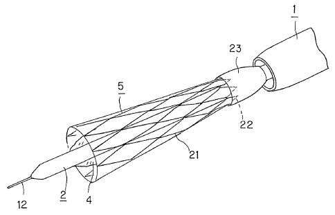
The vascular stent 5, shown in Fig.7, is formed to a tubular shape, with a
yarn 21 of a biodegradable polymer. That is, this vascular stent 5 is prepared
to a
tubular form., in particular a cylindrical form, by bending the yarn 21 of the
biodegradable polymer in a zigzag pattern to form concatenated vee shapes and
by
spirally winding the resulting zigzag shaped yarn into a tube, in particular a
cylinder,
as shown in Fig.7.
The biodegradable polymer, forming the yarn 21, may be exemplified by
aliphatic polyesters, fatty acid anhydrides, aliphatic polycarbonates,
polyphosphasen and copolymers containing at least one of these substances.
Specifically, the biodegradable polymer may be one or more of such
materials as poly-L-lactic acid (PLLA), polyglycolic acid, plyglactin,
CA 02526276 2005-11-17
21
polydioxanone, polyglyconate, s-caprolactam, polylactic acid- c-caprolacton
copolymers and polyglycolic acid-s-caprolacton copolymers.
The vascular stent 5, implanted in the blood vessel using the stent delivery
system of the present invention, is mounted on the balloon 4, mounted on the
catheter 2 in a state contracted in diameter, as shown in Fig.5. At this time,
the
balloon 4 is not expanded and is in a folded state, as shown in Fig.6. The
part of the
catheter 2, mounting the balloon 4, is formed to have an outer diameter
approximately equal to or slightly larger than the inner diameter of the
vascular
stent 5, contracted in diameter, in order that the vascular stent 5, thus
contracted in
diameter, will be mounted on the balloon 4 in close contact therewith.
The vascular stent 5 is mounted in this manner in close contacted state on the
balloon 4, mounted on the catheter 2, so that, when the balloon 4 is expanded,
the
vascular stent may be expanded quickly to follow up with the expansion of the
balloon 4.
The vascular stent 5, mounted on the balloon 4, is inserted, along with the
catheter 2, carrying the balloon 4, in the inner bore opening 3 of the
protective
sheath 1 for the catheter 2, and is kept in a state contracted in diameter by
the
protective sheath 1, as shown in Fig.2. The vascular stent 5, inserted into
the inner
bore opening 3 of the protective sheath 1 for the catheter 2, transitions from
the
state in which the stent is mounted on the balloon 4 and housed within the
protective sheath 1 to the state in which it is protruded from the distal end
of the
CA 02526276 2005-11-17
22
protective sheath 1, by causing movement of the protective sheath 1 relative
to the
catheter 2.
Meanwhile, the vascular stent 5, formed using the yarn 21, formed of the
biodegradable polymer, is given the self-expanding properties, and hence the
force
of expansion in a direction of dilation is conferred to the stent, by the
stent being
inserted into the blood vessel and warmed by body temperature of the living
body,
without the force of expansion being applied from outside. That is, although
the
vascular stent 5, held by the protective sheath 1, is kept contracted in
diameter, the
force of expansion is stored in the stent heated by being warmed up with the
body
temperature.
The vascular stent 5, in which the force of expansion is stored in the
protective sheath 1, is gradually freed from the support by the protective
sheath 1,
such that, when a part of the stent is protruded from the protective sheath 1,
it is
jumped up from within the protective sheath 1, and is dislocated from the
catheter 2.
Hence, it becomes difficult to locate the stent accurately in the targeted
site for
implantation within the blood vessel. Additionally, the position of the stent
mounted on the balloon 4 can scarcely be maintained.
Thus, with the vascular stent 5, used for the stent delivery system according
to the present invention, has its one end retained by a temporary holding
member 23,
in order to prevent the stent 5 from being expanded and jumped up from the
protective sheath, as shown in Figs.5 and 8. That is, as the vascular stent 5
is
CA 02526276 2005-11-17
23
housed and held in the protective sheath 1, the one proximal side end of the
stent
located opposite to the distal end of the protective sheath 1, operating as an
end for
protrusion, is held by the temporary holding member 23.
The temporary holding member 23, temporarily holding the vascular stent 5,
is formed as a tubular member covering up and temporarily holding the outer
rim
portion of the one end of the vascular stent 5, as shown in Figs.5 and S. The
temporary holding member 23 is formed of an elastic material, such as latex.
When formed of an elastic material, the temporary holding member 23 is
preferably formed to a tubular shape of a diameter smaller than the outer
diameter
of the catheter 2. When formed to this shape, the temporary holding member 23
pressures and holds the one end of the vascular stent 5 from the outer rim
portion
thereof to reliably hold the vascular stent 5. The temporary holding member 23
preferably covers up the one end side of the vascular stent 5 mounted on the
balloon
4 from the outer peripheral surface 2a of the catheter 2, as shown in Figs.5
and 8.
By the tubular temporary holding member 23 holding the vascular stent 5,
the protective sheath 1 may be moved smoothly relative to the vascular stent
5,
because the vascular stent 5 is held by the temporary holding member 23 and
thereby maintained in a state contracted in diameter.
With the vascular stent 5, having its one end held by the temporary holding
member 23, the force of expansion is accumulated as long as the vascular stent
is
housed within the protective sheath 1, as shown in Fig.8. When the stent is
CA 02526276 2005-11-17
24
subsequently protruded from the protective sheath 1, the opposite end side of
the
stent, protruded initially from the protective sheath 1, is expanded under the
force of
expansion, accumulated therein, as shown in Fig.9. However, the proximal one
end
side of the vascular stent 5 towards the proximal end of the protective sheath
1
continues to be retained by the temporary holding member 23 and is kept in the
state of close contact with the balloon 4. That is, it is not the vascular
stent 5 in its
entirety, i.e. the stent body from its one end up to its other end, that may
possibly be
expanded and disengaged from the balloon 4.
Meanwhile, the vascular stent 5, mounted on the balloon 4 of the catheter 2,
is promptly expanded with expansion of the balloon 4, brought about by removal
of
the force of support by the protective sheath 1, caused by relative movement
of the
protective sheath 1 with respect to the catheter 2. Since the temporary
holding
member 23 is adapted for holding the outer peripheral surface 2a of the
catheter 2
and the one end side of the vascular stent 5, as shown in Fig.5, the temporary
holding member 23 keeps on to hold the one end side of the vascular stent 5,
in the
initial stage of expansion of the balloon 4 for expanding the vascular stent
5.
However, when the vascular stent 5 is expanded further, the temporary holding
member 23 becomes detached from the vascular stent 5 and is left on the
catheter 2.
That is, the vascular stent 5 is disengaged from the temporary holding member
23,
because the vascular stent 5 is expanded beyond the expansion yield point of
the
temporary holding member 23.
CA 02526276 2005-11-17
The vascular stent 5 may be freed more reliably from the retention by the
temporary holding member 23 by setting the width of the part of the temporary
holding member 23, lying on the outer peripheral surface 2a of the catheter 2,
so as
to be larger than the width of the part of the temporary holding member lying
on the
one end side of the vascular stent 5, or by bonding the temporary holding
member
to the outer peripheral surface of the catheter using an adhesive.
Since the temporary holding member 23 holds only the one end side of the
vascular stent 5, the temporary holding member may readily be detached from
the
vascular stent 5, without obstructing the expansion of the vascular stent 5,
when the
vascular stent 5 is expanded with expansion of the balloon 4. Moreover, since
the
one end side of the vascular stent 5, lying towards the inner side of the
protective
sheath 1, is retained by the temporary holding member 23, the vascular stent
5, once
protruded to outside the protective sheath 1, may subsequently be housed again
within the protective sheath 1.
When the vascular stent 5, formed using the yarn 21 of the biodegradable
polymer, is freed from support by the protective sheath 1, the temporary
holding
member 23 has to hold the vascular stent 5 in such a manner as to prevent the
vascular stent 5 from being jumped up from the protective sheath 1, and in
such a
manner as not to obstruct the expansion of the stent by the balloon 4. Thus,
it is
sufficient for the temporary holding member 23 to hold a small region towards
one
end side of the vascular stent 5, as shown in Figs.8 and 9. It is also
unnecessary for
CA 02526276 2005-11-17
26
the temporary holding member 23 to hold the entire outer rim portion of the
one end
side of the vascular stent 5. In the case of the vascular stent 5, formed by
bending
the yarn 21 in a zigzag pattern to form a pattern of a concatenation of vee
shapes
and by spirally winding the resulting zigzag pattern of the yarn, as shown in
Fig.7,
it is sufficient that part of plural vee-shaped parts 22 of a ring pattern is
retained by
one end of the temporary holding member, as shown in Fig. 11.
The catheter 2, mounting the vascular stent 5 on the balloon 4 as described
above, is inserted from the proximal end of the protective sheath 1 into the
inner
bore opening 3, with the distal end carrying the vascular stent 5 first, as
shown in
Figs.l and 2. By inserting the catheter 2 into the inner bore opening 3 of the
protective sheath 1, the vascular stent 5, mounted on the distal end of the
catheter 2,
is also inserted into the inside of the protective sheath 1. The inner bore
opening 3
for the catheter 2, provided in the protective sheath 1, is formed to an inner
diameter
approximately equal to the outer diameter of the part at the distal end of the
catheter
2 carrying the balloon 4 and the vascular stent 5. The result is that the
vascular stent
5, mounted on the catheter 2, is inserted into the inner bore opening 3 for
the
catheter 2 of the protective sheath 1, so as to be maintained in its
contracted state.
The proximal end side of the catheter 2, opposite to its distal end side
carrying the vascular stent 5, operates as a catheter reciprocation portion
which is
held by the operator's finger when the catheter 2 is protruded from the
protective
sheath 1 and reciprocated relative to the protective sheath 1, as shown in
Fig. 1. This
CA 02526276 2005-11-17
27
catheter reciprocation portion is provided with a bend control part 25 for
preventing
the bending of the catheter 2 formed as a long tube from a readily bendable
synthetic polymer material, such as polyethylene polymer. The bend control
part 25
is formed by providing a tube 24, formed of a highly rigid material,
insusceptible to
deformation or flexure, to the outer periphery of the catheter 2, as shown in
Fig.12.
This tube 24 is formed of metal, such as aluminum or stainless steel.
It should be noted that the proximal end of the catheter 2 is slightly
protruded
from one end of the metal tube 24 of the bend control part 25, for connecting
a first
connection fixture 26, which will be explained subsequently.
This first connection fixture 26 is mounted to the proximal end side of the
catheter 2 provided with the bend control part 25, as shown in Figs. I and 12.
The
first connection fixture 26 is formed of a highly rigid synthetic polymer
material
scarcely susceptible to elastic deformation. The first connection fixture is
made up
by a guide section 27 for guiding a guide wire 12 being inserted into the
inner bore
opening 13 and by a connecting section 28 to which is connected a fluid supply
fitting adapted for delivery of a fluid to the balloon 4 through the fluid
passage 14.
The first connection fixture 26 is generally in a Y-shape by having the
connecting
section 28 branched from the guide section 27, adapted for guiding the guide
wire,
as shown in Fig.12.
The guide section 27 for guiding the guide wire, making up the first
connection fixture 26, is provided with a first through-hole 29 communicating
with
CA 02526276 2005-11-17
28
the inner bore opening 13 for the guide wire of the catheter 2, whilst the
connecting
section 28 is provided with a second through-hole 30 communicating with the
fluid
passage 14 of the catheter 2. The first connection fixture 26 is provided to
the
proximal end part of the catheter 2, with the first through-hole 29
communicating
with the inner bore opening 13 for the guide wire 2, and with the second
through-hole 30 communicating with the fluid passage 14. That is, the first
connection fixture 26 is provided in position, by fitting a tubular connector
32,
provided on its one side, and which is made up by the guide section 27 and the
connecting section 28, unified to each other, to the proximal end of the
catheter 2.
In this case, an end part of the tube 24, forming the bend control part 25 on
the
proximal end part of the catheter 2, is fitted to the tubular connector 32. In
this
manner, the proximal end side of the catheter 2 is protected against flexure
or
bending, by being provided with the bend control part 25, to which is
connected the
first connection fixture 26. This first connection fixture is formed of a
tough
synthetic polymer material, as is the bend control part 25
The catheter 2, formed as described above, is inserted into the inner bore
opening 3 for the catheter 2 of the protective sheath 1, with its distal end
side,
provided with the balloon 4 and fitted with the vascular stent 5, as an
inserting end
side, as shown in Figs.1 and 2.
Meanwhile, the protective sheath 1 is used for the purpose of supporting the
vascular stent 5, formed of a biodegradable polymer, and which has the
properties
CA 02526276 2005-11-17
29
of self-expanding on heating to the state expanded in diameter, in a
contracted state.
Thus, the inner bore opening 3 for the catheter 2, formed in the protective
sheath 1,
has a diameter (inner diameter) approximately equal to or slightly larger than
the
outer diameter of the vascular stent 5, mounted in a state contracted in
diameter to
the distal end of the catheter 2. When the catheter 2, formed to such size, is
inserted
into the inner bore opening 3 for the catheter 2, it may be feared that large
frictional
resistance is generated between the inner peripheral surface of the inner bore
opening 3 for the catheter and the vascular stent 5 mounted to the catheter 2.
However, since the protective sheath 1, used in the present invention, is
provided on
its inner peripheral surface with the inner coating layer 8, the catheter 2,
mounting
the vascular stent 5, may smoothly be inserted into the inside of the
protective
sheath 1, while the protective sheath 1 may smoothly be reciprocated relative
to the
catheter 2.
The vascular stent 5, inserted into the protective sheath 1, formed as
described above, is kept inserted in the protective sheath 1, as shown in
Fig.2, so
that it is kept in the state of being supported in a state contracted in
diameter by the
protective sheath 1.
The vascular stent 5, mounted on the catheter 2, is delivered as far as a
targeted site for implantation in the blood vessel of the living body, and
subsequently is protruded from within the protective sheath 1 so as to be
expanded
in diameter by expansion of the balloon 4. Thus, the catheter 2, mounting the
CA 02526276 2005-11-17
vascular stent 5 thereon, and the protective sheath 1, may be reciprocated
relative to
each other at least between the position in which the vascular stent 5 is
housed in
the protective sheath 1 and the position in which the vascular stent 5 has
been
displaced to outside the protective sheath 1. Thus, if the catheter 2 is
inserted into
the blood vessel, along with the protective sheath 1, and the catheter 2 or
the
protective sheath 1 is moved, there is the risk that the vascular stent 5,
mounted to
the distal end of the catheter 2, is protruded from the distal end of the
protective
sheath 1. If the vascular stent 5, formed of the biodegradable polymer, is
freed from
support by the protective sheath 1, and is heated by body temperature of the
living
body, the vascular stent 5 is expanded from the contracted state to the state
enlarged
in diameter. The result is that the vascular stent 5 is detached from the
balloon 4
provided on the catheter 2. Hence, it may be feared that not only the vascular
stent
ceases to be expandable by the balloon 4 but also it ceases to be deliverable
to the
targeted site for implantation in the blood vessel.
Thus, the protective sheath 1 and the catheter 2, inserted into the inside of
the
protective sheath 1, need to be secured to each other, in order to prevent the
protective sheath or the catheter from being inadvertently moved back and
forth in
the course of delivery of the vascular stent 5 to a site for implantation in
the blood
vessel or during storage, so as not to cause the vascular stent to be
protruded from
the protective sheath 1.
In the inner bore opening 3 for the catheter of the protective sheath 1, into
CA 02526276 2005-11-17
31
which is inserted the catheter 2, there is provided a spacing between the
outer
peripheral surface of the catheter 2 and the protective sheath 1, extending
along the
entire length of the protective sheath 1, as shown in Fig.2. In this spacing
is charged
a liquid, such as physiological saline.
Thus, the proximal end part of the protective sheath 1 is provided with a
fixation unit 3 5, for preventing the catheter 2 inserted into the protective
sheath 1,
and the protective sheath 1, from performing relative movement to each other,
and a
second connection fixture 37 provided with a liquid supply fixture connecting
part
36, fitted with a liquid charging fixture for charging the liquid, such as
physiological saline, into the inner bore opening 3 for the catheter, as shown
in
Figs. I and 13.
The proximal end of a catheter inserting part 40, forming the second
connection fixture 37, is provided with the fixation unit 35 for securing the
catheter
2 inserted into the catheter inserting part 40, as shown in Fig.13. The
fixation unit
35 includes a catheter tightening member 43 of an elastic material, such as
rubber,
inserted within the catheter inserting part 40, and through which is passed
the
catheter 2, a holder for the catheter tightening member 44 for holding the
catheter
tightening member 43 as it is housed therein, and a compressing fixture 45 for
compressing the catheter tightening member 43 housed within the holder for the
catheter tightening member 44, as shown in Fig. 13.
The catheter tightening member 43 is formed as a ring having a catheter
CA 02526276 2005-11-17
32
inserting center opening 46 passed through by the catheter 2. The holder for
the
catheter tightening member 44 is formed as one with and at the proximal end of
the
catheter inserting part 40, in the manner of enhancing the diameter of the
catheter
inserting part 40. The catheter tightening member 43 is housed as it is set on
a
bottom 44a of the holder for the catheter tightening member 44. The
compressing
fixture 45 is mounted for performing reciprocating movement on the holder for
the
catheter tightening member 44 and is mounted by screwing a tubular fitting
part 48
having a tapped portion 47 to the holder for the catheter tightening member 44
having a mating yarned portion 49 on its outer peripheral surface. The
proximal end
part of the tubular fitting part 48 is provided with a rotation part 50 for
causing
rotation of the compressing fixture 45. The compressing fixture 45 is provided
with
a center press-mounting part 51 for compressing the catheter tightening member
43
housed in the holder for the catheter tightening member 44 for press fitting
the
catheter tightening member against the catheter 2 inserted into the catheter
tightening member 43. The press-mounting part 51 is formed as a tube arranged
in a
radially inner area of and coaxially as the fitting part 48. The catheter 2 is
inserted
through this press-mounting part 51. As the compressing fixture 45 is
progressively
screwed to the holder for the catheter tightening member 44, the press-
mounting
part 51 is intruded into the inside of the holder 44 to compress the catheter
tightening member 43 housed within the holder for the catheter tightening
member
44.
CA 02526276 2005-11-17
33
The catheter tightening member 43 is housed within the holder 44 so that the
outer diameter of the outer peripheral part of the catheter tightening member
will be
suppressed from increasing on compression of the catheter tightening member.
Thus, when thrust by the press-mounting part 51, the catheter tightening
member 43
is compressed for reducing the diameter of the catheter inserting center
opening 46
for pressure fitting the catheter tightening member against the catheter 2
inserted
through the catheter inserting center opening 46. The catheter 2 has a press
fit with
the catheter tightening member 43 of the fixation unit 35 provided to the
second
connection fixture 37 mounted to the proximal end of the protective sheath 1,
whereby the catheter 2 is prohibited from performing a reciprocating movement
with respect to the protective sheath 1 to secure the position of insertion
thereof in
the protective sheath 1.
The inside of the holder for the catheter tightening member 44 is
hermetically sealed by the press-fitting the catheter tightening member 43 to
the
catheter 2. By hermetically sealing the proximal end side of the second
connection
fixture 37, the liquid, such as physiological saline charged via liquid supply
fitting
mounted to the liquid supply fixture connecting part 36, provided to a mid
part of
the second connection fixture 37, is charged through a check valve 42 into the
inner
bore opening 3 for the catheter, without leaking out of the catheter inserting
part 40,
and further discharged via inner bore opening 3 for the catheter to outside
the
protective sheath 1.
CA 02526276 2005-11-17
34
When the compressing fixture 45 of the fixation unit 35 is rotated to cause
the press-mounting part 51 to be receded from the catheter tightening member
43 to
decompress the catheter tightening member 43, the catheter 2, whose position
of
insertion relative to the protective sheath 1 has been fixed, is freed of the
tightening
by the catheter tightening member 43 and hence is able to perform relative
movement with respect to the protective sheath 1.
Meanwhile, the bend control part 25, provided to the proximal end side of
the catheter 2, is of such a length that the bend control part remains within
the
inside of the fixation unit 35, provided to the proximal end side of the
second
connection fixture 37, when the vascular stent 5, mounted to the distal end of
the
catheter 2, is moved from the position in which the stent is housed within the
protective sheath 1 as far as the position in which it is protruded from the
protective
sheath 1. The catheter 2, inclusive of the bend control part 25 of a highly
rigid
material, and the second connection fixture 37, mainly composed of a rigid
synthetic polymer material, and into which is inserted the bend control part
25, is
free from flexural deformation and hence may be moved back and forth, as its
linear
state is maintained, thus assuring stabilized reciprocating movement of the
catheter
2.
The state in which the vascular stent 5 is implanted in the blood vessel of
the
living body, using the above-described stent delivery system of the present
invention, will now be explained.
CA 02526276 2005-11-17
First, for implanting the vascular stent 5 in the blood vessel, the vascular
stent 5, mounted in a state contracted in diameter on the balloon 4, similarly
contracted in diameter, is placed and housed in the protective sheath 1, as
shown in
Fig.2. That is, the catheter 2 is fixed as the vascular stent 5, mounted to
its distal
end side, is housed within the protective sheath 1. The catheter 2 is secured
to the
protective sheath 1 by screwing the compressing fixture 45 to the holder 44 in
a
direction indicated by an arrow A in Fig. 13 to compress the catheter
tightening
member 43 by the press-mounting part 51.
The inner bore opening 3 for the catheter of the protective sheath 1 is then
degassed by charging the physiological saline into the inner bore opening 3
for the
catheter from a drug inlet fixture connected to the liquid supply fixture
connecting
part 36 provided to the second connection fixture 37. The physiological
saline,
charged from the drug inlet fixture, is charged into the inner bore opening 3
for the
catheter.
After degassing the inner bore opening 3 for the catheter, the protective
sheath I and the catheter 2 are inserted into the blood vessel of the living
body, with
the distal end side of the protective sheath 1 as an inserting end. The
flexible guide
wire 12 of a fine diameter, inserted into the inner bore opening 13 for the
guide wire
of the catheter 2, is inserted into the blood vessel ahead of the protective
sheath 1
and the catheter 2. The protective sheath 1 and the catheter 2 are inserted
into the
blood vessel with the guide wire 12 as guide. Since the flexible insertion
protecting
CA 02526276 2005-11-17
36
part 10 is provided to the distal end of the protective sheath, the protective
sheath
and the catheter 2 may be inserted such as to protect the inner wall of the
blood
vessel.
The protective sheath 1 and the catheter 2 are inserted into the blood vessel
until the vascular stent 5, housed in the protective sheath 1, reaches the
site for
implantation in the blood vessel.
The position of insertion of the protective sheath 1 into the blood vessel may
be confirmed from outside the living body using the radiopaque section 11
mounted
to its distal end. The position of insertion into the blood vessel of the
vascular stent
5, housed in the protective sheath 1, may be confirmed from outside the living
body
using the radiopaque sections 16, 17 provided to the balloon 4 carrying the
vascular
stent 5. In case the vascular stent 5, provided with the radiopaque section,
is used,
the inserting position may be confirmed from outside the living body using the
radiopaque section provided to the vascular stent 5. If in particular the
radiopaque
sections are provided to both the balloon 4 and the vascular stent 5, not only
the
inserting position in the blood vessel of the vascular stent 5, but also the
relative
position between the balloon 4 and the vascular stent 5 may be confirmed and
hence
it may be correctly determined whether or not the vascular stent 5 has been
mounted in position on the balloon 4, with the result that the vascular stent
5 may
reliably be expanded in diameter, using the balloon 4.
Furthermore, by confirming the positions of the radiopaque section 11
CA 02526276 2005-11-17
37
provided to the protective sheath 1 and the radiopaque section provided to the
balloon 4 or to the vascular stent 5, it is possible to determine the position
of the
vascular stent 5 relative to the protective sheath 1.
After introducing the protective sheath 1 and the catheter 2 until the
vascular
stent 5 has reached the site for implantation in the blood vessel, the
compressing
fixture 45 of the fixation unit 35 provided to the second connection fixture
37 is
rotated to cause movement of the compressing fixture in a direction indicated
by
arrow B in Fig.13 to release the compression of the catheter tightening member
43
by the press-mounting part 51 to release the fixation of the catheter 2
against the
protective sheath 1 by the catheter tightening member 43.
The protective sheath 1 and the catheter 2, freed of tightening by the
catheter
tightening member 43, may now be reciprocated relative to each other. As the
bend
control part 25, provided to the proximal end side of the catheter 2, is
gripped, the
protective sheath 1 is moved relative to the catheter 2 in the direction
indicated by
arrow C in Fig. 13. When the protective sheath 1 is moved along the direction
of
arrow C in Fig.13, the distal end of the catheter 2 is protruded from the
distal end of
the protective sheath 1, so that the vascular stent 5, mounted on the balloon
4, is
protruded along with the balloon 4 to outside the protective sheath 1, as
shown in
Fig.9.
The vascular stent 5, protruded from the protective sheath 1, may be adjusted
as to its site for implantation in the blood vessel by reciprocating the
catheter 2 as
CA 02526276 2005-11-17
38
necessary.
Meanwhile, the vascular stent 5, used in the present invention, is warmed by
body temperature, as the vascular stent, housed in the protective sheath 1, is
transported within the blood vessel of the living body, such that a force of
expansion is accumulated in the vascular stent 5 which will set the vascular
stent 5
from its contracted state to its state expanded in diameter.
When the vascular stent 5, in which the force of expansion has been
accumulated as described above, is protruded from the protective sheath 1, the
vascular stent is freed of the support by the protective sheath 1, and hence
is
expanded in the direction of expanding its diameter.
In the stent delivery system according to the present invention, the vascular
stent 5, mounted on the balloon 4, provided to the catheter 2, has its one end
towards the inner side of the protective sheath 1 carried by the temporary
holding
member 23, so that, when the vascular stent is protruded from the protective
sheath
1, the opposite side end of the stent, protruded first from the distal end of
the
protective sheath 1, is expanded in diameter, while the one end side thereof
remains
mounted on the balloon 4 and is in a state contracted in diameter, as shown in
Fig.9.
That is, the vascular stent 5 has its one end side carried by the temporary
holding
member 23, so that, even if the stent is protruded from the protective sheath
1, it is
not separated from the balloon 4 and is maintained at a certain relative
position with
respect to the balloon 4. Consequently, the vascular stent 5 may reliably be
kept in
CA 02526276 2005-11-17
39
the state enlarged in diameter by the expansion of the balloon 4.
The catheter 2, which has caused the vascular stent 5 to be protruded from
the protective sheath I and has performed the back-and-forth movement to set
the
vascular stent at a targeted site for implantation in the blood vessel, is
secured to the
protective sheath 1. This securing of the catheter 2 to the protective sheath
1 is
carried out by compressing the catheter tightening member 43 of the fixation
unit
35, as described above.
After securing the catheter 2 to the protective sheath 1, the balloon 4 is
expanded by charging a liquid, such as contrast agent, supplied to the fluid
passage
14 of the catheter 2, into the balloon 4 via through-hole 15. The contrast
agent,
adapted for expanding the balloon 4, is supplied to the fluid passage 14 from
a
balloon expanding/ contracting fixture, connected to the connecting section
28,
provided to the first connection fixture 26, and is thence charged via through-
hole
15 into the inside of the balloon 4.
When the balloon 4 is expanded, the vascular stent 5, mounted to the outer
peripheral side of the balloon 4, is expanded with the expansion of the
balloon 4, as
shown in Fig.14. Before and at an earlier stage of expansion of the balloon 4,
the
opposite end side of the vascular stent 5, not retained by the temporary
holding
member 23, has been expanded to its state enlarged in diameter, as shown in
Fig.9.
However, the one end side of the vascular stent 5 is held in its contracted
state by
the temporary holding member 23. Consequently, the vascular stent 5 is
enlarged in
CA 02526276 2005-11-17
diameter with expansion of the balloon 4. The vascular stent 5, expanded in
diameter with the expansion of the balloon 4, is implanted in a lesion of
hyperplasia,
which has caused the stenosis in the blood vessel 100, to support the wall of
the
blood vessel from the inside, as shown in Fig.14.
The balloon 4, expanded for expanding the diameter of the vascular stent 5,
is contracted after expanding the vascular stent 5 in diameter, as shown in
Fig.15.
The balloon 4 thus expanded is contracted by the balloon expanding/
contracting
fixture sucking up the contrast agent charged into the balloon 4.
Since the vascular stent 5, used for the present invention, has the
self-expanding function, the force of expansion which tends to keep the state
of
diameter expansion as at the time of the preparation acts after contraction of
the
balloon 4, and hence the blood vessel 100 is expanded from its inside, as
shown in
Fig. 15.
After the vascular stent 5, mounted on the catheter 2, is expanded and
implanted in the site of stenosis in the blood 100, the protective sheath 1
and the
catheter 2 are extracted from within the vessel.
As described above, with the protective sheath 1 and the catheter 2 thus
extracted from within the blood vessel 100, the vascular stent 5 is ultimately
implanted within the blood vessel 100.
It should be noted that, with the protective sheath 1, used for the delivery
system for the vessel, according to the present invention, the flexible
tubular part 9,
CA 02526276 2005-11-17
41
superior in flexibility to the proximal end side, is provided to the distal
end side,
and hence the protective sheath 1 may readily be inserted into the curved or
sinuous
blood vessel, so as to conform to the shape of the blood vessel. Since the
flexible
tubular part 9 is curved to conform to the shape of the blood vessel, in which
the
protective sheath 1 is to be inserted, the flexible tubular part 9 may be
smoothly
inserted without applying a large load on the aorta curved to a U-shape.
Consequently, the vascular stent 5, mounted on the catheter 2, inserted into
the
protective sheath 1, may reliably and readily be implanted on the targeted
site for
implantation in the intricately bent or sinuous blood vessel.
In the foregoing explanation, the temporary holding member 23 for holding
the vascular stent 5 contracted in diameter is formed using an elastic
material, such
as latex. However, the stent delivery system according to the present
invention is
not limited to using the temporary holding member of the above-described
embodiment.
That is, such a temporary holding member, adapted for releasing the holding
so as not to obstruct the expansion of the stent for a vessel with the
expansion of the
balloon, may be used. For example, such a temporary holding member may be used
which is ruptured with the expansion of the balloon to release the holding of
the
vascular stent.
An instance of a stent delivery system, employing a temporary holding
member ruptured with the expansion of the balloon, will now be explained with
CA 02526276 2005-11-17
42
reference to the drawings.
The parts or components which are common to those of the above-described
stent delivery system are depicted by the common reference numerals, and the
detailed description is dispensed with.
A temporary holding member 123, used for the stent delivery system of the
present embodiment, is formed to a tubular shape from a synthetic polymer
material,
as shown in Fig.16. The synthetic polymer material of the temporary holding
member 123 is a material not readily elastically deformed, such as latex, and
may,
for example, be PTFE (polytetrafluoroethylene).
The temporary holding member 123, formed of PTFE to a tubular shape, is
drawn along the longitudinal direction P1 of the tube axis. This temporary
holding
member 123 is loaded so as to cover up a region extending from one end side of
the
vascular stent 5, mounted on the balloon 4, as far as the outer peripheral
area 2a of
the catheter 2, as shown in Fig.17. In one end towards the vascular stent 5 of
the
temporary holding member 123, there is formed a readily rupturable part 121
for
guiding the cleavage of the temporary holding member 123. This rupturable part
121 is formed by forming a slit from one end of the temporary holding member
123
along the axial direction. The temporary holding member 123, provided with the
rupturable part 121, and drawn axially, may readily be ruptured, with the
rupturable
part 121 as a guide for rupturing, when the balloon 4 is expanded and
subjected to
the diameter expanding force.
CA 02526276 2005-11-17
43
The opposite end side of the temporary holding member 123, lying on the
outer peripheral surface 2a of the catheter 2, is bonded with an adhesive, or
set
fixedly using a yarn. Since the opposite side end of the temporary holding
member
123 is secured to the catheter 2, the temporary holding member 123 may be
prevented from becoming disengaged from the catheter 2 even in case the
temporary holding member is ruptured along the rupturable part 121.
With the stent delivery system, the vascular stent 5 has its end side retained
by the temporary holding member 123, so that, even in case the vascular stent
is
protruded from the protective sheath 1, it is possible, as in the above-
described stent
delivery system, to prevent the entire portions of the vascular stent 5 from
the one
end side up to the opposite end side, from being expanded and detached from
the
balloon 4.
Meanwhile, with the stent delivery system of the present embodiment, when
the catheter 2 is moved back and forth relative to the protective sheath 1,
the
vascular stent 5 is protruded, along with the balloon 4, to outside the
protective
sheath 1, and the balloon 4 is expanded, the force of expanding the diameter
of the
temporary holding member is applied from the balloon 4 to the temporary
holding
member 123. When the force of expanding the diameter, applied to the temporary
holding member 123, exceeds the limit of expansion, the temporary holding
member is ruptured with the rupturable part 121 as the guide for rupturing, as
shown in Fig.18. Since the temporary holding member 123 is drawn along the
CA 02526276 2005-11-17
44
longitudinal direction P, of the tube axis, and is formed with the rupturable
part 121
extending along the drawing direction, it may be ruptured readily along the
axial
direction.
The temporary holding member 123 is ruptured along the rupturable part 121
to release the retention of the vascular stent 5. The vascular stent 5,
released from
the holding by the temporary holding member 123, is expanded to conform to the
expansion of the balloon 4.
With the stent delivery system of the present embodiment, the vascular stent
5, the force of self-expansion is imparted to, may reliably be retained by the
balloon
4, thus assuring reliable retention with use of the balloon 4.
In the foregoing, an instance of employing a vascular stent, implanted in the
blood vessel of the living body, has been explained. However, the present
invention
is not limited to the vascular stent and may extensively be used for stents
for the
vessel caused to remain in vessels, such as trachea, bile duct or urethra of
the living
body, to support the lumen of the vessel from the inside.
The present invention is not limited to the above-described embodiments
explained with reference to the drawings and, as will be apparent to those
skilled in
the art, various changes, substitutions or equivalents may be attempted
without
departing from the scope of the invention as defined in the claims.
Industrial Applicability
With the stent delivery system, according to the present invention, described
CA 02526276 2005-11-17
above, the vascular stent, which is formed of a biodegradable polymer and
given
the self-expanding properties, but which needs expansion using the balloon,
may
reliably be implanted on a targeted site for implantation in the vessel.
Moreover, the
vascular stent may be inserted in safety such as to suppress the damage to the
vessel,
such as blood vessel.