Note: Descriptions are shown in the official language in which they were submitted.
CA 02536947 2006-02-24
WO 2005/020905 PCT/US2004/027458
ECHOGENIC COATINGS WITH OVERCOAT
FIELD OF THE INVENTION
[0001] This invention relates to echogenic coatings for biomedical devices,
and methods of
preparing them. The coatings include echogenic irregularities and dramatically
improve the
visibility of the devices when viewed using ultrasound imaging techniques.
BACKGROUND INFORMATION
[0002] Ultrasonic imaging has many applications. This technology is especially
valuable for
medical imaging applications because diagnostic ultrasound procedures are
safe, very acceptable
to patients and less expensive than other digital imaging technologies such as
CT or MRI. Also,
instruments are widely available and images are produced in real time.
However, currently the
contrast resolution of ultrasound is not as good as the other technologies.
Hence, improvements
in image quality open the door to rapid growth of this technique.
[0003] A variety of ultrasound contrast agents are known. These include porous
uniformly-sized
non-aggregated particles as described in Violante and Parker, S.N. 08/384,193.
Such contrast
agents may enhance the visibility of target tissue into which they are
injected, but they can not
enhance the ultrasound visibility of insertable medical devices.
[0004] In many medical procedures, the ability to accurately place a device
within a tissue or
passageway, especially within a suspected lesion, such as an abscess, cyst,
tumor, or in a
specific organ such as kidney or liver, is very important to complete the
diagnosis or therapy of
a patient. Such devices include needles, catheters, stems, dilators,
introducers, angiography and
angioplasty devices, pacemakers, in-patient appliances such as pumps, and
artificial joints. Fine,
needle biopsy, fluid drainage, catheter placement for angiography,
angioplasty, amniocentesis,
or drug delivery are a few examples of medical procedures requiring accurate
placement of
medical devices. Inaccurate device placement may create a need to repeat a
procedure thereby
adding to medical care costs and patient discomfort or may, in some cases,
result in a false
negative diagnosis for example if a biopsy needle missed a lesion. Worse,
misplacement may
harm a patient directly.
[0005] Most medical devices, including catheters, have an acoustic impedance
similar to that of
the tissue into which the device is inserted. Consequently visibility of the
device is poor and
accurate placement becomes extremely difficult if not impossible. Another
problem affecting
the visibility of devices is the scattering angle. For example, stainless
steel needles have an
-1-
CA 02536947 2006-02-24
WO 2005/020905 PCT/US2004/027458
acoustic impedance significantly different from tissue and are highly visible
under ultrasound
imaging when the needle is in the plane of the ultrasound beam, but if the
needle is moved to
some other angle off axis, the ultrasound beam is scattered in a direction
other than the
transducer and the needle becomes less visible or even invisible under
ultrasound imaging.
[0006] Both of the problems described above have been addressed by efforts to
increase the
scattering power of the device so that the device becomes visible even when it
is not completely
in the plane of the ultrasound beam. U.S. patent 4,401,124 describes enhancing
the scattering
power of a needle by means of grooves in the tip of the device. This approach
improves the
angle of echo scattering, but the intensity of the scattered signal is less
than ideal, and at any
angle other than the optimum, signals are lost into the background speckle.
[0007] Another approach to improve the echogenicity of devices is set forth in
Bosley et al., U.S.
patent 5,201,314. This patent describes a material having an acoustic
impedance different from
that of the surrounding medium, and improved scattering. The material may be
the device itself
or a thin interface layer including hard particles such as metal or glass. The
presence of
spherical indentations formed or embossed on the device surface is said to
produce enhanced
scattering.
[0008] One problem with this approach is that the interface layer is generated
during the extrusion
process for forming a plastic device, or by soldering, or ion beam deposition,
which are
inapplicable to many devices, and are expensive and difficult to control. Also
the differences in
acoustical properties between glass or metal and body cavities are not very
large, so
echogenicity is not greatly enhanced. Further, the described devices are not
smooth since the
echogenicity is produced either by indentations in the surface or the addition
of metal or glass
balls of diameter greater than the thickness of the interface layer. The
presence of the panicles
complicates the manufacturing process, and may weaken the surface of the
device which can
lead to sloughing of panicles, device failure, or instability of the desired
effect. Such coatings
have not found their way into the market.
SUMMARY OF THE INVENTION
[0009] This invention satisfies a long felt need for improving the ultrasound
imaging of
biomedical devices. The coatings of the invention provide highly echogenic
devices which are
readily recognized from surrounding tissue or fluid under ultrasound imaging.
[0010] The invention succeeds at providing a broadly applicable method of
enhancing the
ultrasound visibility of surfaces, an objective which previous efforts have
failed to reach. The
_2_
CA 02536947 2006-02-24
WO 2005/020905 PCT/US2004/027458
invention solves two problems of the prior art -- providing the medical device
with an acoustic
impedance quite different from that of the animal or human tissue into which
it is placed (high
acoustic impedance differential), and increasing ultrasound scattering -- by a
simple,
inexpensive, reproducible means of applying a polymer composite coating that
has acoustical
irregularities. The coatings of the invention are easily made by a variety of
methods. They do
not require solid particles or particle preparations and do not require
machining or extrusion,
elements employed in the prior art. Nonetheless, the coatings of the invention
provide improved
echogenicity.
[0011 ] An adherent, smooth coating employing acoustical irregularities to
provide an increased
acoustical impedance differential and increased ultrasound scattering differs
from prior
approaches, and was not previously known or suggested. Such a coating provides
advantages
that were not previously appreciated, such as broad applicability, the
possibility of applying the
coating after the device is manufactured, low cost, uniformity, and
adaptability to be combined
with other coating technologies such as lubricious coatings and coatings
containing
pharmaceutical agents.
[0012] A coated device prepared according to this invention is easily
discernable under ultrasound
imaging regardless of the angle to the transducer. Since the device is easily
recognized against
the background tissue or fluid, its exact location is easily identified. This
positional certainty can
greatly facilitate medical procedures such as biopsies, abscess drainage,
chemotherapy
placement, etc.
[0013] The coatings of the invention include echogenic features, such as
discrete gas bubbles and
pores, providing acoustically reflective interfaces between phases within or
on the coated
surface. These interfaces provide an acoustical impedance differential that is
large, preferably
several orders of magnitude. The shape of the bubbles or other gaseous spaces
also improves
scattering so that a device may be imaged at virtually any angle.
[0014] The advantages and objectives of the invention may be achieved by
entrapping gas bubbles
in a smooth, thin, biocompatible coating which can be applied to virtually any
biomedical
device. Gas bubbles are desirable to provide an acoustic impedance mismatch
(acoustical
impedance differential) much greater than can be obtained by previous
inventions. Gas bubbles,
especially of small diameter less than about 10 microns, are difficult to
stabilize, and satisfactory
methods for producing them are further advantage of this invention. The
presence of bubbles
entrapped in a thin coating, preferably about 5 to about 50 microns thick,
greatly enhances the
-3-
CA 02536947 2006-02-24
WO 2005/020905 PCT/US2004/027458
echogenicity of the device while leaving the device surface very smooth so as
to be virtually
undetectable by the patient or physician.
[0015] According to the invention, a general method for increasing the
echogenicity of an object
when placed in an ambient material and subjected to ultrasound comprises:
providing a coating
liquid comprising a film-forming constituent; applying the coating liquid to
the object; allowing
the film-forming constituent to form a film comprising a solid matrix and
providing the film
with an echogenic structure presenting echogenicity increasing gas/non-gas
interfaces when the
object is placed in the ambient material. The echogenic features are
preferably discrete
compressible gaseous spaces enclosed within the film, pores capable of
entrapping gas when the
object is placed in the ambient material, or combinations.
[0016] The method preferably comprises including a reactive material in the
coating liquid, and
contacting the reactive material with a reactor to produce gas. In a preferred
embodiment, the
reactive material is a diisocyanate such as toluene diisocyanate or a
diisocyanate prepolymer, the
reactor is a hydrogen donor selected from the group consisting of liquid
water, steam, water
vapor an alcohol, and an amine, and the gas is carbon dioxide. In other
embodiments, the
reactive material is a carbonate or bicarbonate salt, the reactor is an acid,
and the gas is carbon
dioxide; the reactive material is a diazo compound, the reactor is ultraviolet
light, and the gas is
nitrogen; the reactive material is a peroxide compound, the reactor is
selected from the group
consisting of an acid, a metal, thermal energy, and light, and the gas is
oxygen.
[0017] The gas may be chlorine, hydrogen chloride or other gas with a vapor
pressure higher than
air.
[0018] In a preferred embodiment, the film-forming constituent is a reactive
polymer forming
material, the applying step comprises reacting the reactive polymer forming
material to produce
a polymer matrix and gas, and the echogenic features comprise features
selected from the group
consisting of discrete compressible gaseous spaces enclosed within the film,
pores capable of
entrapping gas when the object is placed in the ambient material, and
combinations.
[0019] The method may comprise etching the film by chemical or physical means
to produce the
echogenic features.
[0020] The coating liquid may comprise a compound selected Iftom the group
consisting of
perfluorocarbons, hydrocarbons, halogenated hydrocarbons, and other materials
having a
sufficiently high vapor pressure as to generate gas bubbles upon heating of
the coating liquid to
-4-
CA 02536947 2006-02-24
WO 2005/020905 PCT/US2004/027458
a predetermined temperature, and further comprising heating the coating liquid
or the film to the
predetermined temperature to produce gas bubbles.
[0021] The gaseous space may be produced by including in the coating a solid
compound having
a sublimation pressure sufficient to generate bubbles upon heating to a
predetermined
temperature, and heating the coating liquid or the film to the predetermined
temperature to
produce gas bubbles.
[0022] The coating liquid may be sonicated or otherwise agitated to produce
bubbles from about
0.1 to about 300 microns, preferably from about 1 to about 50 microns, most
preferably from
about 5 to about 10 microns, before applying the coating liquid to the object.
Alternatively, one
may incorporate pre-formed polymer bubbles of a few microns in diameter within
the coating
liquid and hence in the polymer matrix. Another option is to include small
particles with a
diameter of a few microns with micropores on the order of 0.1 micron.
[0023] The film-forming component is preferably a dissolved polymer which is
cast on a surface
and from which the solvent is evaporated; a reactive monomer or pre-polymer
reacted to form a
polymer; or a thermosetting melted polymer solidifying upon cooling. The
coating may involve
reacting the polymerizing monomer or pre-polymer to produce a polymer matrix
and gas, and
trapping the gas in the polymer matrix, and/or allowing it to form micropores
on the surface
capable of entrapping gas when inserted into the target material. Isocyanate
reacted with water
to produce polyurethane and carbon dioxide is one example.
[0024] Another embodiment involves selecting the coating liquid such that the
concentration of
solvent is sufficiently high to dissolve the polymer, and the concentration of
non-solvent is
below the level at which the polymer will precipitate; and after applying the
coating liquid,
increasing the proportion of non-solvent to cause precipitation of a polymer
matrix containing
echogenic interfaces. The step of increasing the proportion of non-solvent may
be evaporating
the solvent, adding a non-solvent, or adding steam.
[0025] Before applying the echogenic polymer layer, a pre-coat and/or a base
coat may be applied
to the object. After the echogenic layer is applied, a top coat layer may be
applied to the object
without eliminating the increased echogenicity of the coating. If the
echogenic layer has
cavities, the top coat may reduce the wetability of the echogenic layer so as
to promote the
entrapment of air in the cavities.
[0026] Another aspect of the invention is a coating liquid for producing an
echogenic coating on a
substrate, comprising a liquid vehicle, a constituent that forms a coating
when the coating liquid
-5-
CA 02536947 2006-02-24
WO 2005/020905 PCT/US2004/027458
is applied to the substrate, and a means for providing gas/non-gas interfaces
in the coating. The
interface-providing means are preferably selected from the group consisting of
gas bubbles in
the coating liquid, a reactive material that. generates gas upon reaction with
a reactor, and a
combination of components that causes precipitation of solids with entrapped
gas during
coating. The film-forming component is preferably selected from the group
consisting of
albumin, carboxylic polymers, cellulose, cellulose derivatives, gelatin,
polyacetates,
polyacrylics, ployacrylamides, polyamides, polybutyrals, polycarbonates,
polyethylenes,
polysilanes, polyureas, polyurethanes, polyethers, polyesters, polyoxides,
polystyrenes,
polysulfides, polysulfones, polysulfonides, polyvinylhalides, pyrrolidones,
rubbers, and thermal-
setting polymers.
[0027] The combination of components that causes precipitation of solids
preferably comprises a
solvent/non-solvent mixture and an inclusion-former, the concentration of
solvent is sufficiently
high to dissolve the inclusion-former in the coating liquid, and the
concentration of non-solvent
is sufficiently high to cause the inclusion-former to precipitate as an
inclusion in the coating
during evaporation of the solvent from the coating liquid, and to entrap gas.
[0028] In a third aspect of the invention, an object comprises a substrate and
an echogenic surface
or coating comprising a solid matrix and an echogenic structure that presents
gas/non-gas
interfaces at or near the surface of the object when the object is placed in
an ambient medium,
the interfaces providing the object with enhanced ultrasound visibility. The
gas/non-gas
interfaces preferably provide an acoustic impedance mismatch at the surface of
the device of at
least a factor of about 25.
[0029] The interfaces are preferably selected from the group consisting of
interfaces between the
matrix and discrete compressible gaseous spaces enclosed within the matrix,
interfaces between
the matrix and gas trapped in pores on the matrix, interfaces between gas
trapped in pores on the
matrix and the ambient medium, and combinations. The matrix preferably
comprises a
precipitate formed in the matrix and presenting echogenic gas/matrix
interfaces. The echogenic
structure preferably comprises gaseous spaces selected from the group
consisting of pores,
bubbles, channels, and cavities having a dimension selected from diameters or
widths between
0.1 micron and about 300 microns, preferably between 1 micron and about 50
microns. More
preferably the gaseous spaces are pores with a diameter of about 1 to about 10
microns, channels
about 5 to about 50 microns wide and about 20 to about 500 microns long. The
echogenic
-6-
CA 02536947 2006-02-24
WO 2005/020905 PCT/US2004/027458
surface preferably consists essentially of the matrix and the gaseous spaces,
or may further
comprise solid precipitated material.
[0030] Preferably less than about 50%, more preferably about 10% to about 20%
of the surface
area of the object is made up of gaseous spaces. So long as the space holds
gas, it appears that
the size distribution of the gaseous spaces does not significantly affect the
echogenicity of the
coating. That is, a surface of many submicron spaces and a surface of a few
multimicron sized
spaces may be equally echogenic. The key features contributing to echogenicity
are the total
percentage of surface area made up by gaseous spaces, the compressibility of
the spaces if they
are enclosed (determined by the polymer, thickness, and diameter of the
space), and the ability
to entrap air when inserted into an ambient material if the spaces are open
(determined by the
diameter, shape, and hygroscopic nature of the space).
[0031 ] The gaseous spaces may be located within the echogenic layer or
between the echogenic
layer and a top layer or the target material. Preferably, the gaseous spaces
must be
compressible. If they are pores or channels with trapped gas exposed directly
to the target
material, they are suitably compressible. If the gaseous spaces are enclosed
within the polymer
matrix or covered by a top coat, the material separating the gaseous space
from the target
material must be thin enough and flexible enough that the gas remains
compressible. A gaseous
space separated from the material to be visualized by a hard or thick film is
not likely to
contribute much echogenicity. Preferably, the flexibility of any covering over
the gaseous space
is such that it does not significantly reduce the compressibility of the
underlaying gas, for
example by no more than one order of magnitude. This effect is best achieved
if there is no
more than several microns of coating material over the gaseous space, such as
less than about 5
microns, preferably between about 1 and about 2 microns.
[0032] In summary, the echogenic structures included within the polymer matrix
according to the
invention may be open pores or channels capable of trapping air at the surface
of the coating,
closed bubbles or channels within the polymer matrix, pores or channels that
are thinly covered
with a topcoat layer, and gas-entrapping intrinsically formed solid or semi-
solid inclusions
precipitated within the polymer matrix.
[0033] The gaslnon-gas interfaces are preferably located within the matrix,
between the matrix
and a top layer, or between the matrix and the ambient material.
[0034] The substrate is preferably a medical device such as a catheter,
needle, stmt,
hydrocephalus shunt, draintube, pacemaker, dialysis device, small or temporary
joint
_7_
CA 02536947 2006-02-24
WO 2005/020905 PCT/US2004/027458
replacement, urinary sphincter, urinary dilator, long term urinary device,
tissue bonding urinary
device, penile prosthesis, vascular catheter port, peripherally insertable
central venous catheter,
long term tunneled central venous catheter, peripheral venous catheter, short
term central venous
catheter, arterial catheter, PCTA or PTA catheter, and pulmonary artery Swan-
Ganz catheter.
The coating may further comprise a contrast agent for non-ultrasound imaging
such as for x-ray
or magnetic resonance imaging.
[0035] Further objectives and advantages will become apparent from a
consideration of the
description and drawings.
BRIEF DESCRIPTION OF THE DRAWINGS
[0036] The invention is better understood by reading the following detailed
description with
reference to the accompanying figures, in which like reference numerals refer
to like elements
throughout, and in which:
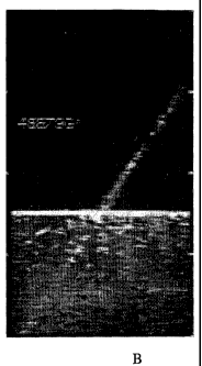
[0037] Figs. 1A and 1B illustrate the enhanced echogenicity of a wire coated
according to the
invention. Figure 1A shows an ultrasound image of a phantom model of blood and
liver, with
an uncoated wire inserted (not visible). Figure 1B shows an ultrasound image
of a wire with an
echogenic coating according to the invention.
[0038] Fig. 2 is a light microscopic image (100X) showing a needle with an
echogenic coating
formed from isocyanate.
[0039] Fig. 3 is an electron micrograph at SOOx magnification showing the same
type of coating as
in Fig. 2, with 30 to 70 micron cavities and 1 to 10 micron pores.
[0040] Fig. 4 is a fight microscopic image (100X) of a needle with a channel
coating formed from
a sonicated albumin solution.
[0041] Fig. 5 illustrates the enhanced echogenicity of a coated 22 gauge
needle in a New Zealand
White rabbit kidney as imaged by a Shimadzu SDU-350A ultrasound system with a
7.5 MHZ
probe. The left column for each rabbit shows the visual rating score for
coated needles
according to Example 1 and the right column shows the much lower rating for
uncoated needles.
[0042] Figs. 6A and 6B further illustrate the enhanced echogenicity of a 22
gauge needle coated
according to the invention. Figure 6A shows an ultrasound image of a breast
phantom with a
simulated cyst. An uncoated needle is not visible. Figure 6B shows the
corresponding
ultrasound image of a needle with an echogenic coating according to the
invention.
_g_
CA 02536947 2006-02-24
WO 2005/020905 PCT/US2004/027458
DETAILED DESCRIPTION OF THE PREFERRED EMBODIMENTS
[0043] In describing preferred embodiments of the present invention
illustrated in the drawings,
specific terminology is employed for the sake of clarity. However, the
invention is not intended
to be limited to the specific terminology so selected, and it is to be
understood that each speciEc
element includes all technical equivalents which operate in a similar manner
to accomplish a
similar purpose. All articles and patents referred to in this application are
incorporated herein by
reference as if each were individually incorporated by reference.
[0044] Echogenicity is the result of backscatter (180 degree reflection back
to the transducer)
caused by a difference in acoustical impedance. The greater the impedance
differential
(mismatch), the greater the echogenicity (backscatter).
[0045] Acoustic impedance of a material decreases as compressibility increases
and as density
decreases. Thus, solids have the highest impedance because they are
uncompressible and dense.
Gases have the lowest impedance because they are compressible and not dense.
Liquids fall in
between. Solids impede sound beams more than liquids, by up to about one order
of magnitude,
and liquids impede sound beams better than gases, by several orders of
magnitude. Thus, an
interface between a solid and a gas produces the highest possible acoustic
impedance mismatch,
due to differences in compressibility and density. Interfaces between
different types of solids,
semi-solids, liquids, and gases can also contribute to echogenicity to a
lesser but significant
degree.
[0046] The inherent acoustic impedance of a coating is very difficult to
measure. However, the
following table demonstrates the vastly different impedance (proportional to
the product of
density and compressibility) for gases compared with liquids and solids. Small
differences from
one gas to another or one solid to another makes very little difference on a
log scale. However,
the orders of magnitude difference between a solid and a gas is easily
distinguished on the (log)
echogenicity scale. This huge difference in acoustic impedance between a gas
and a solid
accounts for one aspect of the advantages of the present invention.
-9-
CA 02536947 2006-02-24
WO 2005/020905 PCT/US2004/027458
Table 1
Material CompressibilityDensity
* (g/cc)
(cm/dyne)
Air 2.3 X 10~ 1.29 X
10-3
Water 4.6 X 10⁻¹¹ 1.00
Erythrocyte3.4 X 10-11 1.09
Aluminum 1.3 X 10⁻¹² 2.7
Nickel 5 X 10-13 8.8
Sources: CRC Handbook of Chemistry and Physics, 64th Edition, R.C. Weast, ed.
(CRC Press,
Inc. Boca Raton, FL 1984); Perry's Chemical Engineers' Handbook, 6th Edition,
D.W. Green,
ed. (McGraw-dill 1984); Practical Handbook of Materials Science, C.T. Lynch,
ed. (CRC Press,
Inc., Boca Raton, FL 1989).
[0047] A comparison of impedances for some common materials demonstrates that
most materials
are at most one order of magnitude different from water (or tissue) except
gases (represented by
air)which are several orders of magnitude different in impedance.
Table 2
Material Characteristic
impedance
c.g.s. Rayl. x
10-5
(g. cm 2 sec 1)
X 10-5
Non-biologicalAir at S.T.P. 0.0004
Castor oil 1.43
Water 1.48
Polythene 1.84
Perspex 3.20
Aluminium 18.0
Mercury 19.7
Brass 38.0
Biological Aqueous humour of 1.50
eye 1.52
Vitreous humour
of eye
-10-
CA 02536947 2006-02-24
WO 2005/020905 PCT/US2004/027458
Brain 1.58
Blood 1.61
Kidney 1.62
Human tissue, mean 1.63
value
Spleen 1.64
Liver 1.65
Muscle 1.70
Lens of eye 1.84
Skull-bone 7.80
Fat 13.8
Source: Table 1.4 from Wells, Physical Principles of Ultrasonic Diagnosis
(Academic Press
London, 1969).
[0048] The acoustic impedance differential (or mismatch) between two objects
is given here as a
factor reflecting the impedance of the object having the higher impedance
divided by the
impedance of the object having the lower impedance. Coatings according to the
invention
preferably provide acoustic impedance differentials at echogenic interfaces of
at least about a
factor of 3, more preferably about a factor of 10, yet more preferably at
least about a factor of 25
(the difference between brass and water), most preferably more than about a
factor of 100.
[0049] An echogenic coating according to the invention is a complex structure
that may have
one or a combination of several physical forms. It is a coating, a material
that forms a thin
essentially continuous layer over the substrate, and could be referred to as a
film. It may be a
complete solid mixture of polymers, and includes intrinsic acoustically
reflective echogenic
features such as gaseous spaces. The spaces may present some discontinuity in
the film without
detracting from the adhesion of the film to the substrate.
[0050] A film-forming component according to the invention is a polymer or
polymer forming
material or similar material that may be dissolved or suspended in a coating
liquid, such that
when the coating liquid is applied to a substrate, the elm-forming component
forms a suitable
thin layer or film upon evaporation of the solvent or suspending liquid. Many
examples are
-11-
CA 02536947 2006-02-24
WO 2005/020905 PCT/US2004/027458
provided. The thin layer or film may be said to comprise a solid matrix of the
dried and/or
reacted film-forming component. The solid matrix of the film has echogenic
features at the
surface or within the matrix.
[0051 ] An echogenic feature of a coating according to the invention means a
structure that is
acoustically reflective in ultrasound applications and thereby increases the
visibility of the
coating and coated objects. Reflective echogenic features according to the
invention may
include gas bubbles, irregular gas pockets, cavities, pores, closed gas-
containing channels, or
microscopically raised and depressed surface regions of the polymer matrix
presenting an
irregular relief capable of trapping air when inserted into a tissue.
[0052] The coating mixture may include additives, and solvent residues blended
together.
Alternatively, the coating may be a complete solution, defined as a mixture
uniformly dispersed
throughout the solid phase with homogeneity at the molecular or ionic level,
or it may be a
combination of dissolved and mixed components, such as a mixture of a polymer
coating
solution and gas bubbles. The coating may take the form of a composite,
defined as a structure
composed of a mixture or combination of polymer and gas bubbles. It may be a
blend, that is a
mixture so combined as to render the components indistinguishable from each
other. It may also
be referred to as a matrix of polymer in which gas bubbles and other
constituents and structures
are dispersed. The coating overall may comprise separate layers, discrete or
intermingled, each
of which may have any or several of these forms.
[0053] The term ultrasound is intended to encompass presently known or
subsequently
developed vibrational signals such as an acoustical signal or beam used to
generate a signal
based on reflection. Utrasound technologies available currently generally have
resolution larger
than that of red blood cells having a diameter of 5 to 10 microns. However,
resolution as low as
about 0.5 microns may be possible, so gas spaces that small or even as small
as 0.01 microns
may be desirable according to the invention. Depending on the coating
structure, gas spaces as
big as 300 microns may be useful to provide enhanced echogenicity. In most
cases, however
gas spaces with a diameter of greater than 1 micron, preferably about 5 to
about 10 microns, up
to about 20 microns, are satisfactory. These are suitable in coatings with an
average thickness of
about 10 to 50 microns. Larger spaces may be appropriate in thicker coatings
up to several
hundred microns thick.
[0054] Increased echogenicity according to the invention may be due to bubbles
or small craters
of these general sizes, preferably about 1 to 10 micron diameter. The
microscopic appearance of
-12-
CA 02536947 2006-02-24
WO 2005/020905 PCT/US2004/027458
these coatings, showing the bubbles and craters, is shown in Figures 2 and 3.
In a thin coating,
despite such craters, the coating still feels smooth. In a thick coating, with
higher relief, the
surface may become rough, so conditions should preferably be controlled to
minimize surface
relief so as to produce a smooth coating for medical applications. For
example, drying a viscous
bubble containing polymer solution at high temperature may lead to a rough
coating. On a
macroscopic scale the coating should remain smooth for medical devices to
avoid discomfort to
the patient and misinterpretation by a physician when attempting to place the
device in a target
site.
[0055] As used here, smooth means sufficiently smooth so as not to cause
difficulty in inserting
a coated object into a target material, which may be determined by a person of
ordinary skill. A
reasonable predictor of smoothness in use is to rub a forger along the surface
of the coating and
determine whether it feels smooth or rough. Smoothness to the touch is
generally adequate
according to the invention.
[0056] When an echogenic surface having small irregularly shaped craters or
pores is
submerged in an aqueous (body) fluid, surface tension prevents these small
craters from being
filled in by water and air may be entrapped on the surface of the device.
Although larger and
rounder or more regular cavities may be present as well, they are more likely
to fill with water ,
leaving a lower level acoustic impedance differential. This feature applies
when the coating is
placed in solid or semi-solid tissue or material, forming a gas/liquid
interface if physiological
fluid contacts the coating, a semi-solid/gas interface if a gel material (such
as coupling gel)
contacts the coating, or a solidlgas interface if solid material contacts the
coating. The
echogenic fethers are stable under conditions of use such as soaking, rubbing,
pressure changes,
and agitation. The coating provides enhanced echogenicity in each of these
situations.
[0057] Thus, in one embodiment of the invention, to take advantage of acoustic
impedance
differential, gas bubbles are entrapped in a smooth thin coating which can be
applied to virtually
any biomedical device. Gas bubbles are desirable to provide an acoustic
impedance mismatch
much greater than could previously be obtained. Generally, in such an
embodiment, craters and
pores are formed at the surface of the coating, and contribute to the
echogenicity of the coating.
[0058] Another embodiment of the invention comprises channels in discontinuous
matrices in a
coating, which also provides a desirable acoustical impedance mismatch and
improved
echogenicity according to the invention. An irregularly shaped channel is
desirable as it may
trap air in pockets and may also have interfaces between materials having
mismatched acoustic
-13-
CA 02536947 2006-02-24
WO 2005/020905 PCT/US2004/027458
impedance to provide echogenicity. The size of the channels should be in the
same general
range as with bubbles and cavities, and should not be so great as to cause
discontinuities in the
coating such that the coating would peel off. Thus, a continuous air channel
or layer would not
be desirable according to the invention.
[0059] The polymer components of the coating may be precipitated together in
such a way as to
generate intrinsic acoustically reflective interfaces, such as solid/gas
interfaces. This approach is
advantageous in that no extraneous solids need to be added to the coating to
provide the
acoustically reflective interfaces.
[0060] The echogenic coating liquid formulations of the invention comprise an
organic solvent
and a polymer system adapted to produce acoustically visible structures when
coated on a
substrate. The polymer system may be a polymer and bubbles having a diameter
between about
1 micron and about 50 microns; a reactive polymerizing monomer that generates
gas during
polymerization; or a polymer solvent/non-solvent mixture wherein the
concentration of solvent
is sufficiently high to dissolve the polymer in the coating liquid, and the
concentration of non-
solvent is sufficiently high to cause the polymer to precipitate during
evaporation of the organic
solvent from the coating liquid entrapping gaseous bubbles with the
precipitate. Such a coating
liquid may be a complete solution, meaning a mixture uniformly dispersed
throughout the liquid
phase with homogeneity at the molecular or ionic level, or it may be a mixture
of a polymer
solution and a gas phase, and possibly gas containing particles dispersed as a
suspension.
[0061] Echogenic coatings can be prepared using a wide variety of polymers.
Bubble trapping
polymers include cellulose esters, polyurethanes, albumin, other proteins,
polyvinyl pyrrolidones
and others that are known to those skilled in the art to be capable of
trapping small bubbles.
Generally a viscous preparation is preferred. Preformed bubbles such as gas
entrapped in
albumin microspheres (for example Albunex~ spheres, Molecular Biosystems,
Inc.) may be
used. Polymer-forming materials that generate gas include isocyanate
prepolymers and diazo
compounds. Preferred materials are isocyanates and albumin.
[0062] The following gas-producing compounds could be used in the practice of
this invention:
polyisocyanates, such as polymethylene polyphenylisocyanate, 4, 4 -
diphenylmethane
diisocyanate, and 2,4-toluene diisocyanate, sodium carbonate, sodium
bicarbonate, aromatic
diazonium salt stabilized compounds, prepolymers or other addition- compounds
such as
oligorners or cooligomers of isocyanates wherein the isocyanate is selected
from toluene
diisocyanate, hexamethylene diisocyanate, cyclohexane diisocyanate, isophorone
diisocyanate,
-14-
CA 02536947 2006-02-24
WO 2005/020905 PCT/US2004/027458
4, 4' diphenylmethane diisocyanate, or a prepolymer, such as trimerized
hexamethylene
diisocyanate biuret. Such prepolymers are available under trade names such as:
Desmodur
(Bayer AG), Tycel (Lord), Hypol (Hampshire), Andur (Anderson Developer
Company), Papi
and Voranate (Dow Chemical Company).
[0063] The isocyanate component of the coating liquid is preferably in a
concentration of between
about 20% and about 40%, and the solvent system for it preferably comprises
about 15 to about
48% dimethylsulfoxide, up to about 35% tetrahydrofuran, up to about 30%
toluene, up to about
32%cyclobexanone, and suitable amounts of hexane, 2-butanone, xylene, ethyl
acetate,
dichloromethane, 1,1,1-trichloromethane, n-methylpyrrofidone, and n-butyl
acetate.
[0064] Solvents for the echogenic, coating layer include ketones, esters,
aromatics, lactones,
amides, halogenated hydrocarbons, alcohols, amines and other common solvents.
Any solvent
system that is capable of dissolving all constituents of the coating into a
homogeneous solution
may be selected. Preferred solvents are tetrahydrofuran, dimethylsulfoxide,
and acetone. For
precipitated coatings, preferred non-solvents include ethanol, isopropanol and
water.
[0065] In most cases, air and gases such as carbon dioxide and nitrogen
generated in situ are
adequate to effect echogenicity enhancement and offer low cost and simplicity
of manufacture.
Any gas could be used, and further echogenicity enhancement might be obtained
using gases
which are more highly compressible, less dense, and are less likely to
dissipate when drying the
coating and are less soluble or slower diffusing in physiological solutions.
[0066] Substrates to which echogenic coatings according to the invention rnay
be applied include
metals such as stainless steel, nickel, gold, chrome, nickel titanium alloy,
platinum and others;
plastics such as silicone, polyurethane, polyethylene, polyamide,
polyvinylchloride, latex and
others; drug particles; and capsules. Preferred devices include needles,
guidewires, catheters,
surgical instruments, equipment for endoscopy, wires, stems, angioplasty
balloons, wound
drains, arteriovenous shunts, gastroenteric tubes, urethral inserts,
laparoscopic equipment,
pellets, or implants.
[0067] Echogenic coatings according to the invention may be applied to a
device as one or more
layers. In a simple embodiment, a matrix polymer is dissolved in an organic
solvent and aerated
to produce a coating liquid having bubbles of the desired size. The coating
liquid is applied to
the substrate, and dried rapidly enough to set the bubbles in place, to
provide an echogenic
coating. However, substrates such as metal guidewires, stainless steel
needles, and silicone,
-15-
CA 02536947 2006-02-24
WO 2005/020905 PCT/US2004/027458
polyethylene, and nylon catheters, and other polyolefin and polyarnide
substrates may not allow
for adequate adhesion of the echogenic layer.
[0068] In a multilayer embodiment, the surface is first treated to enable
strong adhesion of the
echogenic coating to the device. This treatment may include applying a first
layer, or pre-coat
and a second layer, or base coat, if necessary, to effect adhesion of the
echogenic coat to the
surface. The active echogenic layer containing echogenic interfaces is then
applied over the
dried base coat to enhance the echogenicity of the device. Optionally, a
fourth layer, a finish or
top coat, may be applied to improve the durability of the echogenic coating
against abrasive
forces, to enhance lubricity, provide a smoother surface, to protect the:
echocoat from
deleterious effects of exposure to body fluids, or to incorporate
pharmaceutical agents such as
antithrombogenics, antibiotics, andantimicrobials, or to impart other
desirable properties,
according to methods known in the art. For example, lubricious coatings are
described in U.S.
patent no. 5,331,027, and coatings comprising pharmaceutical agents are
described in U.S.
patent no. 5,525,348. A top coat may also be used to reduce the wetting of the
echogenic;
coating layer. Wetting of the echogenic layer should be avoided as
echogenicity may decrease if
this echogenic coating swells excessively and releases the otherwise entrapped
gas.
Additionally, wetting may cause gas entrapped in surface pores or cavities to
be released.
Enhanced mechanical stability can be useful for applications in which the
device may be
subjected to high shear forces as may occur if a coated device is forced to
move against a bone.
[0069] Pre-treatment of the substrate surface may not be required on a
substrate if the adhesion
between the device surface and the echogenic layer is adequate. Alternatively,
a single sub-layer
rather than a pre-coat and base coat may be adequate depending on the degree
of adhesion
between the coating and the device.
[0070] In a pre-coat, useful polymers include acrylics, acrylic copolymers,
polyolefin copolymers,
polyethylene/polyacrylic acid copolymer, chlorinated polyolefin polyacetals,
epoxies, mixtures
and others known to one skilled in the art. Adhesion on polyolefm surfaces is
improved by
using polyolefin acrylic copolymers, and adhesion on silicone polymers is
improved by using
precoat layers which contain polydimethylsiloxane polymers.
[0071] In a base coat, typical polymers include polyurethanes, cellulose
esters, acrylics, acrylic
copolymers, polyacetals, epoxies, others, and mixtures of the above. An
example of a base coat
includes cellulose ester, an acrylic polymer, a polyurethane, and 2-hydroxy-4-
methoxy
-16-
CA 02536947 2006-02-24
WO 2005/020905 PCT/US2004/027458
benzophenone dissolved in a mixture of, solvents including tetrahydrofuran,
cyclohexanone, and
ethyl acetate.
[0072] In a top coat, preferred polymers are hydrophobic polymers such as
polyvinylbutyral,
polyacetals, acrylics, acrylic copolymers, vinyls and others known to one
skilled in the art.
Also, one or more top coats) also may be applied to further improve the
surface smoothness of
the coated device. Polymeric solutions of the appropriate viscosity will flow
into crevices which
may have developed during drying of the echogenic coating and produce a very
smooth outer
surface in the process. Such top coat compositions can include hydrophobic
polymers or hybrid
polymers such as polyvinylacetate, cellulose esters, vinylacetal polymers,
acrylates,
polyurethanes, epoxies, and others. Although the crevices may be filled, there
may still be
solid/gas interfaces sufficient to improve the ultrasound visibility of the
coated device.
[0073] In general, the layers of an echogenic coating may be applied as
polymers dissolved in a
solvent by dipping, spraying or other methods known to those skilled in the
art. Other
compositions will occur to those skilled in the art to improve adhesion on
these or other
substrates as needed.
[0074] The echogenic coating may be referred to as the ultrasonically active
layer and contains
interfaces between different materials, such as between a solid polymer phase
and a gas (air,
carbon dioxide, water vapor, an inert gas, or otherwise). Alternatively, the
active layer contains
echogenic interfaces between different portions of the coating, such as with
the interface
between a solid and a gas internally within the coating layer, or at the
exterior surface of the
product. In general, the most important portion of the coating for
echogenicity is at or near the
surface, and features well beneath the surface are less important. Hence, the
active echogenic
layer coat composition is preferably designed to efficiently entrap gas
bubbles or matrices
containing gas bubbles.
[0075] In one embodiment of the invention, advantages may be found in using
polymer coatings
containing various materials in addition to gas (such as solids, suspensions,
and liquids).
Differences in echogenic responses are known to exist between various
materials, such as
different portions of the coating having a different blend of polymer and
additives, or shapes
such as folds, flaps, or contours of the polymer surface, but most strongly
between gases and
solids or gases and liquids. Surface irregularities, which can be completely
bound within a
coating or on the exterior surface, can further enhance the differences in
echogenic response, and
improve scattering. Thus, in certain embodiments, this invention incorporates
microscopic
-17-
CA 02536947 2006-02-24
WO 2005/020905 PCT/US2004/027458
surface irregularities and/or different materials in coatings for application
to materials, to
achieve enhanced echogenicity.
[0076] In another embodiment, echogenic active coating layers may consist of
one or more
polymer or mixed (hybrid) polymer layers which are applied to a substrate or
pre-coated
substrate as a coating liquid comprising the polymers in a solvent system. The
polymer layer,
once applied, may then be exposed directly to a non-solvent liquid for the
polymer thereby
causing the polymer component to precipitate. This can be done by using a
mixed solvent
system comprising a solvent for the polymer and a liquid that is a non-solvent
for the polymer.
In such cases, the non-solvent liquid may be selected so that it evaporates
more slowly than the
polymer solvent component. Thus, the solvent system changes from one which can
dissolve the
polymer to a mixture which is sufficiently rich in the non-solvent component
so that the polymer
component precipitates onto the substrate. Selected polymers precipitated in
this fashion have
desirable features such as the ability to trap air or gas during the
precipitation process or
otherwise provide an echogenically active layer.
[0077] Such precipitated layers may also be created by casting the polymer
layer to precipitate.
For instance, a water insoluble polymer, dissolved in an organic solvent, may
be cast on a
surface and, while still wet with solvent, be immersed in water or exposed to
water vapor such
as steam to precipitate the polymer layer, or other treatment that leads to
precipitation of the
polymer from solution entrapping gaseous spaces in an irregular fashion. The
irregularity
distinguishes this type of coating from the typical type of polymer coating
which is a continuous
dried, cured film formed by evaporation of the solvent from a polymerlsolvent
solution, and is
transparent and clear. In this embodiment, the curing process is interrupted
by an aqueous
precipitation step that causes the polymer component to fornl a solid having a
different, irregular
structure, one that is white, and reflects light in all directions. The gas
entrapping irregularities
due to precipitating the polymer coating during formation provide an enhanced
echogenic
response.
[0078] In a third embodiment, it is possible to incorporate polymer components
which are capable
of producing gas vapor bubbles upon treatment. Such bubbles can then be
trapped in the
polymer layer. It is also possible to create bubbles in the liquid polymer
solution before casting
the layer. The resulting layers are found to have echogenic responses which
are significantly
different than typical body tissues such that objects coated with them are
rendered more readily
apparent against typical body tissues. One example comprises:
-18-
CA 02536947 2006-02-24
WO 2005/020905 PCT/US2004/027458
a) providing an echogenic coating solution including an isocyanate polymer in
an
organic solvent;
b) applying the echogenic coating solution to a device by dipping or spraying;
c) drying the device coated with the echogenic coating solution for a few
minutes at
room temperature to remove some of the organic solvent;
d) exposing the coated device to water to generate bubbles in situ; and
e) drying the coated device to remove the remaining solvent.
[0079] The reaction of the isocyanate is:
R1NC0+HZO ~ R,NHZ+COZ (slow)
RINHZ+R1NC0 ~ R~ureaR2 (fast)
This method is distinct from prior art methods of using isocyanate to form a
hydrogel as in
Lambent, U.S. 4,585,666. Here, reaction conditions are adjusted to maximize
generation of gas
and entrapment of bubbles within the solid matrix of the coating film. For
example, the reaction
is performed at or near room temperature to prevent gas from escaping too
quickly. In the
Lambent method, the reaction is typically run at an elevated temperature and
with hydrophilic
components such as polyvinylpyrrohdone such that gas is not entrapped. Thus,
an aspect of the
invention is to react the reactive component under temperature and humidity
conditions that
generate and entrap echogenic quantities of gas within the coating film, and
without components
that would interfere in such a process.
[0080] A diazonium salt reacted with ultraviolet light to produce nitrogen is
another example of
this approach. An acid base reaction such as with bicarbonate of soda may be
used. Other ways
to generate gas in situ will be evident to a person of ordinary skill.
[0081] Another embodiment comprises preparing a viscous composition, of e.g.
acrylic latex
polymer, polyvinylpyrrohdone, albumin or other polymer; sonicating with enough
energy to
produce a coating liquid containing bubbles of 5 to 20 micron diameter;
immediately coating the
device with this echogenic coating solution by dipping or other means known to
one skilled-in-
the-art; then drying at, for example, about 80o C or less to harden the
coating without
destabilizing the bubbles. For a given mixture, the time and parameters for
sonication may
readily be determined empirically, e.g. by observing samples under a
microscope, and once
suitable conditions are determined, these may be used repeatedly and
reproducibly. Other
-19-
CA 02536947 2006-02-24
WO 2005/020905 PCT/US2004/027458
means may be used to generate bubbles, too, such as shaking, mixing, blending,
and so forth, so
long as the desired bubble size is achieved.
[0082] Yet another embodiment is to prepare a bubble-containing echogenic
coating solution as in
the preceding paragraph, and apply it to a substrate. The coated device is
then dried slowly at
room temperature or even colder. As a result of the low temperature drying,
bubbles escape and
collapse, and the dried coating appears to consist of a matrix of channels of
5 to 20 microns
width and varying length. Such a coating is shown in Figure 4. These channels
may contain
trapped air, and so enhance the echogenicity of the device. Also, the
irregular shape of the
coating may contribute to increased acoustic impedance differential and
increased scattering.
[0083] A different embodiment involves selective extraction. The method
involves forming a
layer of two components, a matrix component having a relatively lower
solubility in an
extraction solvent and an extractable component with a relatively higher
solubility in the
extraction solvent. The matrix component must have good adhesion to the
substrate after being
deposited. Preferably the matrix component is nitrocellulose, the extractable
components are
camphor and dibutylphthalate, and the extraction solvent is isopropanol. After
forming the film,
the extractable component is selectively extracted in the extraction solvent,
leaving the matrix
component behind as a solid matrix film containing voids. Upon drying, such a
layer is
echogenic. Similar results may be achieved by chemical or physical etching of
a coating
surface.
[0084] Echogenicity can be measured by imaging a device against a suitable
background -- water,
tissue -- and subjectively evaluating image optical densities. Figures 1A and
1B show the
enhanced echogenicity of a coated wire according to the invention using a
water-immersed
"phantom" which simulates the echogenicity of various tissues and is designed
for reproducible
placement of the coated device being tested. The images are qualitatively
reproducible.
[0085] To use this phantom, a sample (such as a needle, wire, catheter) is
inserted into a 2 mm 20
diameter hole drilled into the phantom and off axis from the ultrasound beam.
An image is
produced using an ultrasound transducer focused on the control material in the
phantom.
[0086] As evidenced by Figure 1A, an uncoated wire is not visible at all as
sound waves are
reflected away from the transducer. The bright area on the bottom half of the
image is a plastic
composite material which simulates the echogenic characteristics of liver
tissue. The darker area
on the top of the image is water, which has an echogenicity similar to that of
blood. The wire is
inserted diagonally into a hole drilled in the phantom.
-20-
CA 02536947 2006-02-24
WO 2005/020905 PCT/US2004/027458
[0087] Figure 1B, shows a wire coated with an isocyanate-based coating
according to the
invention in the same phantom. The wire coated according to the invention is
plainly visible
against the water phase, and it is plainly visible against the phantom. This
type of coating had
the best visibility among those tested. These results indicate that the
coating would be visible in
blood and in tissue of similar ultrasound qualities to liver. The uncoated
wire, Figure 1A, is
completely invisible under these imaging conditions.
[0088] These results were confirmed by imaging in a breast phantom and rabbit
kidneys. As
shown in Figure 6A, an uncoated 22 gauge needle is virtually invisible in a
breast phantom
having a simulated cyst (see "uncoated needle" and "Simulated cyst"), while a
similar needle
coated as in example 1 is readily visualized under identical conditions (see
"Needle coated with
ECHO-COATTM). These results were further confirmed when uncoated and coated
needles
were imaged in rabbit kidneys. See Figure 5. Figure 5 shows the average
ratings of three
independent observers of five needles per group, one kidney imaged in each
rabbit. Only the tip
of the uncoated needle may be discerned while almost the entire shaft of the
coated needle is
visible as well as the tip.
[0089] Imaging was done with a Quantum Quad 2000 imaging system at 7.5 MHz, or
a Shimadzu
SDU-350A system with a 7.5 MHz 150 degree convex probe, although other
devices, lower
frequencies, and other settings may be used.
[0090] The microscopic appearance of gaseous coatings according to the
invention is shown in
Figure 2, a light micrograph at a magnification of 100X, and Figure 3, an
electron micrograph at
SOOx magnification. The coatings had a thickness of about 5 to about 20
microns. The coating
has prominent crater like cavities about 30 to 70 microns in diameter at the
surface. The cavities
are not very deep, and the coating feels very smooth.
[0091] Electron microscopy reveals that this coating also contains a large
number of smaller (1 to
micron diameter) cavities and/or bubbles as shown in Figure 3. Without
intending to limit
the scope of the invention, it is believed that these smaller gaseous spaces
contribute more to the
increased echogenicity since surface tension may preclude their being filled
by water when the
device is inserted into a body fluid, whereas the larger cavities are more
easily wetted and filled.
With an embodiment of the invention having channels as in Figure 4, ultrasound
imaging with a
phantom as described above shows an improvement over the signal of an uncoated
wire as in
Figure 1A. This improvement is not as great as with the coatings formed with
isocyanate. The
microscopic appearance of coatings according to this second embodiment reveals
long irregular
-21-
CA 02536947 2006-02-24
WO 2005/020905 PCT/US2004/027458
grooves estimated to be about 30 microns wide and 100 to 300 microns long.
These grooves
apparently are not deep since the coatings feel quite smooth. Such coatings
can be made by
sonicating an albumin solution, coating, and drying at room temperature.
[0092] A precipitated coating also provides an ultrasound image that is an
improvement over the
uncoated wire shown in Figure 1A. The microscopic appearance of a precipitate
coating appears
to be quite smooth, but surprisingly provides improved echogenicity.
[0093] Coatings according to the invention are biocompatible, thin, smooth,
and adhere strongly
to substrates. They are stable in that they retain echogenicity for an
extended period suitable for
medical procedures, preferably at least one minute, more preferably at least
five minutes, most
preferably at least an hour. Preferably, they may be used repeatedly over two
or more hours
without substantial loss of echogenicity.
[0094] Such coatings improve the safety and efficacy of numerous procedures.
For example,
physicians can accurately place coated cardiovascular stems and monitor any
migration by
ultrasound imaging. Coated biopsy needles are easier to place accurately in a
lesion, improving
the diagnostic value of the samples obtained. Amniocentesis can be conducted
more safely by
visualizing the sample needle as well as the fetus. Cutting devices may be
accurately placed
under ultrasound guidance to improve Laparoscopic surgical procedures. The
invention also has
application in non-medical fields as will be apparent to those of ordinary
skill familiar with the
use of ultrasound.
[0095] Open gas-trapping structures that have entrapped air bubbles provide
excellent ultrasound
signal back to the transducer regardless of the angle of the incident beam.
This signal, however,
can degrade with time in an aqueous environment as water or other ambient
liquid gradually
displaces the gas that has been trapped in these structures. Accordingly, an
embodiment of this
invention comprises a very thin topcoat, for~example on the order of
approximately one micron
in thickness that is so flexible that the compressibility of the entrapped gas
is not decreased by
any significant amount, e.g. less than 40 percent, and preferably less than
30, 25, 20, 10, or 5
percent reduction in echogenicity. Such top coatings retain high echogenicity
while
significantly enhancing the longevity of this echogenicity in a fluid
environment. Applying a
relatively inflexible coating over 5 microns in thickness results in a severe
degradation of the
echogenicity and is of little practical value. Overcoats of under about 5
microns, and especially
under 3, 2, or 1 microns, are preferred.
-22-
CA 02536947 2006-02-24
WO 2005/020905 PCT/US2004/027458
[0096] Other advantages of applying a very thin topcoat over the echogemc
layer are to increase
the durability, enhance lubricity, provide a smoother surface, protect the
echogenic layer from
deleterious effects of exposure to body fluids, to incorporate pharmaceutical
agents such as
antithrombogenics, antibiotics, or antimicrobials, or to impart other
desirable properties to the
device surface. Polymers that may be used include those known to one skilled
in the art as
having a flexural modulus in excess of 500 psi and preferably in excess of
1000 psi, elongation
at break greater than 100 percent and preferably greater than 200 percent and
low water
permeability.
[0097] Examples of materials that can be used to form a thin flexible topcoat
that does not
significantly degrade the echogenicity include polyethylene, certain
ethylene/vinyl acetate
copolymers, certain epoxy type resins, polydimethylsiloxane,
polytetrafluoroethylene,
polyvinylbutyral, polyvinylidinechloride and certain polyurethanes,
polyimides, rubbers,
acrylate polyrners/copolymers, polybutadiene, styrene butadiene and styrene
butadiene/styrene
copolymers, and others having the desired characteristics. With any of these
polymers, the film
can be applied as a coating so that it becomes sufficiently thin and flexible
so as to minimize any
effect on compressibility of the entrapped gas.
[009] Prolonging echogenicity is especially useful when the coatings are
applied to long-term
indwelling devices such as stems, central venous catheters, peripherally
inserted catheters,
grafts, shunts, percutaneous transcardiac arterial catheters, and implants
for, e.g., drug delivery
devices. Other devices that may be long indwelling may be selected from the
group consisting
of a catheter, needle, hydrocephalus shunt, draintube, pacemaker, dialysis
device, small or
temporary joint replacement, urinary sphincter, urinary dilator, long term
urinary device, tissue
bonding urinary device, penile prosthesis, vascular catheter port,
peripherally insertable central
venous catheter, long term tunneled central venous catheter, peripheral venous
catheter, short
term central venous catheter, arterial catheter, PCTA or PTA catheter, and
pulmonary artery
Swan-Ganz catheter, guidewire, a surgical instrument, endoscopy equipment, an
angioplasty
balloon, a wound drain, a gastroenteric tube, laparoscopy equipment, a pellet,
and an implant
and combinations thereof.
[0099] Long indwelling will typically mean a prolonged period, at least about
two hours, at least
about one or two days, at least about one week, at least about a month, or
longer.
[0100] In these embodiments, an ultrasonically visible solid device for
inserting into a target
medium comprises a surface and a matrix, wherein the surface has gas-trapping
structures
-23-
CA 02536947 2006-02-24
WO 2005/020905 PCT/US2004/027458
capable of entrapping gas when the device is in the target medium, the
entrapped gas causing the
device to be ultrasonically visible, and the gas-trapping structures are
covered with a thin,
flexible overcoat that does not significantly reduce the compressibility of
the gas trapped in
these structures but does improve the echogenic coating durability, enhance
lubricity, provide a
smoother surface, protect the echogenic layer from deleterious effects of
exposure to body
fluids, or incorporates pharmaceutical agents such as antithrombogenics,
antibiotics, or
antimicrobials, or imparts other desirable properties to the device surface.
[0101] In exemplary embodiments, the overcoat layer has a thickness of between
0.1 and 2
microns, or between 0.1 and 1 micron, or between 0.5 and 1.5 microns, with
examples greater
than 0.1 , 0.2, 0.5, or 1 microns, and less then 0.5, 1, 1.5, 2, 3, or 5
microns thick. The overcoat
may have a flexural modulus greater than 500 psi, or greater than 1000 psi,
and an elongation at
break greater than 100 percent, or greater than 200 percent.
[0102] In further embodiments the overcoat layer results in a decrease of less
than about 10
percent or less than about 20 percent or about SO percent in the
compressibility of the entrapped
gas bubbles. In further embodiments, the overcoat layer has a water
permeability of less than
about 10-10 to about 10-11 [(cm3)(cm)]/(cm2)(s)(cm Hg)].
[0103] In methods according to the invention, a film provided with means for
trapping gas at the
surface is over-coated with another thin, flexible film or top coat in such a
way that the gas
bubbles in the surface pores are trapped underneath the coating, and such a
top coating improves
the echogenic coating longevity and durability, enhances lubricity, provides a
smoother surface,
protects the echogenic layer from deleterious effects of exposure to body
fluids, or incorporates
pharmaceutical agents such as antithrombogenics, antibiotics, or
antimicrobials, and/or imparts
other desirable properties to the device surface. The invention also provides
a coating liquid to
be applied as a thin coating over the echogenic layer, and entrapping bubbles
within the surface
pores, comprising one or more film-forming components.
[0104] The film-forming components) comprises polyethylene, certain
ethylene/vinyl acetate
copolymers, certain epoxy type resins, polydimethylsiloxane,
polytetrafluoroethylene,
polyvinylbutyral, polyvinylidinechloride, certain polyurethanes, polyimides,
rubbers, and/or
acrylate polymerslcopolymers.
[0105] Devices with echogenic surfaces according to the invention may also
have one or more
active agents selected from one or more of the following components. They can
include anti-
thrombogenic agents, anti-inflammatory agents, antineoplastic agents, anti-
proliferative agents,
-24-
CA 02536947 2006-02-24
WO 2005/020905 PCT/US2004/027458
cytostatic agents, cytotoxic agents, antimicrobial agents, anti-restenotic
agents, anti-platelet
agents, and anti-coagulant agents. The active agent may be selected from one
or more of anti-
fibrin and fibrinolytic agents, anti-platelet agents, prostacyclins (and
analogues), glycoprotein
IIb/IIIa agents, thromboxane inhibitors, anti-thrombin and anti-coagulant
agents, anti-mitotic,
antiproliferative and cytostatic agents, antiangiogenic and angiostatic
agents, ACE inhibitors,
growth factor antagonists, antioxidants, vitamins, calcium channel blockers,
fish oil (omega 3-
fatty acid), phosphodiesterase inhibitors, nitric acid donor, Somatostatin
analogues,
immunosuppresives and antiinflamatory agents, antimicrobials, radionuclides
including alpha,
beta and gamma emitting isotopes, COX-2 inhibitors, endothelial promoters,
kinase inhibitors,
epidermal growth factor kinase inhibitors, tyrosine kinase inhibitors, MAP
kinase inhibitors, and
protein transferase inhibitors. The active agent is selected from one or more
of plasmin,
streptokinase, single chain urokinase, urokinase, t-PA (tissue type
plasminogen activator),
aminocaproic acid, aspirin, monoclonal antibodies, peptides, ReoPro,
Cilastagel, eptifibatide,
tirofiban, ticlopidine, Vapiprost, dipyridamole, forskolin, angiopeptin,
argatroban, dextan,
heparin, LMW heparin, Enoxaparin, Dalteparin, hirudin, recombinant hirudin,
anti-thrombin,
synthetic antithrombins, thrombin inhibitors, Warfarin, other coumarins,
vincristine, vinblastine,
paclitaxel and its analogues, methotrexate, cisplatin, fluorouracil,
rapamycin, azathioprine,
cyclophosphamide, mycophenolic acid, corticosteroids, colchicine,
nitroprusside, paclitaxel,
angiostatin and endostatin; genetic materials, oligonucleotides, Cilazapril,
Lisinopril, Captopril,
VEGF, FGF, Probucol, Tocopherol, nifedipine, dipyridamole, Molsidomine,
angiopeptin,
prednisolone, glucocorticoid, dexamethasone, rifamycin, Re-185, Re-156, I-125,
Y-90
celecoxib, Vioxx, dipyridamole, and theophylline.
[0106] A method according to the invention increases the echogenicity of an
object when
subjected to ultrasound in an ambient material, by providing a coating liquid
comprising a film-
forming constituent; applying the coating liquid to the object; allowing the
film-forming
constituent to form a (film comprising a solid matrix; and providing the film
with an echogenic
structure presenting echogenicity-increasing gas/non-gas interfaces
activatable when the gas is
entrapped in the echogenic structure by a very thin, flexible, top coating
applied over gas-
trapping spaces. The echogenic structure may be provided by including in the
coating liquid (a)
gas bubbles, and/or (b) a reactive material that generates gas upon reaction
with a reactor and
further contacting the reactive material with the reactor to produce gas.
-25-
CA 02536947 2006-02-24
WO 2005/020905 PCT/US2004/027458
[0107] The invention thus provides several embodiments of an ultrasonically
visible solid device
for inserting into a target medium, the device comprising a surface, wherein
the surface has an
echogenic structure presenting echogenicity increasing compressible gas/non-
gas interfaces
activatable when the object is in the ambient material.
[0108] The following examples are offered to illustrate the practice of this
invention but are not
intended to be limiting.
EXAMPLE 1
[0109] A steel wire was dip-coated in a precoat solution consisting of an
acrylic polymer, a
polyolefin/acrylic co-polymer, and isocyanate, dissolved in a mixture of
tetrahydrofuran and
cyclohexanone, and cured. The wire was then dip-coated in a base coat solution
consisting of
cellulose ester, an acrylic polymer, and a polyurethane resin, dissolved in a
mixture of solvents
including cyclohexanone, tetrahydrofuran, ethyl acetate, and benzyl alcohol,
and cured. This
device was then coated with an echogenic coating solution comprising 20%
isocyanate pre-
polymer dissolved in a mixture of 50 percent (w/w) dimethylsulfoxide in
tetrahydrofuran. The
coating was then partially dried at room temperature for 3 to 5 minutes to
allow some of the
THF (which is the more volatile solvent) to evaporate. The isocyanate pre-
polymer polymerizes
on exposure to water and gives off carbon dioxide. The device was dipped in
water at room
temperature for three minutes to cause the polymerization reaction to occur
quickly, trapping
bubbles of carbon dioxide and forming pores and craters ranging from about 1
to about 70
microns diameter in the coating. The coating was then dried. Echogenicity
increased as
compared to uncoated steel wire, as shown in Figures 1A and 1B.
EXAMPLE 2
[0110] A steel wire was coated with a precoat and basecoat as in Example 1.
The wire was then
coated with a 20% isocyanate prepolymer dissolved in tetrahydrofuran with 1%
surfactant
(silicone). Polymerization was brought about by applying steam to the coated
device for two
minutes. An echogenic coating of the bubble/cavity/pore type was formed.
EXAMPLE 3
[0111] An echogenic coating solution contained 90% acrylic polymer in water.
This liquid was
sonicated for 40 seconds to provide the desired bubble size. A wire was coated
with the coating
liquid and dried in air at room temperature. An echogenic coating with
channels was formed.
-26-
CA 02536947 2006-02-24
WO 2005/020905 PCT/US2004/027458
EXAMPLE 4
[0112] A wire was given a precoat as in Example 1. The coating of Example 3
was applied. An
echogenic coating with channels was formed.
EXAMPLE 5
[0113] A steel wire was coated first with a precoat and basecoat as in Example
1. This device was
then coated with an echogenic coating solution containing 7.8% cellulose
acetate butyrate
combined with 8% of a polyvinyl pyrrolidone-vinyl acetate copolymer in a
solvent mixture of
acetone, isopropanol, and 3.4 percent (w/w) water. The coating solution was
allowed to dry at
room temperature, and a precipitate formed. The coated device showed an
increase in
echogenicity when compared to a similar uncoated wire.
EXAMPLE 6
[0114] A device was dipcoated in an ethanolic solution of 5 percent
polyvinylpyrrolidone/vinyl
acetate copolymer and dried for 20 minutes at 80° C. An echogenic
coating solution consisting
of 25 percent serum albumin was prepared and sonicated at high intensity for
20-30 seconds for
a four milliliter volume. The pre-coated device then was coated with this
echogenic coating and
dried for 20 minutes at 80° C. The device was finally coated with a
topcoat consisting of 10
percent polyvinylbutyral which was dried for 20 minutes at 80° C.
Coated devices demonstrated
to 25 times increased echogenicity compared to the corresponding uncoated
device.
EXAMPLE 7
[0115] A device was pre-coated as in example 1. Then, a 2 percent solution,
w/v, of
tridodecylmethylannnonium heparinate in toluene was diluted 1 part to 3 parts
of ethanolic 10
percent polyvinyl butyral, and mixed by vortex mixer to introduce air and form
bubbles
stabilized by the polymer. The pre-coated device was immediately coated with
this solution and
dried for 20 minutes at 80° C. Echogenicities approximately double
those of the uncoated
device were observed.
EXAMPLE 8
[0116] Devices were pre-coated as in example 1, then coated with a 45 percent
aqueous dispersion
of acrylic polymers, sonicated at high intensity for 12 seconds per milliliter
of echogenic coating
solution, and dried for 20 minutes at room temperature. Echogenicities of
these coated devices
were 6 to 7 times greater than the uncoated devices.
-27-
CA 02536947 2006-02-24
WO 2005/020905 PCT/US2004/027458
EXAMPLE 9
[0117] A steel wire was given a precoat and a basecoat as in Example 1. 20
percent isocyanate
prepolymer was dissolved in a mixture of 49.5 percent tetrahydrofuran, 49.5
percent DMSO, 1
percent water. The device was coated and allowed to air dry at room
temperature. A coating
was formed which contained bubbles and cavities in the size range of 30-70
microns.
EXAMPLE 10
[0118] The coating material and steps were the same as in Example 1. The wire
was tested for
echogenicity, soaked in water for 60 minutes and retested. The echogenic
coating retained its
echogenicity, demonstrating the stability of the echogenic coating during an
extended period.
EXAMPLE 11
[0l 19] A polyethylene substrate was coated first with a precoat consisting of
acrylic and cellulose
polymers and then coated with an echogenic coating solution comprising 20%
isocyanate pre-
polymer dissolved in a 50:50 mixture of dimethylsulfoxide and tetrahydrofuran.
The coating
was then partially dried at room temperature for 3 to 5 minutes and then
dipped in water at room
temperature for 3-5 minutes to cause the polymerization reaction to occur
rapidly. An echogenic
coating was formed.
EXAMPLE 12
[0120] A polyamide substrate was coated with an echogenic coating solution
comprising 20%
isocyanate prepolymer dissolved in a 50:50 mixture of dimethylsulfoxide and
tetrahydrofuran.
The coating was then partially dried at room temperature for 3 to 5 minutes
and then dipped in
water at room temperature for 3-5 minutes to cause the polymerization reaction
to occur rapidly.
An echogenic coating was formed.
EXAMPLE 13
[0121 ] A polyurethane substrate was coated as in Example 12, also forming an
echogenic coating.
EXAMPLE 14
[0122] A wire was coated as in Example l, and then was coated with a top coat
solution of
polyvinylbutyral in ethanol. The wire was echogenic even though the echogenic
coating was
covered by another coating layer.
-28-
CA 02536947 2006-02-24
WO 2005/020905 PCT/US2004/027458
EXAMPLE 15
[0123] A wire coated with a precoat and base coat as in Example 1 was then
coated with a
solution containing 20 percent isocyanate prepolymer dissolved in a 48 percent
dimethyl
sulfoxide, 32 percent tetrahydrofuran solvent mixture, The coated wire was
exposed to water at
room temperature for 10 minutes and then dried for 15 minutes at 85° C.
The coated wire was
tested for echogenicity initially and after 10 and 30 minutes in the liver
phantom. The coating
maintained its echogenicity over the 30 minute test period.
EXAMPLE 16
[0124] A wire was coated as in Example 15, and then was coated with a top coat
of 3 percent
polyvinyl butyral in ethanol. The coated wire was tested for echogenicity in
the liver phantom
as in Example 15. A smooth echogenic coating was formed which maintained its
echogenicity
over the30 minute test period.
EXAMPLE 17
[0125] A wire was coated as in Example 15 and then was coated with a top coat
containing
polyvinylpyrrofidone (K-90) and cellulose, ester in a solvent mixture of
ethanol and 4-
butyrolactone. A smooth echogenic coating was formed that was also slippery
when wet with
water.
EXAMPLE 18
[0126] A wire was coated with precoat and base coat as in Example 1. It was
then coated with a
solution containing 40 percent isocyanate prepolymer in a 50:50 solvent
mixture of
dimethylsulfoxide and tetrahydrofuran. The coated wire was exposed to water at
room
temperature for 10 minutes and then dried for 15 minutes at 85° C. An
echogenic coating was
formed.
EXAMPLE 19
[0127] A wire was coated with precoat and base coat as in Example 1. It was
then coated with a
solution containing 20 percent isocyanate prepolymer and 2 percent silicone
surfactant in a
mixture of dimethylsulfoxide and tetrahydrofuran. An echogenic coating was
formed.
EXAMPLE 20
[0128] A wire was coated with a precoat containing a polyolefin/acrylic
copolymer and an epoxy
resin in a solvent mixture of tetrahydrofuran and cyclohexanone. The wire was
then coated with
a solution containing 20 percent isocyanate prepolymer dissolved in a 48
percent dimethyl
sulfoxide, 32 percent tetrahydrofuran solvent mixture. The coated wire was
exposed to water at
-29-
CA 02536947 2006-02-24
WO 2005/020905 PCT/US2004/027458
room temperature for 10 minutes and then dried for 15 minutes at 85° C.
An echogenic coating
was formed.
EXAMPLE 21
[0129] Stainless steel 22 gauge needles were coated with a precoat, a base
coat and an echogenic
coating as described in Example 15, and inserted into the kidneys of New
Zealand white rabbits
and imaged with a Shimadzu SDU-350A ultrasound imaging system using a
150° convex probe
at 7.SMHz. Five needles were coated, five were not. Each needle was inserted
through
ultrasound coupling gel, into the rabbits' skin and into each of the four
kidneys of two rabbits.
The echogenic coated needles had enhanced visibility in the rabbit kidneys
when compared to
uncoated needles, as seen in Fig. 5. Even after use in all four kidneys, and
being coated with
coupling gel for about two hours, the echogenic coated needles retained almost
all their
enhanced echogenicity.
EXAMPLE 22
[0130] A glass slide was coated with a composition containing ethylene acrylic
copolymer and
epoxy resin in tetrahydrofuran and cyclohexanone, and dried at 85° C
for 30 minutes. Next, the
coated glass slide was overcoated with a composition containing
nitrocellulose, camphor and
dibutylphthalate in tetrahydrofuran, toluene, butyl acetate, ethyl acetate and
ethanol, and dried
for 30 minutes at 85° C. Next, the coated glass slide was soaked in
isopropanol for 10 minutes
to extract camphor and dibutylphthalate which are alcohol soluble and leave
behind a porous
matrix film of nitrocellulose which is not soluble in isopropanol. The sample
was echogenic.
EXAMPLE 23
[0131] Polyurethane tubing was coated with a solution containing
polyvinylpyrrolidone (K-90), a
polyamide resin and an epoxy in a solvent mixture of n-methylpyrrolidone,
tetrahydrofuran,
dimethylacetamide, ethanol and xylene. The coating was dried at 85° C
for 45 minutes. A
coating containing bubbles was formed.
EXAMPLE 24
[0132] A large number of isocyanate based coatings were made with varying
concentrations of
solvent and other components. For each of 47 samples, a wire was coated with a
pre coat and
basecoat as in Example 1. The wire was then dip coated in a solution
containing the
corresponding echogenic coating solvent mixture with or without added
surfactant. The wire
was then exposed to water for 10 minutes, then dried at 85° C for 15
minutes.. All coatings gave
-30-
CA 02536947 2006-02-24
WO 2005/020905 PCT/US2004/027458
an echogenic response as compared to an uncoated wire which gave no echogenic
response.
This demonstrates the range of conditions at which the inventive method may be
performed.
The following materials were used.
A: isocyanate prepolymer, H: xylene, 0-20%
20-30%
B: dimethylsulfoxide, 16-48%I: ethyl acetate, 0-20%
C: tetrahydrofuran, 0-35% J: dichloromethane,
0-32%
D: toluene, 0-30% K: 1,1,1-trichloroethane,
0-16%
E: cyclohexanone, 0-32% L: n- methylpyrrolidone,
0-20%
F: hexane, 0-16% M: n-butyl acetate,
0-16%
G: 2-butanone, 0-20% N: silicone surfactant,
0-2%
EXAMPLE 25
[0133] A wire was coated with a precoat and base coat solution as in Example
1. The wire was
then dip coated in a solution consisting of sodium bicarbonate (25% w/w),
cellulose ester, an
acrylic polymer, and a polyurethane resin dissolved in a mixture of solvents
containing
cyclohexanone, tetrahydrofuran, ethyl acetate, and benzyl alcohol. The wire
was dried at 85° C
for 60 minutes. Next, the wire was immersed in glacial acetic acid and water.
An echogenic
surface was formed.
[0134] The embodiments illustrated and discussed in this specification are
intended only to teach
those skilled in the art the best way known to the inventors to make and use
the invention.
Nothing in this specification should be considered as limiting the scope of
the present invention.
Modifications and variations of the above described embodiments of the
invention are possible
without departing from the invention, as appreciated by those skilled in the
art in light of the
above teachings. It is therefore to be understood that, within the scope of
the claims and their
equivalents, the invention may be practiced otherwise than as specifically
described.
-31-