Note: Descriptions are shown in the official language in which they were submitted.
CA 02540761 2003-08-27
WO 02/074052 PCT/US02/08430
APPLICATOR AND METHODS FOR PLACING A TRABECULAR SHUNT FOR GLAUCOMA TREATMENT
Background of the Invention
The present invention generally relates to medical devices and methods for
reducing intraocular
pressure in the animal eye by permitting aqueous humor to flow out of the
anterior chamber through a
surgically implanted pathway. More particularly, the present invention relates
to an applicator and methods
for placing a trabecular shunt for glaucoma treatment.
The human eye is a specialized sensory organ capable of light reception and
able to receive visual
images. The trabecular meshwork serves as a drainage channel and is located in
anterior chamber angle
formed between the iris and the cornea. The trabecular meshwork maintains a
balanced pressure in the
anterior chamber of the eye by draining aqueous humor from the anterior
chamber.
About two percent of people in the United States have glaucoma. Glaucoma is a
group of eye
diseases encompassing a broad spectrum of clinical presentations, etiologies,
and treatment modalities.
Glaucoma causes pathological changes in the optic nerve, visible on the optic
disk, and it causes
corresponding visual field loss, resulting in blindness if untreated. Lowering
intraocular pressure is the major
treatment goal in all glaucomas.
In glaucomas associated with an elevation in eye pressure (intraocular
hypertension), the source of
resistance to outflow is mainly in the trabecular meshwork. The tissue of the
trabecular meshwork allows the
aqueous humor ("aqueous") to enter Schlemm's canal, which then empties into
aqueous collector channels in
the posterior wall of Schlemm's canal and then into aqueous veins, which form
the episcleral venous system.
Aqueous humor is a transparent liquid that fills the region between the
cornea, at the front of the eye, and the
lens. The aqueous humor is continuously secreted by the ciliary body around
the lens, so there is a constant
flow of aqueous humor from the ciliary body to the eye's front chamber. The
eye's pressure is determined by
a balance between the production of aqueous and its exit through the
trabecular meshwork (major route) or
uveal scleral outflow (minor route). The trabecular meshwork is located
between the outer rim of the iris and
the back of the cornea, in the anterior chamber angle. The portion of the
trabecular meshwork adjacent to
Schlemm's canal (the juxtacanilicular meshwork) causes most of the resistance
to aqueous outflow.
Glaucoma is grossly classified into two categories: closed-angle glaucoma,
also known as angle
closure glaucoma, and open-angle glaucoma. Closed-angle glaucoma is caused by
closure of the anterior
chamber angle by contact between the iris and the inner surface of the
trabecular meshwork. Closure of this
anatomical angle prevents normal drainage of aqueous humor from the anterior
chamber of the eye. Open-
angle glaucoma is any glaucoma in which the angle of the anterior chamber
remains open, but the exit of
aqueous through the trabecular meshwork is diminished. The exact cause for
diminished filtration is
unknown for most cases of open-angle glaucoma. Primary open-angle glaucoma is
the most common of the
glaucomas, and it is often asymptomatic in the early to moderately advanced
stage. Patients may suffer
-1-
CA 02540761 2003-08-27
WO 02/074052 PCT/US02/08430
substantial, irreversible vision loss prior to diagnosis and treatment.
However, there are secondary open-
angle glaucomas which may include edema or swelling of the trabecular spaces
(e.g., from corticosteroid
use), abnormal pigment dispersion, or diseases such as hyperthyroidism that
produce vascular congestion.
Current therapies for glaucoma are directed at decreasing intraocular
pressure. Medical therapy
includes topical ophthalmic drops or oral medications that reduce the
production or increase the outflow of
aqueous. However, these drug therapies for glaucoma are sometimes associated
with significant side
effects, such as headache, blurred vision, allergic reactions, deafh from
cardiopulmonary complications, and
potential interactions with other drugs. When drug therapy fails, surgical
therapy is used. Surgical therapy
for open-angle glaucoma consists of laser trabeculoplasty, trabeculectomy, and
implantation of aqueous
shunts after failure of trabeculectomy or if trabeculectomy is unlikely to
succeed. Trabeculectomy is a major
surgery that is widely used and is augmented with topically applied anticancer
drugs, such as 5-flurouracil or
mitomycin-C to decrease scarring and increase the likelihood of surgical
success.
Approximately 100,000 trabeculectomies are performed on Medicare-age patients
per year in the
United States. This number would likely increase if the morbidity associated
with trabeculectomy could be
decreased. The current morbidity associated with trabeculectomy consists of
failure (10-15%); infection (a
life long risk of 2-5%); choroidal hemorrhage, a severe internal hemorrhage
from low intraocular pressure,
resulting in visual loss (1 %); cataract formation; and hypotony maculopathy
(potentially reversible visual loss
from low intraocular pressure).
For these reasons, surgeons have tried for decades to develop a workable
surgery for the trabecular
meshwork.
The surgical techniques that have been tried and practiced are
goniotomy/trabeculotomy and other
mechanical disruptions of the trabecular meshwork, such as trabeculopuncture,
goniophotoablation, laser
trabecular ablation, and goniocurretage. These are all major operations and
are briefly described below.
GoniotomylTrabeculotomy: Goniotomy and trabeculotomy are simple and directed
techniques of
microsurgical dissection with mechanical disruption of the trabecular
meshwork. These initially had early
favorable responses in the treatment of open-angle glaucoma. However, long-
term review of surgical results
showed only limited success in adults. In retrospect, these procedures
probably failed due to cellular repair
and fibrosis mechanisms and a process of "filling in." Filling in is a
detrimental effect of collapsing and closing
in of the created opening in the trabecular meshwork. Once the created
openings close, the pressure builds
back up and the surgery fails.
Trabeculopuncture: Q-switched Neodynium (Nd) YAG lasers also have been
investigated as an
optically invasive technique for creating full-thickness holes in trabecular
meshwork. However, the relatively
small hole created by this trabeculopuncture technique exhibits a filling-in
effect and fails.
GoniophotoablationlLaser Trabecular Ablation: Goniophotoablation is disclosed
by Berlin in U.S.
Pat. No. 4,846,172 and involves the use of an excimer laser to treat glaucoma
by ablating the trabecular
-2-
CA 02540761 2003-08-27
WO 02/074052 PCT/US02/08430
meshwork. This was demonstrated not to succeed by clinical trial. Hill et al.
used an Erbium;YAG laser to
create full-thickness holes through trabecular meshwork (Hill et al., Lasers
in Surgery and Medicine 11:341-
346, 1991). This technique was investigated in a primate model and a limited
human clinical trial at the
University of California, Irvine. Although morbidity was zero in both trials,
success rates did not warrant
further human trials. Failure was again from filling in of surgically created
defects in the trabecular meshwork
by repair mechanisms. Neither of these is a viable surgical technique for the
treatment of glaucoma.
Goniocurrefage: This is an ab interno (from the inside), mechanically
disruptive technique that uses
an instrument similar to a cyclodialysis spatula with a microcurrette at the
tip. Initial results were similar to
trabeculotomy: it failed due to repair mechanisms and a process of filling in.
Although trabeculectomy is the most commonly performed filtering surgery,
viscocanulostomy (VC)
and non-penetrating trabeculectomy (NPT) are two new variations of filtering
surgery. These are ab externo
(from the outside), major ocular procedures in which Schlemm's canal is
surgically exposed by making a
large and very deep scleral flap. In the VC procedure, Schlemm's canal is
cannulated and viscoelastic
substance injected (which dilates Schlemm's canal and the aqueous collector
channels). In the NPT
procedure, the inner wall of Schlemm's canal is stripped off after surgically
exposing the canal.
Trabeculectomy, VC, and NPT involve the formation of an opening or hole under
the conjunctiva and
scleral flap into the anterior chamber, such that aqueous humor is drained
onto the surface of the eye or into
the tissues located within the lateral wall of the eye. These surgical
operations are major procedures with
significant ocular morbidity. When trabeculectomy, VC, and NPT are thought to
have a low chance for
success, a number of implantable drainage devices have been used to ensure
that the desired filtration and
outflow of aqueous humor through the surgical opening will continue. The risk
of placing a glaucoma
drainage device also includes hemorrhage, infection, and diplopia (double
vision).
Examples of implantable shunts and surgical methods for maintaining an opening
for the release of
aqueous humor from the anterior chamber of the eye to the sclera or space
beneath the conjunctiva have
been disclosed in, for example, U.S. Pat. No. 6,059,772 to Hsia et al., and
No. 6,050,970 to Baerveldt.
All of the above surgeries and variations thereof have numerous disadvantages
and moderate
success rates. They involve substantial trauma to the eye and require great
surgical skill in creating a hole
through the full thickness of the sclera into the subconjunctival space. The
procedures are generally
performed in an operating room and have a prolonged recovery time for vision.
The complications of existing filtration surgery have prompted ophthalmic
surgeons to find other
approaches to lowering intraocular pressure.
The trabecular meshwork and juxtacanilicular tissue together provide the
majority of resistance to
the outflow of aqueous and, as such, are logical targets for surgical removal
in the treatment of open-angle
glaucoma. In addition, minimal amounts of tissue are altered and existing
physiologic outtlow pathways are
utilized.
-3-
CA 02540761 2003-08-27
WO 02/074052 PCT/US02/08430
As reported in Arch. Ophthalm. (2000) 118;412, glaucoma remains a leading
cause of blindness,
and filtration surgery remains an effective, important option in controlling
the disease. However, modifying
existing filtering surgery techniques in any profound way to increase their
effectiveness appears to have
reached a dead end. The article further states that the time has come to
search for new surgical approaches
that may provide better and safer care for patients with glaucoma.
Therefore, there is a great clinical need for a method of treating glaucoma
that is faster, safer, and
less expensive than currently available modalities.
Summary of the Invention
Glaucoma surgical morbidity would greatly decrease if one were to bypass the
focal resistance to
outflow of aqueous only at the point of resistance, and to utilize remaining,
healthy aqueous outflow
mechanisms. This is in part because episcleral aqueous humor exerts a
backpressure that prevents
intraocular pressure from going too low, and one could thereby avoid hypotony.
Thus, such a surgery would
virtually eliminate the risk of hypotony-related maculopathy and choroidal
hemorrhage. Furthermore, visual
recovery would be very rapid, and the risk of infection would be very small,
reflecting a reduction in incidence
from 2-5% to about 0.05%.
Co-pending applications, Ser. No. 09/549,350, filed 4/14/2000, entitled
APPARATUS AND METHOD
FOR TREATING GLAUCOMA, and Ser. No. 09/704,276, filed 11/1/2000, entitled
GLAUCOMA TREATMENT
DEVICE, disclose devices and methods of placing a trabecular shunt ab inferno,
i.e., from inside the anterior
chamber through the trabecular meshwork, into Schlemm's canal. Both co-pending
patent applications are
incorporated herein by reference.
Techniques performed in accordance with aspects herein may be referred to
generally as "trabecular
bypass surgery." Advantages of this type of surgery include lowering
intraocular pressure in a manner which
is simple, effective, disease--site-specific, and can potentially be performed
on an outpatient basis.
Generally, trabecular bypass surgery (TBS) creates an opening, a slit, or a
hole through trabecular
meshwork with minor microsurgery. TBS has the advantage of a much lower risk
of choroidal hemorrhage
and infection than prior techniques, and it uses existing physiologic outflow
mechanisms. 1n some aspects,
this surgery can potentially be performed under topical or local anesthesia on
an outpatient basis with rapid
visual recovery. To prevent "filling in" of the hole, a biocompatible
elongated device is placed within the hole
and serves as a stent. U.S. Patent Application No. 09/549,350, filed April 14,
2000, the entire contents of
which are incorporated herein by reference, discloses trabecular bypass
surgery.
Summary of the Invention
As described in U.S. Patent Applications Ser. No. 09/549,350, filed 4/14/00,
and Ser. No.
09/704,276, filed 11/1/00, a trabecular shunt for transporting aqueous humor
is provided. The trabecular
shunt includes a hollow, elongate tubular element, having an inlet section and
an outlet section. The outlet
-4-
CA 02540761 2003-08-27
WO 02/074052 PCT/US02/08430
section may optionally include two segments or elements, adapted to be
positioned and stabilized inside
Schlemm's canal. In one embodiment, the device appears as a "T" shaped device.
One aspect of the invention includes a delivery apparatus for placing a
trabecular shunt through a
trabecular meshwork of an eye, the shunt having an inlet section and an outlet
section, the delivery apparatus
including a handpiece having a distal end and a proximal end; an elongate tip
connected to the distal end of
the handpiece, the elongate tip having a distal portion and being configured
to be placed through a corneal
incision and into an anterior chamber of the eye; a holder attached to the
distal portion of the elongate tip, the
holder configured to hold and release the inlet section of the trabecular
shunt; and an actuator on the
handpiece that actuates the holder to release the inlet section of the
trabecular shunt from the holder.
In some embodiments, the holder comprises a clamp. In some embodiments, the
apparatus further
comprises a spring within the handpiece that is configured to be loaded when
the shunt is being held by the
holder, the spring being at least partially unloaded upon actuating the
actuator, allowing for release of the
shunt from the holder. ,
In various embodiments, the clamp comprises a plurality of claws configured to
exert a clamping
force onto the inlet section of the shunt. The holder may also comprise a
plurality of flanges.
In some embodiments, the distal portion of the elongate tip is made of a
flexible material. This can
be a flexible wire. The distal portion can have a deflection range, preferably
of about 45 degrees from the
long axis of the handpiece.
The delivery apparatus can further comprise an irrigation port in the elongate
tip.
Some aspects include a method of placing a trabecular shunt through a
trabecular meshwork of an
eye, the shunt having an inlet section and an outlet section, including
advancing a delivery apparatus holding
the trabecular shunt through an anterior chamber of the eye and into the
trabecular meshwork, placing part of
the shunt through the trabecular meshwork and into a Schlemm's canal of the
eye; and releasing the shunt
from the delivery apparatus.
In various embodiments, the method includes using a delivery apparatus that
comprises a
handpiece having a distal end and a proximal end; an elongate tip connected to
the distal end of the
handpiece, the elongate tip having a distal portion and being configured to be
placed through a corneal
incision and into an anterior chamber of the eye; a holder attached to the
distal portion of the elongate tip, the
holder configured to hold and release the inlet section of the trabecular
shunt; and an actuator on the
handpiece that actuates the holder to release the inlet section of the
trabecular shunt from the holder.
In one aspect, the trabecular shunt is removably attached to a delivery
apparatus (also known as
"applicator"). When the trabecular shunt is deployed from the delivery
apparatus into the eye, the outlet
section is positioned in substantially opposite directions inside Schlemm's
canal. In one embodiment, a
deployment mechanism within the delivery apparatus includes a push-pull type
plunger. In some
-5-
CA 02540761 2003-08-27
WO 02/074052 PCT/US02/08430
embodiments, the delivery applicator may be a guidewire, an expandable basket,
an inflatable balloon, or the
like.
Among the advantages of trabecular bypass surgery is its simplicity. The
microsurgery may
potentially be performed on an outpatient basis with rapid visual recovery and
greatly decreased morbidity.
There is a lower risk of infection and choroidal hemorrhage, and there is a
faster recovery, than with previous
techniques.
For purposes of summarizing the invention, certain aspects, advantages, and
novel features of the
invention are described herein. It is to be understood that not necessarily
all such advantages may be
achieved in accordance with any particular embodiment of the invention. Thus,
the invention may be
embodied or carried out in a manner that achieves or optimizes one advantage
or group of advantages as
taught herein without necessarily achieving other advantages as may be taught
or suggested herein.
Brief Description of the Drawings
Figures 1 and 2 are schematic cross sections of a trabecular shunt and
applicator.
Figure 3 is a schematic cross section of a fluid-pressure or pneumatic release
embodiment of the
trabecular shunt applicator.
Figure 4 is a schematic cross section of a trabecular shunt applicator with a
hinge-release
mechanism.
Figure 5 is an oblique elevational view of a trabecular shunt applicator with
a retractable blade
mechanism.
Figure 6 is an oblique elevational view of a trabecular shunt retrieval device
with a claw grasp
mechanism.
Figures lA and 7B are schematic cross sections of a trabecular punch device.
Figures 8A and 8B are close-up elevational views of the trabecular shunt
retrieval device utilizing a
claw grasp mechanism.
Figures 9A through 9D illustrate an adhesive mechanism for release of the
trabecular shunt from the
applicator.
Figures 10A and 10B are schematic cross sections of a plunger release
mechanism for the
trabecular shunt applicator.
Figures 11A and 11B show a hook-and-eye mechanism for release of the
trabecular shunt from its
applicator.
Figure 12A and 12B are elevational views of a magnetic release mechanism for
the trabecular shunt
applicator.
Figures 13A and 13B are schematic cross sections of a screw release mechanism
for the trabecular
shunt applicator.
-6-
CA 02540761 2003-08-27
WO 02/074052 PCT/US02/08430
Figures 14A and 14B are elevational views of a release mechanism for the
trabecular shunt
applicator utilizing an elastic band.
Figures 16A and 16B are schematic cross sectional views of a pin release
mechanism for the
trabecular shunt applicator.
Figures 17A through 17B demonstrate several breakaway mechanisms for the
trabecular shunt
applicator.
Figure 18 is a schematic cross section view of a wedge configuration for the
trabecular shunt and
applicator.
Figure 9 is a schematic cross section of a spring loaded release mechanism for
the trabecular shunt
applicator.
Figures 20A, 20B and 21 are elevational views of a catch-release mechanism for
the trabecular
shunt applicator.
Figures 22A and 22B demonstrate a suction release mechanism for the trabecular
shunt applicator.
Figure 23 is an oblique elevational view of an articulating arm embodiment of
the trabecular shunt
retrieval device.
Figures 24 and 24B are elevational views of a control arm and trabeculotomy
device for the
trabecular shunt applicator.
Figures 25A through 25C are schematic oblique elevational views of various
trabecular meshwork
punching and drilling devices.
Detailed Description ofi the Preferred Embodiment
Figure 1 illustrates one embodiment of a trabecular shunt applicator 2. The
applicator 2 comprises
an outer tube 4 and inner tube 6, and two or more flanges 8 at the distal end
of the inner tube 6. These
flanges 8 can hold the inlet section of trabecular shunt 10 in place while the
inner tube 6 is in a retracted
position within the outer tube 4 of the applicator 2. When the inner tube 6 is
pushed distally (in the direction
of the arrows) relative to the outer tube 4, the flanges 8 hold less tightly
to the shunt 10, allowing it to be
dislodged from the inner tube 6.
Figure 2 demonstrates another embodiment of the trabecular shunt applicator 2.
In this
embodiment, the trabecular shunt 10 is held by the flanges 8 of the inner tube
6. A plunger 9 can move
forward and backward (arrows) within the inner tube 6. When the plunger 9 is
advanced distally, towards the
trabecular shunt 10, the trabecular shunt 10 may be dislodged from the flanges
8 and left in position in the
trabecular meshwork of the patient's eye.
Another embodiment of the trabecular shunt applicator is illustrated in Figure
3. In this embodiment,
the shunt 10 is held in place by a pneumatic tube 12. The pneumatically-
actuated clamp utilizes a fluid (gas
or liquid) to channel the actuation force rather than the mechanical linkage
used in some other embodiments.
This pneumatic tube 12 comprises an inner wall 16 and an outer wall 14.
Between the inner wall 16 and
-7-
CA 02540761 2003-08-27
WO 02/074052 PCT/US02/08430
outer wall 14 lies an inner cavity 18. Within the inner cavity 18 fluid can
flow (arrows). When fluid flows into
the inner cavity 18 under pressure, inner wall 16 and outer wall 14
straighten, causing the distal ends 20 of
the pneumatic tube 18 move away (curved arrows) from the shunt 10.
Pressurizing the lumen causes the
end-effectors (the distal ends 20) to open (Bourdon Tube type of actuator) and
releases the shunt 10. In this
case, the spring loading is in the closing direction and it is forced open by
pneumatic pressure to release the
shunt 10. Pressurization could be accomplished by a variety of methods,
including pressing a small bladder
with a fingertip. When the distal ends 20 of the pneumatic tube 18 do so, they
can release the shunt 10
within the patients eye.
Another embodiment of the trabecular shunt applicator is shown in Figure 4. In
this embodiment two
or more holders 24 hold the shunt 10 in place. Rods 22 extend from the outer
tube 4 to the holders 24.
When the outer tube 4 is retracted proximally relative to the inner tube 6
(straight arrows), the rods 22 exert
traction on the holders 24, pulling them outwardly (curved arrows), away from
the shunt 10. As the outer tube
4 is retracted further relative to the inner tube 6, the holders 24 release
the trabecular shunt 10, leaving the
trabecular shunt 10 in place in the eye. The holders 24 may be attached to the
inner tube via hinges 26,
pivots, or any other acceptable means known to those skilled in the art.
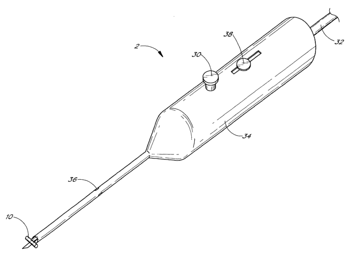
Figure 5 illustrates one embodiment of the trabecular shunt applicator 2,
holding the trabecular shunt
10 in place. Additionally, a trabecular meshwork blade 28 extends from the
distal end of the applicator 2. In
this embodiment, the blade 28 may be extended by spring action from the distal
end of the applicator 2 when
the operator pushes a button 30 or similarly actuates extension of the blade
28. The blade 28 can be
retracted within the applicator 2 by means of a slide button 38, which the
operator can move proximally to
retract the blade 28. Alternatively, a plunger 32 may move the blade 28
forward and backward within the
applicator 2. Also shown is the outer tube 34 of the applicator 2, as well as
holes 36 in the applicator 2.
These holes 36 may be used for aspiration or irrigation of the anterior
chamber of the eye during the
performance of trabecular meshwork surgery.
Figure 6 illustrates one embodiment of a trabecular shunt retrieval device 29.
To reacquire a shunt
that is dropped in the anterior chamber requires the ability to grasp the
shunt in a variety of orientations and
from a variety of positions in the eye. Extending from the end of the
retrieval device 29 is one or more claws
40 that can grasp the shunt 10. These claws may be extended from or retracted
into the retrieval device 29.
Actuation of these retractable claws 40 may be effected by an operator's push
of a button 30 or engagement
of any of a variety of other similar actuating devices that are known to those
skilled in the art.
Figure 7A shows one embodiment of a trabecular meshwork trephine, or punch 42.
An inner tube 6
resides within an outer tube 4. The inner tube 6 is in communication with an
inner plunger 46. The proximal
end 50 of the inner plunger 46 is acted upon by a hammer 52 that is attached
to a spring 48. The spring 48
may be recoiled or loaded, storing potential energy, and the hammer 52 is then
held in place by an actuator
54 or other similar member in communication with the actuator 54. When the
actuator 54 is acted upon by an
.g.
CA 02540761 2003-08-27
WO 02/074052 PCT/US02/08430
operator, the spring 48 releases its potential energy, causing the hammer 52
to move forward, contacting the
proximal end 50 of the inner plunger 46. This in turn causes the punch 44 to
move forward, contacting the
trabecular meshwork.
Figure 7 view is a close-up, cross-sectional view of the punch 44. Again seen
as the outer tube 4,
the inner tube 6, and the punch 44 of the device. This trephine or punch may
comprise a circular blade 56 or
other similar configuration known to those skilled in the art for making a cut
or punch hole in the trabecular
meshwork of an eye.
Figures 8A and 8B demonstrate one embodiment of a trabecular shunt retrieval
device 29. Again
seen are the claws 40, which may hold the shunt 10 when the claw is partially
retracted within the retrieval
device 29. As illustrated in Figure 8B, when the claws are extended from the
retrieval device 29, a spring
action within the claws 40 causes them to move away from the shunt 10 (curved
arrows).
Figures 9A through 9D illustrate an adhesive mechanism for attaching and
detaching the shunt 10 to
the applicator 2.
In Figure 9A, the adhesive 60 holds the shunt 10 to the applicator 2, in the
sense that the adhesive
60 adheres to both the shunt 10, on one side, and the applicator 2 on another
side. Once the adhesive is
broken by various means, including traction, heat, and/or light, the shunt 10
moves away from the applicator
2, as illustrated in Figure 9C.
Figure 9B shows another embodiment of the adhesive mechanism. A protrusion 58
extending from
the applicator 2 helps adherence of the applicator 2 to the shunt 10 by means
of the adhesive 60. Once the
adhesive bond between the shunt 10 and the applicator 2 is broken, as
illustrated in Figure 9D, the shunt may
be left in place within the eye of the patient.
Figures 10A and 10B illustrates another embodiment of the applicator 2. In
this embodiment, an
inner plunger 46 is attached to a distal pusher 60. When the inner plunger 46
and distal pusher 60 move
distally (left arrows) within outer tube 4, the distal pusher 60 comes in
contact with the shunt 10 causing it to
be pushed away from the outer tube 4. The shunt 10 may thence be left in the
eye of the patient.
Figures 11A and 11B illustrate a hook-and-eye embodiment of a detachment
mechanism for a
trabecular shunt applicator 2. A hook-and-eye fastener 62 (such as VeIcroT"'
or a miniaturized version of
same) may be attached to a protrusion 58 on the applicator 2. When the
applicator 2 is pulled away from the
shunt 10 the two sides of the hook-and-eye fastener 62 come apart; leaving one
side of the hook-and-eye
fastener 62 attached to the shunt 10, in the other side of the hook-and-eye
fastener 62 attached to the
protrusion 58 of the applicator 2. In this fashion, the shunt 10 may be left
within the eye of the patient, and
the applicator 2 withdrawn from the eye.
Figures 12A and 12B illustrate a magnetic detachment mechanism for the
trabecular shunt
applicator 2. The applicator 2 and the shunt 10 are held together at a
junction 64 by magnetic attraction (the
magnetic fields shown stylistically by curved arrows), as illustrated in
Figure 12B. When the applicator 2 is
.g.
CA 02540761 2003-08-27
WO 02/074052 PCT/US02/08430
moved away from the shunt 10, the magnetic "seal" between the applicator 2 and
the shunt 10 at the junction
64 is broken, allowing the shunt 10 to be left behind in the patient's eye,
when the applicator 2 is withdrawn
from the eye.
Figures 13A and 13B illustrate another embodiment of the applicator 2. In this
embodiment, the
shunt 10 has screw threads 66 along one of its portions. These screw threads
66 fit into complementary
threads in the applicator 2. When the surgeon desires to leave the shunt 10 in
place within the eye of the
patient, the surgeon may unscrew the applicator 2 from the shunt 10 by turning
the applicator 2 in a
counterclockwise or clockwise fashion (curved arrows).
Figures 14A and 14B illustrate another detachment mechanism for the trabecular
shunt applicator 2.
In this embodiment, an elastic band 68 holds the shunt 10 in place on the
applicator 2 by wrapping around
the shunt 10 and a protrusion 58 on the applicator 2. The surgeon may cut the
elastic band 68, as illustrated
in Figure 14B, using a scissors 66 or similar cutting device as known to those
skilled in the art. When the
elastic band 68 is cut by the cutting instrument, such as the scissors 66, the
elastic band breaks away from
the protrusion 58 on the applicator 2 as well as the shunt 10. This allows the
shunt 10 to be left in place in
the eye and the applicator 2 to be withdrawn from the eye.
Another embodiment of a detachment mechanism is shown in Figures 15A and 15B.
In this
embodiment, a thread 70 or other tying device, such as a suture or string, is
wrapped around the shunt 10
and the protrusion 58 on the applicator 2. The surgeon can cut the thread 60
using a scissors 66 or other
similar cutting instrument, as illustrated in Figure 15B. When the thread 70
is so cut, the applicator 2 may be
withdrawn from the eye, leaving the shunt 10 in place within the eye.
Figures 16A and 16B demonstrate another detachment mechanism for the
trabecular shunt 10 and
the applicator 2. A pin 72 holds the shunt 10 in place within the outer tube 4
of the applicator 2. As illustrated
in Figure 16B, when the pin 72 is withdrawn from the outer tube 4 (upward
arrow), the pin is removed from a
hole 74 in the outer tube 4, as well as a shunt hole 76 in the shunt 10. This
allows the applicator 2 to be
moved away from the shunt 10, allowing the applicator 2 to be withdrawn from
the eye while the shunt 10
remains in place within the eye.
Figures 17A through 17D illustrate various embodiments of detachment
mechanisms for the
trabecular shunt applicator 2. Figure 17A illustrates an attachment to the
shunt 10 of a protrusion 58
extending from the applicator 2. This protrusion 58 may connect to the shunt
10 via various means, such as
by glue, welding or plastic fusion, or the molding or fabrication process. In
Figure 17B, the protrusion 58 has
been broken, allowing the applicator 2 to move away from the shunt 10. The
protrusion 58 may be broken in
a variety of means, including, as shown in Figure 17C, energy transfer from an
energy source 78, such as a
laser or thermal energy transferring device, as is well known to those skilled
in the art. In figure 17D, a light
source 80 can use ultraviolet light or other spectral frequencies to effect a
chemical or electrochemical
-10-
CA 02540761 2003-08-27
WO 02/074052 PCT/US02/08430
change in the protrusion 58 causing it to break. Once the light source 80 or
other energy source 78 has
broken the protrusion 58, the applicator 2 may be withdrawn from the eye,
leaving the shunt 10 in place.
Figure 18 illustrates a wedge-fit mechanism for the applicator 2. The outer
tube 4 of the applicator 2
has a wedge configuration 84 within its lumen, and a similar wedge
configuration in the inlet portion of the
shunt 10 allows for a tight, "wedged," fit for the shunt 10 within the
applicator 2. Once the shunt 10 is in place
within the eye, the applicator 2 may be moved away from the shunt 10, causing
the shunt 10 to be dislodged
from the outer wall 4 of the applicator 2 by virtue of the aforementioned
wedge configuration 84 of the
applicator 2 and shunt 10.
Figure 19 illustrates a spring release mechanism for the applicator 2. In this
embodiment, a hammer
52 is attached to a base 82 by a spring 48. When the spring 48 is loaded with
energy, the hammer is then
trapped in placed by an actuator 54 or other member in communication with the
actuator 54. When the
actuator 54 is actuated by an operator, the spring 48 is released, unloading
its energy and driving the
hammer 54 away from the base 82, toward the shunt 10. This drives the shunt 10
away from the outer wall 4
of the applicator 2, allowing it to be left in place within the eye. The
applicator 2 may then be withdrawn from
the eye.
Figures 20A and 20B illustrate another embodiment of a detachment mechanism
for the trabecular
shunt applicator 2. In this embodiment, one or more protrusions 58 extend from
the applicator 2. One or
more protuberances 86 extend from the protrusion 58. These protuberances 86
are preferably made of
flexible plastic or rubber and can fit within one or more indentations 88 in
the shunt 10. These protuberances
86 cause the shunt 10 to be held in place within the applicator 2 because the
protuberances 86 fit within the
indentations 88 in the shunt 10. When the surgeon pulls the applicator 2 away
from the shunt 10 after the
shunt 10 has been placed through the trabecular meshwork, the protuberances 86
are pulled out of the
indentations 88 on the shunt, allowing the shunt 10 to break free of the
applicator 2. Once the protuberances
86 slide out of the indentations 88 in the shunt 10, the applicator 2 may be
withdrawn from the eye, while the
shunt 10 remains in place within the eye.
Figure 21 illustrates a similar embodiment of a detachment mechanism to that
shown in Figures 20A
and 20B. In this embodiment, the protrusions 58 are more rigid than that shown
in Figures 20A and 20B,
being made of semi-rigid plastic or metal, and the protrusions 58 extend from
the applicator 2. One or more
protuberances 86 extend from the protrusion 58. These protuberances 86 can fit
within one or more
indentations 88 in the shunt 10. These protuberances 86 cause the shunt 10 to
be held in place within the
applicator 2 because the protuberances 86 fit within the indentations 88 in
the shunt 10. When the surgeon
pulls the applicator 2 away from the shunt 10 after the shunt 10 has been
placed through the trabecular
meshwork, the protuberances 86 are pulled out of the indentations 88 on the
shunt, allowing the shunt 10 to
break free of the applicator 2. Once the protuberances 86 slide out of the
indentations 88 in the shunt 10, the
applicator 2 may be withdrawn from the eye, while the shunt 10 remains in
place within the eye.
-11-
CA 02540761 2003-08-27
WO 02/074052 PCT/US02/08430
Figures 22A and 22B illustrate a suction detachment mechanism for the
trabecular shunt applicator
2. In this embodiment, the shunt 10 is held in place within the applicator 2
by negative pressure, i.e., suction
(right arrows). The suction may be provided by any suitable suction device as
is well known to those skilled
in the art. In Figure 22B, the suction has been turned off and oxygen, air, or
other suitable gas is allowed to
flow into the applicator 2 (left arrows). This gas influx and consequent
pressure change causes the shunt 10
to breakaway from the applicator 2, allowing the shunt 10 to break free of the
applicator 2. This allows the
shunt 10 to be left in place in the eye.
Figure 23 illustrates one embodiment of an articulating applicator or
retrieval device 90. In this
embodiment, a proximal arm 92 is attached to a distal arm 94 at a joint 96.
This joint 96 is movable such that
an angle formed between the proximal arm 92 and the distal arm 94 can change.
One or more claws 40 can
extend from the distal arm 94, in the case of a shunt retrieval device.
Similarly, this articulation mechanism
may be used for the trabecular shunt applicator, and thus the articulating
applicator or retrieval device 90 may
be either an applicator for the trabecular shunt, a retrieval device, or both,
in various embodiments.
Figures 24A and 24B illustrate embodiments of a control arm 98 which is
attached to a mechanism
for performing trabeculotomy. In Figure 24A, a blade 100 extends from an end
of the control arm 98. In
some embodiments, the long axis of the control arm 98 runs parallel or
semiparallel to the long axis of the
applicator 2. The blade 100 may be used to make a trabeculotomy in preparation
for placing the trabecular
shunt 10 through the trabecular meshwork and into Schlemm"s canal.
Figure 24B shows a "hot tip" 102 at the end of the control arm 98. This hot
tip may be a cautery,
laser, or other energy transfer device for making a hole in the trabecular
meshwork in preparation for placing
the shunt 10 through the trabecular meshwork and into Schlemm's canal.
Figures 25A through 25C illustrate various embodiments of devices, such as
trephines, that can
punch holes in the trabecular meshwork. In Figure 25A, a trabecular meshwork
punch 104 is illustrated. This
punch 104 can make holes 112 in the trabecular meshwork 110. These holes 112
can be of various
configurations, depending on the shape of the distal blade of the trabecular
meshwork punch 104.
In Figure 25B, a blade 107 extends from the end of a trabecular meshwork
cutter 106. This blade
107 can make various punch holes 114 in the trabecular meshwork 110, as
illustrated.
Figure 25C illustrates a trabecular meshwork drill 108. The drill 108 has a
distal drill bit 111, which
can make a drill hole 112 in the trabecular meshwork 110.
There are many alternatives for maintaining the anterior chamber during the
installation of the
trabecular shunt 10, including the irrigating, irrigating side port, over-
fill, viscoelastic, and air bubble.
Additionally, there are many alternatives for creating a trabecular meshwork
incision. Of these, the
punch, stab, drill, and shunt alternatives are likely to create surgeon-
independent, repeatable incisions. The
ideal size of the shunt 10 is based on the size of the Schlemm's canal that it
is inserted into and on the size of
the hole in the trabecular meshwork. A surgeon-independent incision would help
ensure that the shunt fits
-12~
CA 02540761 2003-08-27
WO 02/074052 PCT/US02/08430
well despite who is performing the surgery. Of these surgeon-independent
alternatives, the punch and drill
remove material that will leave room for the outlet portion of the shunt
without having to create overlaps or
folds in the trabecular meshwork tissue. The drill alternative creates debris
and is therefore perhaps less
desirable than the punch. The sharp shunt alternative is enticing, since it
removes the need to cross the
anterior chamber twice; however, the sharp tip may potentially do damage to
the inside of Schlemm's canal
or may lead to inappropriate placement of the shunt.
There are multiple alternatives for creating a corneal incision, including the
micro-knife.
Due to the anatomy of trabecular meshwork being in a curved ring configuration
inside the eye, and
in view of the ab interno approach within the confined space of the anterior
chamber, the tip section of the
trephine for creating an opening within the trabecular meshwork may be angled.
An angled-tip trephine may,
in some circumstances, more easily enable creating an opening in the
trabecular meshwork suitable for
inserting a the glaucoma shunt more easily into Schlemm's canal.
While inserting a glaucoma shunt through the trabecular meshwork into
Schlemm's canal in an ab
interno procedure, it is desirable to cause minimal injury to Schlemm's canal.
Therefore, one consideration
for creating an opening using a trephine is to limit its penetrating distance
in Schlemm's canal. The
trabecular meshwork is generally about 200 to 400 microns. Some embodiments
provide a depth-limited
microtrephine adapted for cutting through at least a major portion of the
trabecular meshwork, while not
injuring the back (outer) surface of Schlemm's canal.
To further simplify the operation of creating an opening in the trabecular
meshwork, one aspect
provides an automated microtrephine, which, by a touch of a button at the
handpiece, permits a
predetermined cutting force and/or cutting distance, thereby eliminating much
of an operator's chance for
error in creating an opening.
While certain aspects and embodiments of the invention have been described,
these have been
presented by way of example only, and are not intended to limit the scope of
the invention. Indeed, the novel
methods and systems described herein may be embodied in a variety of other
forms without departing from
the spirit thereof. The accompanying claims and their equivalents are intended
to cover such forms or
modifications as would fall within the scope and spirit of the invention.
-13-