Note: Descriptions are shown in the official language in which they were submitted.
CA 02596204 2007-08-07
Atty. Dkt. No.: 084122-0600
METHOD AND SYSTEM FOR
DETERMINING AN OPTIMAL DILUTION OF A REAGENT
BACKGROUND OF THE INVENTION
100011 The present invention relates generally to the field of biological
tissue
s analysis. More specifically, the present invention relates to a system
and method for
determining an optimal dilution of a reagent for use in a quantitative
immunoassay.
There are a variety of different immunoassays for which the claimed invention
is
applicable including but not limited to tissue-based immunohistochemical, cell-
based
immunohistochemical analysis such as flow cytometry, and high content
screening
(HCS) immunohistochemical analysis, enzyme linked immunosorbent assay (ELISA),
and western blot assays.
10002] The determination of optimal dilutions of a reagent for a given
biological
specimen is beneficial to quantitative immunoassays. By way of example, if a
reagent
is not concentrated enough, then the analysis with the reagent is likely to
produce
under-detection along with loss of sensitivity at the upper range of the
assay.
Correspondingly, if the reagent is too concentrated, the reagent is likely to
produce
over-detection along with loss of sensitivity in the lower range of the assay.
10003] Existing approaches for determining an optimal dilution of a reagent
are
purely qualitative. The qualitative nature of such an optimization
considerably
reduces reproducibility of a particular dilution and analysis. Further, such
qualitative
approach fails to account for a variety of pertinent factors causing reduced
reliability
of results obtained utilizing such a qualitative optimization.
[0004] Immunohistochemistry (IHC) is an immunoassay method for detection of
analytes in tissue sections. Traditional IHC assay results have been
qualitative in
nature, often done by a manual visual assessment through a microscope using a
subjective scoring system to indicate a relative amount of analyte present in
the tissue
sample. In contrast, qualitative IFIC analytically measures the amount of one
or more
analytes of interest in a tissue section. Analytical systems have been
developed for
quantitative 1HC analysis. For example one such system is the AQUA technology
described in US Patent application 7,219,016 and in Camp et al 2002 Nature
-2-
CA 02596204 2007-08-07
Atty. Dkt. No.: 084122-0600
Medicine 8(11)1323-1327. However, even with the introduction of such
quatitative
analysis for IHC, typically preliminary assays are run to determine optimal
concentrations of reagents to be used in the analytical assay and these
results are
assessed qualitatively, not quantitatively. There is a need for methods for
quantitatively determining optimal reagent concentrations for use in
quantitative
immunoassays, including IHC.
SUMMARY OF THE INVENTION
100051 The present invention addresses the above-identified considerations by
= quantitatively determining an optimal dilution of a reagent for use in
quantitative
lo immunoassays. In one embodiment, multiple dilution sets are received,
where each of
the dilution sets consist of a different respective dilution value and a
respective
arrangement of immunoassay staining intensity values. A respective dynamic
range
metric is determined for each of the multiple dilution sets relative to the
respective
arrangement of immunoassay staining intensity values. Having found the
respective
is dynamic range metric, a dilution set having the numerically optimal
dynamic range
metric is selected and the dilution value of that dilution set is selected as
being
representative of an optimal dilution level of the reagent for use in a
quantitative
immunoassay.
[0006] In another embodiment of the present invention, a system for
determining an
20 optimal dilution of a reagent for use in a quantitative immunoassay has
means for the
reception of multiple dilution sets, where each of the dilution sets consist
of a
different respective dilution value and representative arrangement of
immunoassay
staining intensity values. Further, the system has means for determining a
respective
dynamic range metric for each of the multiple dilution sets relative to the
respective
25 arrangement of immunoassay staining intensity values. The system is
configured with
a means for identifying a dilution value which is representative of an optimal
dilution
value for use in the quantitative immunoassay from the dilution set have the
numerically optimal dynamic range metric.
[0007] According to another embodiment of the present invention, a system for
30 determining an optimal dilution of a reagent for use in a quantitative
immunoassay
-3-
CA 02596204 2007-08-07
Atty. Dkt. No.: 084122-0600
=
has an optical microscope configured to magnify at least a portion of a slide-
mounted
tissue sample as well as an image sensor which is in optical communication
with the
microscope such that the image sensor is configured to obtain a digitized
image of the
magnified portion of the slide-mounted tissue sample inserted in the
microscope. The
5 system is also equipped with a processor module in communication with the
image
sensor. The processor is configured to (i) automatically receive multiple
dilution sets,
each dilution set having a different respective dilution value and comprising
a
respective arrangement of immunoassay staining intensity values, (ii)
determine for
each of the multiple dilution sets a respective dynamic range metric related
to the
io respective plurality of irrununoassay staining intensity values, and
(iii) identify the
dilution set having the numerically optimal dynamic range metric, the dilution
value
= of the identified dilution set being representative of an optimal
dilution level of the
reagent for use in the quantitative iminunoassay.
100081 According to yet another embodiment of the present invention, a
computer
is readable medium is loaded with computer readable instructions for
execution by a
processor for the purpose of performing a method for determining an optimal
dilution
of a reagent for use in a quantitative immunoassay. hilhat method, multiple
dilution
sets are received, where each of the dilution sets consists of a different
respective
dilution value and a respective arrangement of immunoassay staining intensity
values.
20 A respective dynamic range metric is determined for each of the multiple
dilution sets
relative to the respective arrangement of immunoassay staining intensity
values.
Having found the respective dynamic range metric, a dilution set having the
numerically optimal dynamic range metric is selected and the dilution value of
that
dilution set is selected as being representative of an optimal dilution level
of the
25 reagent for use in a quantitative immunoassay.
100091 - In still another embodiment of the present invention, an
electromagnetic
signal carries computer-readable instructions for determining an optimal
dilution of a
reagent for use in a quantitative immunoassay. The instructions are such that
multiple
dilution sets are receivedõ where each of the dilution sets consist of a
different
30 respective dilution value and a respective arrangement of immunoassay
staining
intensity values. A respective dynamic range metric is determined for each of
the
-4-
CA 02596204 2007-08-07
Atty. Dkt. No.: 084122-0600
multiple dilution sets relative to the respective arrangement of immunoassay
staining
intensity values. Having found the respective dynamic range metric, a dilution
set
having the numerically optimal dynamic range metric is selected and the
dilution
value of that dilution set is selected as being representative of an optimal
dilution
s level of the reagent for use in a quantitative immunoassay.
[0010] It is to be understood that both the foregoing general description and
the
following detailed description are exemplary and explanatory only, and are not
restrictive of' the invention as claimed. These and other features, aspects
and
advantages of the present invention will become apparent from the following
to description, appended claims, and the accompanying exemplary embodiments
shown
in the drawings, which are briefly described below.
BRIEF DESCRIPTION OF THE DRAWINGS
[0011] The foregoing and other objects, features and advantages of the
invention
will be apparent from the following more particular description of preferred
15 embodiments of the invention, as illustrated in the accompanying
drawings in which
like reference characters refer to the same parts throughout the different
views. The
drawings are not necessarily to scale, emphasis instead being placed upon
illustrating
the principles of the invention.
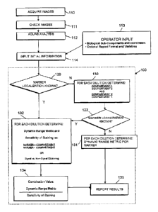
10012] FIG. I is a flow diagram of a process for determining an optimal
dilution of
20 a reagent according to an embodiment of the present invention.
100131 FIG. 2 is a flow diagram illustrating how results of the optimization
analysis
are presented in an exemplary embodiment of the present invention.
[0014] FIG. 3A through FIG. 311 together illustrate a set of exemplary
histograms
for a variety of different reagent dilution levels plotting frequency of an
immunoassay
as staining intensity values.
[00151 FIG. 4 is a graph illustrating a relationship between an average
absolute
deviation obtained from histograms of collected immunoassay staining intensity
values for a variety of different reagent dilution levels and the respective
dilution
levels.
-5-
CA 02596204 2007-08-07
Atty. Dkt. No.: 084122-0600
[0016] FIG. 5A and FIG. 5B are a set of graphs illustrating a relationship
between a
dilution level and a sensitivity of staining determined according to an
embodiment of
the present invention .
[00171 FIG. 6 is a graph illustrating determination of an optimal dilution of
a
s reagent based on a combination of specificity of staining and a dynamic
range metric
according to an embodiment of the present invention.
[0018] FIG. 7A is a graph illustrating determination of an optimal dilution of
a
reagent based on a combination of specificity of staining and a dynamic range
metric
according to an embodimentof the present invention.
to 100191 FIG. 78 is a graph illustrating determination of an optimal
dilution of a
reagent based on a combination of specificity of staining and a dynamic range
metric
according to another embodiment of the present invention.
[0020] FIG. 8 is a graph illustrating determination of an optimal dilution of
a
reagent based on a combination of specificity of staining, a dynamic range
metric, and
15 a signal to noise metric according to an embodiment of the present
invention.
[0021] FIG. 9 is a diagram of an exemplary embodiment of a system for
determining an optimal dilution of a reagent for use in a quantitative
immunoassay
according to an embodiment of the present invention.
[0022] FIG. 10 is a table illustrating utilization of a parametric Pearson
regression to
20 examine data according to one embodiment of the present invention.
[0023] FIG. ii is a table illustrating utilization of a non-parametric
Spearman
regression to examine data according to one embodiment of the present
invention.
[0024] FIG. 12 is a table illustrating utilization of a parametric Pearson
regression to
examine data according to one embodiment of the present invention.
25 [0025] FIG. 13 is a table illustrating utilization of a non-parametric
Spearman
regression to examine data according to one embodiment of the present
invention.
[0026] FIG. 14 is a table illustrating a combination of numerical factors to
determine an optimal dilution for a reagent for use in a quantitative
immunoassay
according to an embodiment of the present invention.
30 100271 FIG. 15 is a table illustrating additional exemplary data used in
calculating
the factors shown in FIG. 14.
-6-
CA 02596204 2007-08-07
Atty. Dkt. No.: 084122-0600
100281 FIG. 16 is a table illustrating a combination of numerical factors for
determining an optimal dilution for a reagent for use in a quantitative
immunoassay
according to an embodiment of the present invention.
100291 FIG. 17 is a table illustrating additional exemplary data used in
calculating
the factors shown in FIG. 16.
DETAILED DESCRIPTION OF THE PREFERRED EMBODIMENTS
[0030] As is known in the art, reagents are designed to detect particular
biological
sub-components, for example a protein in a biological specimen. Typically, a
reagent
is detected by an secondary reagent and/or a detection reagent. By way of an
example, a reagent may be detected by a secondary reagent comprising a
fluorescent
dye or an enzyme. As such, the images acquired of the stained samples consist
of
pixels, each pixel having a power or intensity level normalized for time of
exposure.
Optimally, the power or intensity level correlates with the concentration of
the
biological sub-components of the specimen as detected by the detection
reagents. A
preferred reagent for detecting a protein of interest in a sample is an
antibody capable
of binding to that protein, that in the context of the assay is known as a
primary
antibody. The secondary reagent can also be an antibody, known as a secondary
antibody that is specific for the species of the primary antibody. The
secondary
antibody typically has a detectable label. Antibodies within the scope of the
present
invention include, e.g., but are not limited to, monoclonal, polyclonal,
chimeric,
humanized, diabody, and human monoclonal and human polyclonal antibodies which
specifically bind the target polypeptide, a homolog, derivative or a fragment
thereof.
The antibodies useful as binding agents of the present invention include,
e.g., but are
not limited to, IgG (including IgG I, IgG2, IgG3, and IgG4), IgA (including
lgAl and
IgA2),IgD, IgE, or 1gM, and IgY.
[00311 In general, an optimal concentration for use of a primary antibody in
an
immunoassay is determined in an initial experiment that evaluates a plurality
of
antibody titrations on a biological specimen. For example, a dilution series
of an
antibody such as a 1:10, 1:25, 1:100, 1:500 is tested, each dilution on a
separate
sample of the same biological specimen. For example the biological specimen
may
-7-
CA 02596204 2016-06-13
be a tissue sample and each dilution is tested on a histological tissue
section of the
specimen, ideally serial sections of the specimen. In a preferred embodiment,
the
each dilution of reagent is tested on a plurality of biological specimens
representative
of those intended for the analytical assay. In a preferred embodiment the
plurality of
biological specimens may be a tissue microarray (TMA) comprised of cored
tissue
samples from numerous different histological tissue blocks. A separate TMA
section
on a slide is stained with one of the dilution series of the primary antibody
utilizing
common IHC techniques. The resulting stained specimens are each imaged using a
system for viewing the detectable signal and acquiring a digital image of the
staining.
io The images thus obtained are then used by the method of the invention
for
quantitatively determining the optimal concentration of the reagent for
quantitative
immunoassay studies.
[0032] An overview of a system and method of the invention for determining an
optimal dilution of a reagent is shown in the flow diagram in FIG 1. In one
embodiment of the present invention, a set of images of stained samples are
acquired
as shown in step 110. Each of the images portrays a respective sample stained
with a
particular concentration of a reagent to be optimized.
100331 Following the acquisition of a set of images, those images are
preferably
quality checked in step 111. The quality of an image being suitable for
quantitative
immunoassay can be determined manually or automatically,
Following the quality check of step
111, the images are subjected to a quantification analysis such as an AQUA
analysis
as shown in step 112 (AQUA is a registered trademark of HistoRx Corp. of New
Haven, CT). AQUA analysis is further described in U.S. Patent No. 7, 219,016
and in Camp et al 2002
Nature Medicine 8(11)1323-1327. The results of a quantification analysis is
the
production of a plurality of staining intensity values corresponding to the
plurality of
biological specimens assay with each of the plurality of dilution sets. Each
of these
dilution sets are inputted into the optimization analysis resulting in an
identification of
-8-
CA 02596204 2007-08-07
Atty. Dkt. No.: 084122-0600
one of the dilution sets representing an optimal dilution of a reagent for use
in a
quantitative immunoassay 100 by way of an initial input step as shown by 114.
1110341 Additionally, an operator may input initial information into the
analysis 100
through step 113. For example, in step 113 an operator may enter information
indicating what reagent dilution level of the reagent was used for each
particular
image of stained sample. In another embodiment, more than one dilution level
of the
reagent may be utilized, each for a separate stained sample. In such an
embodiment,
an operator in step 113 may enter information indicating what reagent dilution
level
was used for a particular location on an image of a particular stained sample.
An
lo operator may also enter information corresponding to the style of the
report following
the optimization analysis the operator desires as well as a set of pre-
determined
variables useful in the analysis, in step 113. Further, in step 113 an
operator may also
enter information indicating where particular stain specific compartments are
located
in a stained image if those compartments are known. A stain specific
compartment
contains biological sub-components that the reagent is designed to detect,
such as
nuclei, cytoplasm, or cellular walls. Correspondingly, an operator in step 113
may
enter information indicating where particular non-stain specific compartments
are
located in a stained image if those compartments are known. A non-stain
specific
compartment does not contain biological sub-components that the reagent is
designed
to detect. By way of example, an operator may indicate where certain tissue
sub-
groups (such as breast or colon cancer samples) are located in a TMA on a
slide to be
evaluated, and their coordinates. Furthermore, an operator may indicate that
the
reagent is known to specifically stain a stain specific compartment (such as
tumor
tissue as opposed to stroma), or a stain specific sub-cellular compartment
(such as cell
nuclei). Therefore, the expectation is that if such information is available
the
reagent's marker will be expressed in the stain specific compartments while
not being
expressed in the non-stain specific compartments, this differential being
utilized in
particular aspects of the invention. In yet another embodiment of the present
invention, knowledge of the stain specific and non-stain specific compartments
is
known following the quantification analysis in step 112 or through step 130.
-9-
CA 02596204 2007-08-07
Atty. Dkt. No.: 084122-0600
[00351 For each dilution set, the existence of such reagent or marker
localization
information is determined in a decision step 120. If the marker localization
information is not available for a particular dilution set, the optimization
analysis 100
attempts to discern such information in step 130, otherwise step 132
(calculation of a
6 specificity of staining which requires knowledge of marker localization
and
calculation of a.dynamic range metric) is performed. Ratios for each of the
different
compartments are formulated, the largest ratio indicative of marker
compartment
localization (step 130). In more detail, the marker localization information
is
determined by comparing staining intensity values of two compartments of the
to biological specimen(s) of a particular dilution set By way of example,
there may be a
first compartment 1 and a second compartment 2 as indicated in step 130. An
average
staining sensitivity value is calculated for compartment 1 from the set of
immunoassay staining sensitivity values determined to be associated with
compartment 1. Also, an average staining sensitivity value is calculated for
15 compartment 2 from the set of immunoassay staining sensitivity values
determined to
be associated with compartment 2. Once these two numbers have been calculated,
a
ratio of compartment I divided by compartment 2 and a ratio of compartment 2
divided by compartment 1 are each calculated. If either of these ratios are
greater
than or equal to an upper threshold quantity, e.g., 1.5, or less than or equal
to a lower -
zo threshold quantity, e.g., 0.666, then it indicates that the marker
specific reagent
localizes to the compartment with the numerically largest average staining
intensity
value.
10036) In some embodiments of the present invention, the optimization analysis
performs multiple comparisons to attempt to identify multiple stain specific
and non-
25 stain specific compartments. If after step 130 the optimization analysis
is able to
determine at step 122 the marker localization information step 132 is
performed,
otherwise step 131 is performed. In particular, step 132 performs the
calculation of a
dynamic range metric as well as a sensitivity of staining, whereas step 131
only
performs the calculation of a dynamic range metric. Following the completion
of
so either steps 131 or 132, the results of the optimization analysis 100
are reported in
step 135.
-10-
CA 02596204 2007-08-07
Atty. Dkt. No.: 084122-0600
100371 In step 132, for each dilution set of the multiple dilution sets, a
dynamic
range metric and a sensitivity of staining are each calculated. Each of these
quantities
are discussed in more detail below. Following the calculation of the dynamic
range
metric and sensitivity of staining for each of the dilution sets, the dynamic
range
metric and sensitivity of staining are combined with one another to generate a
combination value for each dilution set. The resulting combination values are
used to
select the dilution set with the most numerically optimal combination value.
Associated with the selected dilution set is a dilution value representative
of an
optimal dilution of a reagent. The results of the selection are reported in
step 135.
le 100381 A dynamic range metric is always optimal as a numerically large
number. In
some embodiments as discussed further on in this detailed description a large
sensitivity of staining is optimal, while in other embodiments a numerically
small
sensitivity of staining is optimal. As a consequence of these varied
embodiments for
the optimality of a sensitivity of staining an optimal combination value is
the
numerically greatest value in some embodiments, while in other embodiments an
optimal combination value is the numerically smallest value.
100391 FIG. 6 graphically illustrates a combination of a specificity of
staining value
and a sensitivity of staining value, such as a Dynamic Range Metric, to
provide a
combined metric (e.g., a sensitivity specificity metric) for each of several
different
exemplaiy dilutions of a reagent identified along the horizontal axis. In this
example,
the two features in combination, the sensitivity specificity metric resulted
in the 1:600
dilution of a reagent having the greatest numeric value resulting in that
dilution being
selected as the most optimal.
100401 FIG. 7A and FIG. 7B also illustrate the combination of a specificity of
staining value, signal to noise metric (N/ER ratio) and a Dynamic Range Metric
(AAD) to provide a sensitivity specificity metric (N/ER Ratio/Avg Dev Nuclear
(which is AADJ) for each of several different exemplary dilutions of a
reagent.
FIG. 7A was generated from one set of samples, while FIG. 78 was generated
from
another set of samples. FIG. 7A graphically illustrates both the specificity
of staining
value (N/ER Ratio) as well as the dynamic range metric (Average Deviation
Number). In FIG. 7A, the dynamic range metric was fairly consistent, i.e.,
flat, but
-11-
CA 02596204 2007-08-07
Atty. Dkt. No.: 084122-0600
the specificity of staining value indicated that either of the 1:250 or 1:500
dilution of a
reagent could be optimal. However, when the two features are combined
(N/ER/Avg
Dev Nuclear) as shown in the top curve of FIG. 7A, the 1:250 dilution of a
reagent
had the greatest numeric value resulting in that dilution being selected as
the optimal
dilution.
10041] In FIG. 7B, the dynamic range metric indicated that a dilution of 1:250
may
be optimal, while the specificity of staining indicated that the dilution of
1:2000 may
be optimal. However, when the two features are combined as shown in FIG. 7B,
the
1:250 once again had the greatest numeric value resulting in that dilution
being
io selected as the most optimal. FIG. 7A and FIG. 78 all together
illustrate how the two
features can be utilized effectively in combination to balance the specificity
of
staining with the dynamic range metric to ultimately select an optimal
dilution of a
reagent.
[(1042] Referring now again to FIG 1. in step 131, for each dilution set of
the
multiple dilution sets the optimization analysis calculates a dynamic range
metric for
each one of the plurality of dilution sets. Following the calculation of the
dynamic
range metrics for each one of the multiple dilution sets, a dilution set is
selected with
the most numerically optimal dynamic range metric. Associated with the
selected
dilution set is a dilution value which is representative of an optimal
dilution of the
zo reagent. A dynamic range metric is optimal as a numerically large
number. The
results of selecting the most numerically optimal dilution set along with the
optimal
dilution value of the reagent are reported in step 135.
[0043] Referring now to FIG. 1 in combination with FIG. 2, the flow diagram of
PIG. 2 illustrates how the results of the optimization analysis 100 can be
reported 135.
A report may include: a dynamic range metric, such as a copy of the histogram
or
AAD graph (as illustrated); a signal to noise metric, such as a ratio (as
illustrated) or a
cluster analysis; and 3) the sensitivity specificity metric combining both
(e.g., as
shown in FIG. 6¨can use the AAD graph, the nucleus cytoplasm ratio graph and
the
FIG. 6 combined graph as a report output)] In particular, the dilution level
of the
selected optimal dilution can be reported at step 140. In one embodiment of
the
present invention data from the analysis supporting the selection of a
particular
-12-
=
CA 02596204 2007-08-07
Atty. Dkt. No.: 084122-0600
optimal dilution may also be reported at step 141. In yet a further
embodiment, sets
of graphs and histograms can be reported at step 142 to support the selection
of a
particular optimal dilution. Additionally, as previously discussed, a user may
indicate
at step 113 how that user desires a report from step 135 to be presented.
s 100441 Referring now to the calculation of a dynamic range metric in more
detail,
the dynamic range metric can be used as a proxy of the spread of a particular
diluted
reagent's detection pattern. For instance, FIG. 3A is a histogram illustrating
a
frequency of particular immunoassay staining intensity values for a sample, in
this
case a collection of samples in a TMA format treated with a 1:50 dilution. As
further
illustrated by the histogram of FIG. 3A, the 1:50 dilution has a wide spread
of stain
intensity values, wherein the spread represents the ability of the assay to
utilize the
reagent at that concentration to accurately detect a marker across this range
of
expression in tissue sections. Note the impact of antibody concentration on
the
dynamic range of the provided data. For instance in comparison with the
histogram
of FIG, 3G, illustrating the frequency of particular immunoassay staining
intensity
values for a sample (the TMA) treated with a 1:10,000 dilution, the 1:10,000
dilution
has a narrower and therefore less than optimal dynamic range spread.
Consequently,
the 1:10,000 dilution of the reagent likely caused fairly severe under-
detection as
previously discussed in this detailed description. Indeed, as one progresses
along
through the varying dilutions of FIG. 3A (1:50 dilution), FIG. 3B (1:100
dilution),
FIG. 3C (1:300 dilution), FIG. 3D (1:600 dilution), FIG. 3E (1:1000 dilution),
FIG. 3F (1:5000 dilution), FIG. 3G (1:10,000 dilution), and FIG. 3H (1:20,000
dilution), the dynamic range of the data tends to decrease substantially. This
feature
of the dilution values is captured in the graph FIG. 4, showing how the
average
deviation increases substantially as the reagent becomes more diluted. In one
embodiment of the present invention, the dynamic range metric is an average
absolute
deviation (AAD), and an optimal dynamic range metric is a numerically small
value
which represents the best spread of data. In another embodiment of the present
invention, the dynamic range metric is a weighted combination of a standard
deviation, a variance, and a swing ratio, and an optimal dynamic range metric
is a
numerically large value which represents the best spread of data.
-13-
CA 02596204 2007-08-07
Atty. Dkt. No.: 084122-0600
10045] Referring now to the calculation of a dynamic range metric in more
detail,
the dynamic range metric can be used as a proxy of the spread of a particular
diluted
reagent's detection pattern. For instance, FIG. 3A is a histogram illustrating
a
frequency of particular immunoassay staining intensity values for a sample
treated
with a 1:50 dilution. As further illustrated by the histogram of FIG. 3A, the
1:50
dilution has a wide spread of stain intensity values, wherein the spread
represents the
ability of the reagent to accurately express itself in areas where there are
instances of
the biological sub-components that the reagent is designed to detect. Note the
impact
of antibody concentration on the dynamic range of the provided data. For
instance in
io comparison with the histogram of FIG. 3E, illustrating the frequency of
particular
immunoassay staining intensity values for a sample treated with a 1:10,000
dilution,
the 1:10,000 dilution has a narrower and therefore less than optimal spread.
Consequently, the 1:10,000 dilution of the reagent likely caused fairly severe
under-
detection as previously discussed in this detailed description. Indeed, as one
.. progresses along through the varying dilutions of FIG. 3A (1:50 dilution),
FIG. 3B
(1:100 dilution), FIG. 3C (1:300 dilution), FIG. 3D (1:600 dilution), FIG. 3E
(1:1000
dilution), FIG. 3F (1:5000 dilution), FIG. 3G (1:10,000 dilution), and FIG. 3H
(1:20,000 dilution), the dynamic range of the data tends to decrease
substantially.
This feature of the dilution values is captured in the graph FIG. 4, showing
how the
zo average deviation increases substantially as the reagent becomes more
diluted. In one
embodiment of the present invention, the dynamic range metric is an average
absolute
deviation. In another embodiment of the present invention, the dynamic range
metric
is a weighted combination of a standard deviation, a variance, and a swing
ratio
100461 Referring to embodiments of the present invention in which the dynamic
range metric is an average absolute deviation, the calculation initially
involves the
calculation of a mean of all of the plurality of immunoassay staining
intensity values
of a dilution set. Once the mean has been calculated, each immunoassay
staining
intensity value from the plurality is subtracted from the mean and an absolute
value is
taken resulting in an absolute deviation from the mean. All of the absolute
deviations
from the mean for each plurality of immunoassay staining intensity values are
summed together and divided by the total number of the plurality of
immunoassay
-14-
CA 02596204 2007-08-07
Atty. Dkt. No.: 084122-0600
staining intensity values. The resulting value is an average absolute
deviation. While
standard deviation calculations could also be used, the average absolute
deviation
method does not square the distance between the mean and therefore is less
affected
by extreme values (i.e., the tails of the data distribution). In this
particular
embodiment, the most optimal dynamic metric range is the numerically greatest
number. In traditional mathematical formula terms the average absolute
deviation is
calculated as:
11
ADD = E(ly -y1)/ N
Where, 11- is the mean of the arrangement of immunoassay staining intensity
values,
IYI is the absolute value of Y, N is the number of immunoassay staining
intensity
values and i is an integer varying between 1 and n.
[0047] Referring now to alternative embodiments of the present invention, the
data
is is log transformed and the Dynamic Range Metric can be formulated as a
weighted
combination of a standard deviation, a variance, and a swing ratio. A standard
deviation is a measure of the spread of a multi-set of values, such as the
arrangement
of staining sensitivity values. A standard deviation can be calculated by
mathematically comparing the value of a number with the expected value of that
number. The values compared can be those of the arrangement of immunoassay
staining intensity values, such that the expected value is the mean of the an-
angement
of immunoassay staining intensity values. The calculation of a standard
deviation for
an arrangement of immunoassay staining intensity values begins by calculating
the
mean of the arrangement of staining intensity values. For each one of the
plurality of
staining intensity values, the mean can be subtracted from the value and the
square of
the result taken to produce a deviation value. All of the deviation values for
each of
the different arrangements of immunoassay staining intensity values can be
summed
together and divided by the total number of the different arrangements of
staining
intensity values and a square root of the result can be taken to produce a
standard
-15-
CA 02596204 2007-08-07
Atty. Dkt. No.: 084122-0600
deviation. In traditional mathematical formulaic terms, a standard deviation
is
calculated as follows:
X)2 li
Where a is the standard deviation, xi is each individual one of the
arrangements of
immunoassay staining intensity values, x "bar" is the mean of the arrangements
of
immunoassay staining intensity values, N is the number of different
arrangements of
immunoassay staining intensity values and i is an integer varying between 1
and n.
100481 The variance of a multi-set, such as the arrangement of immunoassay
staining intensity values, is a non-negative number which provides an
indication as to
how widely spread the values of a multi-set are likely to be. The larger a
variance, the
more scattered the members of a multi-set are on average. The variance is
calculated
as the square of the standard deviation as discussed above. Therefore, the
calculation
of the variance for a plurality of immunoassay staining intensity values
begins by
calculating the mean of the plurality of staining intensity values. For each
one of the
plurality of staining intensity values, the mean is subtracted from the value
and the
square of the result is taken to produce deviation value. All of the deviation
values
for each of the different arrangements of iinmunoassay staining intensity
values are
summed together and divided by the total number of different arrangements of
staining sensitivity values resulting in the variance. In traditional
mathematical
formula terms a variance is calculated as follows:
1 a _
2
62
N11
Where a2 is the variance, xi is each individual one of the plurality of
immunoassay
staining intensity values, x "bar" is the mean of the plurality of immunoassay
staining
intensity values, N is the number of plurality of immunoassay staining
intensity
values, and i is an integer varying between 1 and a.
100491 The swing ratio of plurality of immunoassay staining intensity values
is
determined as an average of a selected number of the highest-valued
immunoassay
-16-
,
CA 02596204 2007-08-07
Atty. Dkt. No.: 084122-0600
staining intensity values divided by an average of a selected number of the
lowest-
valued immunoassay staining intensity values. In some embodiments of the
present
invention, a number of the highest and lowest numbers are utilized to
calculate the
swing ratio. In some embodiments of the present invention, the arrangement of
immunoassay staining intensity values are logarithmically transformed prior to
further
manipulation as in calculating the swing ratio. In some embodiments of the
present =
invention, the logarithm utilized is to the second base.
10050l As mentioned previously, the embodiment currently tieing discussed is
such
that the dynamic range metric is a weighted combination of a standard
deviation, a
variance, and a swing ratio. In one embodiment of the present invention, each
of the
factors of a standard deviation, a variance, and a swing ratio are each
weighted by
multiplying each of the factors by the integer one and all three weighted
results
summed together to produce the dynamic range metric. In some embodiments of
the
present invention, each of the factors of a standard deviation, a variance,
and a swing
ratio are weighted by multiplying one or more of the factors by a respective
weighting
value and summing all three weighted results together to produce a dynamic
range
metric. In still other embodiments of the present invention, each of the
factors of a
standard deviation, a variance, and a swing ratio are weighted by multiplying
one or
more of the factors by a respective weighting value and multiplying all three
results
together to produce a dynamic range metric.
(00511 The specificity of the immunoassay staining intensity associated with a
stain
specific compartment in a dilution set is evaluated in one embodiment by the
calculation of a specificity of staining which involves comparing a first set
of
immunoassay staining intensity values measured for a stain specific
compartment to a
second set of immunoassay staining intensity values associated with a non-
stain
specific compartment in a dilution set. The purpose of such a comparison is to
determine the optimal reagent titer that maximizes specific signal while
minimizing
noise. In one embodiment the specificity of staining is computed by summing
each of
a set of immunoassay staining intensity values associated with a stain-
specific
compartment and then computing a stain specific average, and also summing each
of
a set of immunoassay staining intensity values associated with a non-stain
specific
-17-
CA 02596204 2007-08-07
Atty. Dkt. No.: 084122-0600
compartment and then computing a non-stain-specific average. Following the
calculation of these two averages, the stain specific average can be divided
by the
non-stain specific average to produce the specificity of staining, or a Signal
to Noise
Metric. In such an embodiment, a numerically large sensitivity of staining
value is
optimal. In another embodiment the non-stain specific average is divided by
the stain
specific average to produce the sensitivity of staining. In such an
embodiment, a
numerically small sensitivity of staining value is optimal. FIG. 5A and FIG.
5B
illustrate the specificity of staining feature. In FIG. 5A, the stain specific
biological
sub-component was a nucleus whereas the non-stain specific biological sub-
io component was the cytoplasm of a cells in the tissue sample. The ratio
shown in FIG.
5A is the stain specific average divided by the non-stain specific average,
therefore an
optimal value is a numerically large value. As illustrated by FIG. 5A then,
the 1:600
dilution demonstrated the most specificity. FIG. 5B provides more detail by
illustrating a frequency distribution illustrating the specificity of staining
values for
multiple treated samples and each dilution. The ratio in FIG. 5B is the stain
specific
average divided by the non-stain specific average, therefore an optimal value
is a
numerically large value. The dilution of the reagent showing the greatest
shift to the
right of the graph has the best sensitivity of staining. In FIG. 5B, a
dilution of 1:600
is shown as having the best sensitivity of staining.
zo [00521 Referring again to FIG. 1, in some instances the quality of the
data to be
input into the optimization analysis resulting in an optimal dilution of a
reagent for
use in a quantitative immunoassay 100 can be checked at step 114. In step 114,
in
one embodiment the plurality of staining sensitivity values for each dilution
set are
initially logarithmically transformed. In one embodiment of the present
invention a
base 2 logarithm is used. Following the transformation, each of the
arrangements of
immunoassay staining intensity values are subjected to a regression analysis.
Following the regression analysis the results are compared against a
regression
criteria indicative of an established quality. If the results are such that
they do not
meet the established quality the dilution set is removed from the optimization
analysis
at step 114. In one embodiment of the present invention the regression
analysis
performed is parametric, such as a Pearson's R regression analysis. In still
another
-18-
CA 02596204 2007-08-07
Atty. Dkt. No.: 084122-0600
embodiment of the present invention, the regression analysis performed is non-
parametric, such as a Speanuan's Rho regression analysis. FIG. 10 illustrates
the
utilization of a parametric Pearson regression to examine data in one
embodiment of
the present invention. In the table of exemplary values illustrated in FIG.
10, all of
s the data correlated well and so all the data was used for the analysis.
The table of
exemplary values illustrated in FIG. 12 also demonstrates the utilization of a
parametric Pearson regression to examine data. The data in FIG. 12 all
correlated
well so all the data was also used for the analysis. The table of exemplary
values
illustrated in FIG. 11 demonstrates the utilization of a non-parametric
Spearman
io regression to examine data in one embodiment of the present invention.
In FIG. 11,
all of the data correlated well and so all the data was used for the analysis.
FIG. 13
also illustrates the utilization of a non-parametric Spearman regression to
examine
data. The data in FIG. 13 all correlated well so all the data was used for the
analysis.
10053) In addition to the application of a logarithmic transform and
regression test,
15 step 114 a quality analysis can also be achieved by examining the
skewness of a
particular set of immunoassay staining intensity values. The skewness of a
particular
set of immunoassay staining intensity values aids in determining whether the
values
assume a normal or near-normal distribution. That is, skewness is a measure of
the
asymmetry of a probability distribution of a collection of random values, such
as
20 those in the plurality of immunoassay staining intensity values
associated with each
dilution set. A skewness value of '0' indicates a completely normal
distribution. A
negative value indicates a left-sided distribution tail with most values
having a higher
value. A positive value indicates a right-side distribution tail with most
values having
a lower value. It is generally thought that skewness values that fall outside
the range
25 of about -2 to 2 are significantly deviating from normal. In another
embodiment, a
skewness in the range of about -1.5 to 1.5 is also thought to comprise an
unacceptable
range. Embodiments typically include ranges of between about -2 and -1.5 and
between about 2 and 1.5 as an acceptable range. If a skewness falls outside of
the
acceptable range, the dilution set can be discarded or at least not analyzed
further by
30 the optimization analysis Ma
-19-
CA 02596204 2007-08-07
Atty. Dkt. No.: 084122-0600
100541 The skewness value is calculated at step 114 by first calculating the
mean
value of the arrangement of sensitivity staining values. Following the
calculation of
the mean, for each of the different arrangements of immunoassay staining
intensity
values the mean is subtracted from the immunoassay staining intensity value
resulting
in a difference. The difference is then taken to the 3'1 power and each of the
results of
this calculation for each immunoassay staining intensity value are added
together in to
a top sum. The top sum is then multiplied by the square root of the total
number of
values in the arrangement of staining sensitivity values. Taking again the
differences
previously calculated, those differences are squared and each of the results
are added
.. together to produce yet another result which is taken to the 1.5 power
which results in
a bottom sum. The skewness value can be calculated as the top sum divided by
the
bottom sum. In traditional mathematical formula terms a standard deviation is
calculated as follows:
Ari--1E(x._X)3
i=1
g1= õ
(1 (xi _x)2)312
i=1
Where gi is the skewness value, xi is each individual one of the plurality of
immunoassay staining intensity values, x "bar" is the mean of the plurality of
immunoassay staining intensity values, n is the number of plurality of
immunoassay
staining intensity values and i is an integer varying from I to n.
100551 Referring now to the calculation of the signal to noise metric in step
134, this
calculation only occurs if the optimization analysis 100 is to include this
factor in its
selection of the optimal dilution set. Preferably, a signal to noise metric
fora dilution
set is a numerically large number indicating a substantial value of signal
presence
with respect to noise. The signal to noise metric can be calculated by taking
advantage of the dynamic range of the image pixels as represented in the
arrangement
of immunoassay staining intensity values. An optimal dilution set will have
the
greatest dynamic range of the different arrangements of immunoassay staining
intensity values. An optimal dynamic range exists after two clusters have been
-20-
CA 02596204 2007-08-07
Atty. Dkt. No.: 084122-0600
formulated in a sub-group of immunoassay staining intensity values in the
different
arrangements of immunoassay staining intensity values. A first cluster
represents a
signal cluster indicative of the reagent's marker being the most frequently
and most
intensely expressed among the immunoassay staining intensity values of
interest.
This cluster likely represents specific staining of biological sub-components
that the
reagent is designed to detect (i.e., tumor-specific immunoassay staining
intensity
values such as those defined by an anti-cytokeratin antibody). Additionally, a
second
cluster represents a noise cluster indicative of the reagent's marker being
not as
frequently expressed nor as intense and represents instances where the reagent
io resulted in inaccurate identification or noise. Once these clusters have
been defined, a
distance between the center of the signal cluster and the center of the noise
cluster is
calculated for each set of clusters calculated for the different arrangements
of
immunoassay staining intensity values, resulting a signal to noise metric.
Optimally,
a dilution set's arrangement of staining intensities had the greatest average
distance
between clusters, signifying that a numerically large signal to noise metric
is optimal.
100561 Referring again to FIG. I, the signal to noise metric that is combined
with
the Dynamic Range Metric in the results of step 132 to create a signal
mathematical
relation. Following this combination, the dilution set having the optimal
value of a
signal mathematical relation determined as a combination of the Signal to
Noise
Metric and the Dynamic Range Metric (step 132) is selected, the associated
dilution
value determined, and the results reported at step 135. A signal mathematical
relation
is optimal when it is a numerically large number. As discussed previously, the
dynamic range metric calculated in step 131 can be optimally either a
numerically
small value or numerically a large value.
100571 In one embodiment where the marker localization is not known and can
not
be calculated (130), the Dynamic Range Metric may be combined with a Signal to
Noise Metric calculated by the cluster analysis above resulting in a Signal
Specificity
Metric.
10058] In one embodiment, where the optimal dynamic range metric calculated in
step 131 is optimally a small value, the dynamic range metric from step 131 is
inverted and then mathematically related to the signal to noise metric 134 to
create the
-21-
CA 02596204 2007-08-07
= Atty. Dkt. No.: 084122-0600
signal mathematical relation. In another embodiment in which the optimal
dynamic
range metric calculated in step 131 is optimally a large value, the dynamic
range
metric from step 131 is mathematically related to the signal to noise metric
to create
the signal mathematical relation.
[00591 In yet another embodiment in which the combination value of step 132 is
optimally a numerically large value, the initial combination is mathematically
related
to the signal-to-noise ratio to create the signal mathematical relation. In
yet another
embodiment in which the combination value of step 132 is optimally a
numerically
small value, the combination value is inverted and then mathematically related
to the
io signal to noise metric to create a signal mathematical relation. In some
embodiments,
it is possible to combine the specificity by the compartment ratio together
with
specificity by the signal to noise metric. FIG. 8 graphically illustrates the
combination of a sensitivity of staining value (signal to noise) by cluster
analysis
with a dynamic metric value by Sat-VI-ratio analysis for different dilutions.
In
combining all these features, the 1:50 dilution is identified as the optimal
dilution of a
reagent, because it has the largest combined value.
100601 The tables of FIG. 14 and FIG. 16 illustrate how all the numerical
factors
discussed as one embodiment in this detailed description can be combined to
determine an optimal reagent dilution for use in a quantitative immunoassay.
In
particular, in FIG. 14 the factors of standard deviation, variance, and the
swing ratio
are combined to produce a dynamic range metric for each of the sample
dilutions
analyzed. Further, in FIG. 14 the skewness of the data for each of the sample
dilutions was analyzed to determine if the data was viable. As illustrated in
FIG. 14,
the 1:50 dilution had the optimal standard deviation and variance, yet the
1:100
dilution has the optimal swing ratio. However, as illustrated by the table of
FIG. 14,
when the features are combined the 1:50 dilution provides the optimal dilution
based
on the test results. As a result, FIG. 14 illustrates the novelty of using the
features in
combination to determine an optimal dilution of a reagent. The table of FIG.
15
represents additional data which was used in the calculation of the factors in
FIG. 14.
Similarly, FIG. 16 shows the same features for a different reagent and shows
that for
all three of the factors of standard deviation, variance, and swing ratio the
1:250
-22-
=
CA 02596204 2016-06-13
dilution was the optimal dilution. Consequently, when all the factors were
combined
FIG. 16 illustrates that the 1:250 dilution was selected as the optimal
dilution. The
table of FIG. 17 represents additional data which was used in the calculation
of the
factors in FIG. 15.
[0061] Referring now to FIG. 9, which illustrates an exemplary embodiment of a
system for determining an optimal dilution of a reagent for use in a
quantitative
immunoassay 200. As illustrated, the system 200 includes a microscope 201
configured to magnify a portion of slide-mounted tissue sample 1000. The
microscope
typically consists of a housing light source 220, a mounting means 240, a lens
250,
to filter wheels 230 and 260, a mount 210, and an image sensor 270. The
image sensor
270 is in optical communication with the microscope and is configured to
obtain
digitized images of the magnified portions of the slide-mounted tissue sample
100. In
the illustrated embodiment, there is also a processor module 290 which is in
communication with at least the image sensor 270. The processor module 290 is
configured to (i) automatically receive a plurality of dilution sets, each
dilution set
having a different respective dilution value of the reagent and comprising a
respective
plurality of immunoassay staining intensity values, (ii) determining for each
of the
plurality of dilution sets a respective dynamic range metric related to the
respective
plurality of immunoassay staining intensity values, and (iii) identifying the
dilution
zo set having the numerically optimal dynamic range metric, the dilution
value of the
identified dilution set being representative of an optimal dilution level of
the reagent
for use in the quantitative immunoassay.
[0062] The IHC staining performed to select an optimal titer is ideally done
on
serial sections of tissue microarrays (TMA's) or whole tissue sections
(WTS's). The
staining procedure includes all antibody dilutions of interest in the same
staining
batch, in order to minimize inconsistencies.
[0063] The staining protocol involved deparafinization in xylene, rehydration
through a series of decreasing amounts of ethanol to pure water, and antigen
retrieval
in Tris EDTA. After endogenous peroxidase blocking and blocking with
background
sniper, primary antibodies (mouse) specific for the marker of interest and
cytokeratin
-23-
CA 02596204 2007-08-07
Atty. Dkt. No.: 084122-0600
(Rabbit, Dako) primary antibodies were applied and rinsed off after 1 hour.
Dako
Envision anti-mouse and lnvitmgen alexa 555 GAR were then applied. After
extensive washing, cy 5 tyramide was applied. The slides were then washed in
TBS/Tween 20. Finally, a mounting media with DAP1 was applied and the slides
were dried.
Images of stained tissue sections were acquired on the PM-2000 and analyzed
using
AQUA analysis resulting in an AQUA score correlating to protein
concentration.
AAD Method:
1. The AAD method was used to determine the optimal titration for use for an
le antibody specific for ERCC I (mouse AB-2(8F1) LabVision). The following
dilutions
were tested: 1:50, 1:100, 1:300, 1:600, 1:1000, 1:5000, 1:10,000 and 1:20,000
on a
lung cancer TMA of 40-50 samples. Results are shown in Figures 1-4 and
indicate a
titration of 1:600 is optimal.
2. The AAD method was used to determine the optimal titration for use for an
is antibody specific for HSP70. The following dilutions were tested: 1:100,
1:250,
1:500, 1:1000, and 1:2000 on heart tissue samples. Results are shown in
Figures 5
and indicate a titration of :250 is optimal.
Combined Titer Metric Method:
1. The Combined Titer Metric Method was used to determine the optimal
titration for
20 use for an anti-integrin beta 1 antibody (LabVision mouse Mah 7F10). The
following
dilutions were tested: 1:25, 1:50, 1:100, 1:250, and 1:1000.
100641 The data correlates (by Pearson correlation Table 1 and Spearman's rho
Table 2) across all dilutions tested so all data was used for analysis.
100651 While this invention has been particularly shown and described with
25 references to preferred embodiments thereof, it will be understood by
those skilled in
the art that various changes in form and details may be made therein without
departing from the scope of the invention encompassed by the appended claims.
-24-