Note: Descriptions are shown in the official language in which they were submitted.
CA 02630061 2013-05-27
AN ACTIVE CANNULA FOR BIO-SENSING AND SURGICAL INTERVENTION
BACKGROUND OF THE INVENTION
Field of the Invention
[0002] The present invention generally relates to surgical cannulas and bio-
sensors for minimally invasive surgery. More particularly, the present
invention
relates to devices and techniques for guiding surgical instruments, injectable
matter,
diagnostic devices, and/or bio-sensors through complex trajectories.
Discussion of the Related Art
[0003] Minimally invasive surgical (MIS) techniques have revolutionized
medicine in recent years by enabling surgical treatment without the massive
trauma
typically resulting from traditional open surgery. MIS techniques have enabled
physicians to gain access to and perform interventions in anatomical regions
previously unreachable under open surgical techniques. Further, MIS techniques
have
greatly reduced the trauma associated with surgery, thereby reducing surgery-
related
complications and expediting post-surgery recovery. Without viable MIS
alternatives, surgery in confined spaces within the body (especially the head
and
2
CA 02630061 2008-05-15
WO 2007/059233 PCT/US2006/044386
neck) require large incisions and destructive dismantlement of healthy bone,
skin, and
muscle structure simply to enable tool access to the surgical site.
[0004] Related art MIS tools include rigid laparoscopic devices, which require
a great deal of open space both inside and outside the body to perform
dexterous
motions in surgery. This requirement for open space generally precludes the
use of
laparoscopic devices in many types of surgery. Other related art MIS tools
include
flexible shape memory alloy devices, in which the shape of the device can be
changed
be applying heat to the shape memory alloy as the device is guided within a
patient.
One problem with such a device involves the unintended application of heat to
the
surrounding tissue. Another problem is that the thermal time constants of the
shape
memory alloy require considerable time (as long as several seconds) for
appropriate
heat to be applied and subsequently dissipated. The delays imposed by these
thermal
time constants limit the applicability of such MIS devices.
[0005] Other related art MIS devices include teleoperated surgical robots that
typically have 5-10 mm diameter straight and rigid tools, which have a wire-
actuated
or push rod-actuated wrist. A problem with such related art surgical robots is
that
they are constrained to pivot at the body entry point and do not have the
dexterity to
maneuver through curved trajectories and around obstacles once within the
body. By
being constrained to pivoting at the body entry point, such surgical robots
are
generally unsuitable for complex surgical procedures, such as fetal surgery
within the
womb. In the case of fetal surgery, at least two pivot points are required:
one at the
mother's skin, and another at the wall of the uterus.
3
CA 02630061 2008-05-15
WO 2007/059233 PCT/US2006/044386
[0006] Surgical interventions involving the head and neck are particularly
challenging, even with the advent of MIS techniques. For example, treatment of
lesions at the base of the skull typically involve MIS devices being
endoscopically
inserted through the nose. Because related art MIS devices lack the dexterity
to bend
around and through small openings in the sinus cavities, many healthy tissue
and bone
structures, such as the nasal turbinates, must be removed to enable the MIS
devices to
access various surgical sites, including the base of the skull. Regarding
nasal
turbinates, their normal functions are to purify air and to aid in olefaction.
Once
removed for the purposes of gaining access to surgical sites, they cannot be
reconstructed in such a way that their function is restored. Two exemplary
surgical
sites that cannot be reached using related art straight MIS devices include
areas
behind the carotid arteries (near the base of the eye) and the frontal sinus
cavities,
which involve reaching around a bone located directly behind the bridge of the
nose.
[0007] Other examples of a surgical procedures in which related art MIS
devices lack dexterity is lung surgery and throat surgery. Regarding lung
surgery, a
related art bronchoscope generally can only reach about 1/3 of the lung's
interior.
Currently, there are no low-risk methods of removing biopsy samples or
directly
treating cancer deeper within the lung. Further other related art methods of
lung
biopsy and treatment involve inserting needles, which incurs a substantial
risk of
complications, including lung deflation. Regarding throat surgery, lesions
located
deep within the throat are very difficult to access without large incisions.
The large
incisions are typically made to enable suturing. The throat itself as an
avenue for
suturing would mitigate the need for large incisions. However, related art MIS
4
CA 02630061 2008-05-15
WO 2007/059233 PCT/US2006/044386
devices lack the dexterity to travel long distances through a laryngoscope,
which
typically has an 11 mm diameter.
[0008] Accordingly, what is needed is a surgical tool that has the dexterity
to
be maneuvered around anatomical features in order to gain access to otherwise
unreachable surgical sites. Further, what is needed is a surgical device that
can be
guided through free space within a cavity, such as the sinuses, throat, and
lungs, as
well as through a tissue medium.
SUMMARY OF THE INVENTION
[0009] The present invention provides an active cannula for bio-sensing and
surgical intervention that obviates one or more of the aforementioned problems
due to
the limitations of the related art.
[0010] Accordingly, one advantage of the present invention is that it provides
a physician with better access to areas within the body that are typically
unreachable.
[0011] Another advantage of the present invention is that it reduces the
collateral trauma imposed on tissues in the course of gaining access to a
tissue region
of interest.
[0012] Still another advantage of the present invention is that it enables
novel
treatment methods.
[0013] Still another advantage of the present invention is that increases the
accessibility of anatomical features to needles for the purposes of therapy
and
diagnostics.
CA 02630061 2008-05-15
WO 2007/059233 PCT/US2006/044386
[0014] Still another advantage of the present invention is that it provides
better maneuverability for surgical instruments through both free space and
tissue
media.
[0015] Still another advantage of the present invention is that enhances the
miniaturization of surgical carmulas.
[0016] Still another advantage of the present invention is that it enables
safer
guiding of surgical instruments in the presence of sensitive tissue.
[0017] Additional advantages of the invention will be set forth in the
description that follows, and in part will be apparent from the description,
or may be
learned by practice of the invention. The advantages of the invention will be
realized
and attained by the structure pointed out in the written description and
claims hereof
as well as the appended drawings
[0018] To achieve these and other advantages, the present invention involves
a surgical carmula. The surgical cannula comprises a first flexible tube
having a first
pre-formed curvature; a second flexible tube having a second pre-formed
curvature,
wherein the second flexible tube is disposed within the first flexible tube; a
first
actuator coupled to the first flexible tube, wherein the first actuator
controls a
translation and a rotation of the first flexible tube; and a second actuator
coupled to
the second flexible tube, wherein the second actuator controls a rotation and
translation of the second flexible tube independently of the translation and
rotation of
the first flexible tube.
[0019] In another aspect of the present invention, the aforementioned and
other advantages are achieved by a computer readable medium encoded with
software
6
CA 02630061 2013-05-27
for guiding a surgical cannula, which comprises a program that receives a
desired
cannula path; a program that computes a configuration of a plurality of
overlapping
flexible tubes that substantially matches the desired cannula path; a program
that
computes a plurality of intermediate configurations corresponding to the
desired
cannula path; and a program that commands a plurality of actuators according
to the
plurality of intermediate configurations.
[0020] In another aspect of the present invention, the aforementioned and
other advantages are achieved by a method for guiding a surgical cannula
having a
plurality of overlapping flexible tubes. The method comprises determining a
desired
needle path; selecting the plurality of flexible tubes, wherein each of the
flexible
tubes within the plurality has a pre-formed curvature and a flexibility;
determining a
final overlap configuration of the plurality of flexible tubes such that a
resulting
curvature of the overlap configuration substantially corresponds to the
desired
needle path; and determining a plurality of intermediate overlap
configurations of
the plurality of flexible tubes, wherein each of the intermediate
configurations
correspond to the desired needle path.
[0020a] In a further embodiment of the invention there is provided a
method for planning a shape of a surgical cannula having a plurality of
overlapping flexible tubes. The method comprises determining a desired cannula
path, selecting the plurality of flexible tubes, wherein each of the flexible
tubes
within the plurality has a pre-formed curvature and a flexibility; determining
a
final overlap configuration of the plurality of flexible tubes such that a
resulting
curvature of the overlap configuration substantially corresponds to the
desired
7
CA 02630061 2013-05-27
cannula path; and determining a plurality of intermediate overlap
configurations
of the plurality of flexible tubes, wherein each of the intermediate
configurations
correspond to the desired cannula path.
100211 It is to be understood that both the foregoing general description
and the following detailed description are exemplary and explanatory and are
intended to provide further explanation of the invention as claimed.
BRIEF DESCRIPTION OF THE DRAWINGS
100221 The accompanying drawings, which are included to provide a further
understanding of the invention and are incorporated in and constitute a part
of this
7a
CA 02630061 2008-05-15
WO 2007/059233
PCT/US2006/044386
specification, illustrate embodiments of the invention and together with the
description serve to explain the principles of the invention.
[0023] FIG. 1 illustrates an active cannula, and a system for controlling it,
according to the present invention;
[0024] FIG. 2A illustrates an exemplary outer tube of the active cannula;
[0025] FIG. 2B illustrates an exemplary middle tube of the active cannula;
[0026] FIG. 2C illustrates an exemplary inner tube of the active cannula;
[0027] FIG. 2D illustrates an exemplary active cannula that includes the three
tubes illustrated in FIGs. 2A¨C;
[0028] FIG. 3; further illustrates the active cannula of FIG. 2B, including
degrees of freedom of each tube;
[0029] FIG. 4A illustrates a set of two-axis actuators according to the
present
invention;
[0030] FIG. 4B illustrates an exemplary mechanism for a two-axis actuator;
[0031] FIG. 5 illustrates a set of manual actuators;
[0032] FIG. 6 is an exemplary process for controlling an active cannula;
[0033] FIG. 7 illustrates a kinematic frame for controlling a tube; and
[0034] FIG. 8 illustrates how strain relates to the side lengths and curvature
of
a tube.
DETAILED DESCRIPTION OF THE ILLUSTRATED EMBODIMENTS
[0035] The present invention involves an active cannula, (also referred to as
a
surgical cannula) through which a surgical needle may be deployed. The active
cannula may also be referred to as a snake-like surgical robot. The active
cannula has
8
CA 02630061 2008-05-15
WO 2007/059233
PCT/US2006/044386
a plurality of concentric flexible hollow tubes, wherein each tube has a
predetermined
flexibility and a pre-formed curvature. The tip of the active cannula is
advanced by
selectively translating and rotating each of the flexible tubes. Depending on
the
flexibility, pre-formed curvature, angular orientation, and translational
position of
each of the flexible tubes, the active cannula can be manipulated to take a
planned
complex shape that enables it to maneuver through free space (e.g., navigating
through sinus passages or within bronchial airways) and/or through tissues of
various
resistances. The shape of the active cannula will also be affected by the
resistance of
the tissue medium in such a way that the resistance of the tissue medium may
be taken
advantage of in guiding the active cannula. Continuous actuation of the active
cannula is derived from the elastic energy stored in each of the flexible
tubes as each
of the flexible tubes slide within each other during translation and rotation.
[0036] Further, the active carmula may take a complex shape as it is guided,
either through free space or through a tissue medium, by "pushing against
itself' via
the interacting forces of the concentric flexible tubes. This contrasts with
related art
approaches of guiding needles by having them push against the tissue medium,
wherein the tissue medium may be a soft tissue, or an anatomical feature such
as an
arterial wall.
[0037] FIG. 1 illustrates an exemplary system 100 for controlling an active
cannula according to the present invention. System 100 includes an active
cannula
102 having an outer flexible tube 110, a middle flexible tube 115, and an
inner
flexible tube 120. Inner flexible tube 120 may have an end effector 125 at its
end.
System 100 further includes an inner drive module 140, which is coupled to
inner
9
CA 02630061 2008-05-15
WO 2007/059233 PCT/US2006/044386
flexible tube 120; a middle drive module 135, which is coupled to middle
flexible
tube 115; and an outer drive module 130, which is coupled to outer flexible
tube 110.
Inner drive module 140, middle drive module 135, and outer drive module 130
are
connected to control computer 145.
[0038] Control computer 145 is connected to a host computer 150 over a
control network connection 146a. Control network connection 146a may be a
local
area network (LAN) if host computer 150 and control computer 145 are co-
located.
Alternatively, host computer 150 and control computer 145 may be separated by
great
distances, in which case control network connection 146a may include the
internet.
[0039] Host computer 150 includes a memory 152, which is encoded with
software (hereinafter "the software") for implementing processes associated
with the
present invention. Host computer 150 is connected to a user interface 155.
Host
computer 150 may be a single computer or may include multiple computers that
may
be connected over a network, including the intemet. Memory 152 may include a
single memory device, such as a hard drive, or it may include multiple memory
devices and databases that are distributed over multiple computers. One
skilled in the
art will readily appreciate that many such architectures for host computer
150,
memory 152, and user interface 155, are possible and within the scope of the
invention.
[0040] System 100 may further include a medical imaging system 160, which
includes an image processor 165. Image processor 165 may be connected to host
computer 150 over imaging network connection 146b, which may be the same type
of
network connection as control network connection 146a.
CA 02630061 2008-05-15
WO 2007/059233
PCT/US2006/044386
[0041] FIG. 1 illustrates active cannula 102 being deployed within a patient's
anatomy 170, both of which are within the field of view of medical imaging
system
160. Patient's anatomy 170 includes an entry point 175, where active cannula
102
enters the patient; and surgical site 180, which is the target site of
interest within the
patient at which the surgical intervention or diagnostic is to be performed.
[0042] Medical imaging system 160 may include one or more medical
imaging modalities, such as fluoroscopy, MRI, ultrasound, and the like. The
particular imaging modality of medical imaging system 160 may depend on the
material used for active cannula 102 and the nature of the patient's anatomy
170 in
which active cannula 102 is being deployed. Medical imaging system 160 may be
of
a type that provides 3-dimensional images with sufficient timeliness and
sufficient
frame rate to enable image-based feedback control of active cannula 102 by the
software running on host computer 152.
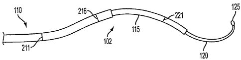
[0043] FIGs. 2A-2D illustrate active cannula 102 and its constituent flexible
tubes. FIG. 2A illustrates an exemplary outer flexible tube 110. Outer
flexible tube
110 may have an outer tube straight section 210, an outer tube curved section
212,
and an outer tube transition point 211 defining the boundary between outer
tube
straight section 210 and outer tube curved section 212. Outer flexible tube
110 may
have an inner diameter that is sufficiently wide to allow middle flexible tube
115 and
inner flexible tube 120 to slide independently within the inner surface of
outer flexible
tube 110. The thickness of outer flexible tube 110 may be a function of the
tube's
desired flexibility, which is described herein further below. Accordingly, the
thickness of outer flexible tube 110 may be tailored to provide a specified
flexibility.
11
CA 02630061 2008-05-15
WO 2007/059233
PCT/US2006/044386
The illustrated circular curvature of outer flexible tube 110 is exemplary,
and many
different curved shapes are possible, given the tube's material, its
thickness, and the
intended use of active cannula 102.
[0044] Outer flexible tube 110 may be made of a shape memory alloy, such as
nitinol, although other materials may be used provided that they are suitable
for
surgical use and have a flexibility that can be predetermined by, for example,
material
properties or by specifying the thickness of the tube walls.
[0045] FIG. 2B illustrates an exemplary middle flexible tube 115. Middle
flexible tube 115 may have a middle tube straight section 215, a middle tube
curved
section 217, and a middle tube transition point 216 defining the boundary
between
middle tube straight section 215 and middle tube curved section 217. Middle
flexible
tube 115 may have an inner diameter that is sufficiently wide to allow inner
flexible
tube 120 to slide within the inner surface of middle flexible tube 115. The
thickness
of middle flexible tube 115 may be a function of the tube's desired
flexibility, which
is described herein further below. Accordingly, the thickness of middle
flexible tube
115 may be tailored to provide a specified flexibility. The illustrated
curvature of
middle flexible tube 115 is exemplary, and many different curvatures are
possible,
given the tube's material, its thickness, and the intended use of active
cannula 102.
[0046] As in the case of outer flexible tube 110, middle flexible tube 115 may
be made of a shape memory alloy, such as nitinol, although other materials may
be
used provided that they are suitable for surgical use and have a flexibility
that can be
predetermined by, for example, specifying a certain thickness for the tube.
Further,
middle flexible tube 115 may or may not be made of the same material as outer
12
CA 02630061 2008-05-15
WO 2007/059233 PCT/US2006/044386
flexible tube 110, depending on the intended shape, thickness, and overall
flexibility
of active cannula 102.
[0047] FIG. 2C illustrates an exemplary inner flexible tube 120. Inner
flexible
tube 120 may have an inner tube straight section 220, an inner tube curved
section
222, and an inner tube transition point 221 defining the boundary between
inner tube
straight section 220 and inner tube curved section 222. Timer flexible tube
120 may
have an inner diameter that is sufficiently wide to serve as a cannula for
passing
fluids, etc. Further, the inner diameter may be sufficiently wide to enable a
cable,
such as a wire, needle, elastic push-rod, or fiberoptic cable, to be carried
to end
effector 125. The thickness of inner flexible tube 120 may be a function of
the tube's
desired flexibility, which is described herein further below. Accordingly, the
thickness of inner flexible tube 120 may be tailored to provide a specified
flexibility.
The illustrated curvature of inner flexible tube 120 is exemplary, and many
different
curvatures are possible, given the tube's material, its thickness, and the
intended use
of active cannula 102.
[0048] As in the case of outer flexible tube 110, inner flexible tube 120 may
be made of a shape memory alloy, such as nitinol, although other materials may
be
used provided that they are suitable for surgical use and have a flexibility
that can be
predetermined by, for example, specifying a certain thickness for the tube.
Further,
inner flexible tube 120 may or may not be made of the same material as outer
flexible
tube 110 and middle flexible tube 115.
[0049] End effector 125 may be one of many devices suitable for the intended
surgical intervention. For example, end effector 125 may be a thermal ablation
probe,
13
CA 02630061 2008-05-15
WO 2007/059233 PCT/US2006/044386
a fiber-optic camera, a tip for injecting radioactive seeds, a needle for
performing a
biopsy, and the like. Further, end effector 125 may be used for acquiring
tissue or
fluid samples for external analysis. Still further, end effector 125 may be a
bio-sensor
to be deployed within a site of interest. Such bio-sensors may include
stereotactic
positioners (e.g., magnetic trackers), molecular sensors, electrical impedance
sensors,
contactless mechanical impedance sensors, optical luminescent sensors, and the
like.
It will be readily apparent to one skilled in the art that end effector 125
may take
many forms and perform many different functions, all of which are within the
scope
of the invention.
[0050] FIG. 2D illustrates active cannula 102, including each of the tubes
illustrated in FIGs. 2A¨D. Inner flexible tube 120 is illustrated as inserted
into middle
flexible tube 115, and the combination of inner flexible tube 120 and middle
flexible
tube 115 are inserted within outer flexible tube 110.
[0051] FIG. 3 illustrates active cannula 102, including inner flexible tube
120,
middle flexible tube 115, and outer flexible tube 110. As illustrated, each
flexible
tube has two degrees of freedom: one around an axial rotational axis, and
another
along a linear translational axis. For example, outer flexible tube 110 has an
outer
rotational degree of freedom 305 and an outer translation degree of freedom
310.
Outer rotational degree of freedom 305 and outer translational degree of
freedom 310
apply to outer flexible tube 110 independently of the other tubes. Middle
flexible
tube 115 has a middle rotational degree of freedom 315 and a middle
translation
degree of freedom 320, both of which apply only to middle flexible tube 115
independently of the other tubes. Inner flexible tube 120 has an inner
rotational
14
CA 02630061 2008-05-15
WO 2007/059233 PCT/US2006/044386
degree of freedom 325 and an inner translation degree of freedom 330, both of
which
apply to inner flexible tube 120 independently of the other tubes.
[0052] Referring again to FIG. 3, active cannula 102 has a plurality of
overlap
transition points T1¨T5. Each overlap transition point T1--1'5 defines a
boundary of a
region in which the each of outer flexible tube 110, middle flexible tube 115,
and
inner flexible tube 120 (or some subset of the three) have a substantially
constant
degree of curvature, or lack of curvature. For example, the region between
overlap
transition points 1"1 and T2 includes outer tube curved section 212, middle
tube straight
section 215, and inner tube straight section 220. Overlap transition point T2
is
coincident with middle tube transition point 216. Accordingly, the region
between T2
and 7'3 includes outer tube curved section 212, middle tube curved section
217, and
inner tube straight section 220.
[0053] Each region bounded by at least one of overlap transition points T1--
2'5
has a curvature that is a function of the curvatures and flexibilities of each
of outer
flexible tube 110, middle flexible tube 115, and outer flexible tube 120, as
well as the
resistance of the surrounding tissue medium. One will note that some regions
have
only middle flexible tube 115 and inner flexible tube 120. In this case, the
curvature
of that region is a function of the curvature of those two tubes within the
region. In
the simplest case, the curvature of the region from 1'5 to end effector 125 is
a function
of the curvature of inner flexible tube 120 and the resistance of the
surrounding tissue
medium.
[0054] FIG. 4A illustrates a set of two-axis actuators according to the
present
invention. The two-axis actuators include outer drive module 130, which is
coupled
CA 02630061 2008-05-15
WO 2007/059233
PCT/US2006/044386
to outer flexible tube 110; middle drive module 135, which is coupled to
middle
flexible tube 115; and inner drive module 140, which is coupled to inner
flexible tube
120. Each of these drive modules independently drive their respective flexible
tube.
For example, outer drive module 130 drives outer flexible tube 110 about outer
rotational degree of freedom 305 and along outer translational degree of
freedom 310.
Middle drive module 135 drives middle flexible tube 115 about middle
rotational
degree of freedom 315 and along middle translational degree of freedom 320.
And
inner drive module 140 drives inner flexible tube 120 about inner rotational
degree of
freedom 325 and inner translational degree of freedom 330.
[0055] FIG. 4B illustrates an exemplary two-axis actuator 405 according to
the present invention. Two-axis actuator 405 may be used for any of outer
drive
module 130, middle drive module 135, and inner drive module 140. Two-axis
actuator 405 includes a lead screw 410, which may be rigidly attached to a
flexible
tube (outer flexible tube 110 is illustrated as an example); a nut 415 that is
threaded
onto lead screw 410; and a linear translation motor 435, which is coupled to
nut 415
via translation gear 425. Two-axis actuator 405 further includes a belt drive
440,
which is coupled to lead screw 410 via sprocket 437. Belt drive 440 is also
coupled to
rotation motor 450 via rotation gear 445. Two axis actuator 405 may also
include
translational and rotational encoders (not shown) that respectively provide
linear
translation position and angular orientation signals to control computer 145.
[0056] Two-axis actuator 405 may operate as follows. In the case of linear
translation, linear translation motor 430 receives commands from control
computer
145 to translate its flexible tube according to a particular translation
distance. In
16
CA 02630061 2008-05-15
WO 2007/059233 PCT/US2006/044386
response, linear translation motor 430 rotates translation gear 425, which
engages nut
415. The subsequent rotation of nut 425 engages lead screw 410, which
translates the
flexible tube.
[0057] In the case of rotation, rotation motor 450 receives commands from
control computer 145 to rotate according to a particular rotation angle. In
response,
rotation motor 450 rotates rotation gear 445, which engages belt drive 440.
Belt drive
440 engages sprocket 437, which in turn rotates lead screw 410. Note, this
rotation of
lead screw 410 causes a translation of lead screw 410 due to the presence of
nut 415.
Accordingly, to prevent a parasitic translation, linear translation motor 430
compensates by rotating nut 415 in the opposite direction. As such, pure
rotation of
the flexible tube may require coordinated motion by rotation motor 450 and
linear
translation motor 430.
[0058] As illustrated in FIG. 4B, lead screw 410 may be hollow. In this case,
if two-axis actuator 405 serves as outer drive module 130, then outer flexible
tube 110
is coupled to lead screw 410, and middle flexible tube 115 and inner flexible
tube 120
may independently translate and rotate within the hollow portion of lead screw
410.
In this way, outer flexible tube 110, middle flexible tube 115, and inner
flexible tube
120 may be translated and rotated independently.
[0059] FIG. 5 illustrates a set of manual two-axis actuators 505a¨c. Here,
manual two-axis actuator 505a may drive outer flexible tube 110 in place of
outer
drive module 130; manual two-axis actuator 505b may drive middle flexible tube
115
in place of middle drive module 135; and manual two axis actuator 505c may
drive
inner flexible tube 120 in place of inner drive module 140. Each of manual two
axis
17
CA 02630061 2008-05-15
WO 2007/059233
PCT/US2006/044386
actuators 505a¨c may include translational and rotational encoders, which
provide
linear position and angular orientation signals to control computer 145.
[0060] Variations to the two-axis drive modules are possible. For example,
two-axis actuator 405 may include manual controls, such as knobs, which
respectively
override linear translation motor 430 and rotational motor 450. Further,
system 100
may include a combination of motor-driven and manual actuators. Further, two-
axis
actuator 405 is exemplary. As such, there may be other ways of achieving
linear
translation and rotation of each of the flexible tubes apart from the ways
shown here.
One skilled in the art will readily appreciate that many such variations are
possible
and within the scope of the invention.
[0061] FIG. 6 illustrates an exemplary process 600 for controlling an active
cannula associated with the present invention. All or part of process 600 may
be
performed by the software stored on memory 152 and executed on host computer
150
and/or control computer 145 and/or imager processor 165. Process 600 may be
divided into two sub-processes: path planning (steps 605-625) and path plan
execution (steps 630-655).
[0062] In step 605, medical imaging system 160 acquires an image of
patient's anatomy 170. Medical imaging system 160 may be configured to have a
field of view than encompasses entry point 175 and the surgical site 180.
Depending
on its imaging modality (e.g. MRI, ultrasound, etc.), medical imaging system
160 may
acquire a 3-D image of patient's anatomy, whereby each pixel or voxel of the
image is
registered to an image coordinate frame. Imager processor 165 may provide the
18
CA 02630061 2008-05-15
WO 2007/059233 PCT/US2006/044386
image, as well as image registration information, to host computer 150 over
imaging
network connection 146b.
[0063] In step 610, the physician determines a desired path from entry point
175 to surgical site 180. In doing so, the physician may identify a path
through which
active cannula 102 will travel, along with an error boundary around the path.
Depending on the location of surgical site 180, and the presence of
intervening tissue
or organs, the path may involve a complex path having variable error
boundaries.
[0064] The physician may use user interface 155 to define the path and its
error boundaries. In doing so, the physician may use a cursor to tag points
within the
registered image acquired in step 605. The software identifies the location of
these
selected points in the registered image and stores these locations in memory
152.
[0065] In step 615, the software computes a final configuration of active
cannula 102 that will achieve the path selected in step 610. In doing so, the
software
may determine the translational position and rotational orientation of each of
outer
flexible tube 110, middle flexible tube 115, and inner flexible tube 120, that
will make
active cannula 102 conform to the path.
[0066] In computing a final configuration that conforms to the path, the
software divides active cannula 102 into a set of regions defined by overlap
transition
points T1-2"5. In doing so, the software may select an initial set of
translational
= positions and rotational orientations for each of outer flexible tube
110, middle
flexible tube 115. The locations of overlap transition points T1¨T5 depends on
the
overlap of the three flexible tubes. Then for each region bounded by overlap
19
CA 02630061 2008-05-15
WO 2007/059233
PCT/US2006/044386
transition points T1--T5, the software computes the instantaneous equilibrium
curvature
(in x and y components) in that region according to the following relation:
E EiI cos(Oi ¨ )K
= ____________________________ "
E E
and
EEiIisin(Oi
i=1
EEiIi
where n is the number of flexible tubes (n=3 in this example); K1 is the
instantaneous
curvature of the ith flexible tube in that region; Et is the Modulus of
Elasticity
(Young's Modulus) of the material in the ith flexible tube; i is the cross
sectional
moment of inertia of the id, flexible tube; Oi is the angular orientation of
the ith
flexible tube at the closest overlap transition point Tin the direction toward
the
actuators; and 0 is the equilibrium angle of combined flexible tubes given
their
individual angular orientations, wherein 0 is determined at the base of the
region. In
other words, for example, for a region bounded by overlap transition points T3
and
0 is pertains to the equilibrium angle at T3.
[0067] Of these terms, lc , Ei, and I are known. The remaining terms are
solved for by (1) computing the torsional energy in the straight sections
between the
actuators and the first transition point and the bending energy (as a function
of
flexible tube orientations) stored in active cannula, and (2) solving for the
shape that
provides the minimum energy. In doing so, the software computes the torsional
CA 02630061 2008-05-15
WO 2007/059233 PCT/US2006/044386
energy stored in straight sections 210, 215, and 220 of outer flexible tube
110, middle
flexible tube 115, and inner flexible tube 120, respectively; and the software
computes
the bending energy stored in curved sections 212, 217, and 222 of outer
flexible tube
110, middle flexible tube 115, and inner flexible tube 120, respectively. The
software
does this by computing the combined stored energy according to the following
relation:
1 _______________________________
E(q)= E " (ai 2 + LL
k(Kx¨Kicos(Ou ¨ j))2 + (K isin(0 _0))2)
j=1 I .1=1 i=12
where ai is the angle input at inner drive module 140, middle drive module
135, and
outer drive module 130; Oij is the angle of the ill, flexible tube at the jth
transition
point Ti; 65 +
r '02'= = = qiin are the equilibrium planes of each of the in regions of
overlap
between overlap transition points T; and q= (9
1,1,- 1,2 ,* , 01,
02,= = =,0m) = Solving
for the minimum value of E(q) yields the rotational orientations 01,1, 00, = =
=,(91,,, at Th
and the equilibrium planes
01 O2 = =Oln of each region of overlap between transition
points T. These values can also be used in the equations for xi, and icy above
to
compute the curvatures in each overlap region between transition points T of
active
cannula 102.
[0068] FIG. 7 illustrates a kinematic frame for controlling a flexible tube.
As
illustrated, 0 refers to the equilibrium angle of flexible tube 710 at an
overlap
transition point 7, and a refers to the input rotation angle imparted by the
rotational
motor of two-axis actuator 405.
21
CA 02630061 2008-05-15
WO 2007/059233
PCT/US2006/044386
[0069] Further to step 615, the software may select different tubes from
among an inventory of tubes for outer flexible tube 110, middle flexible tube
115, and
inner flexible tube 120. In this case, a plurality of each flexible tube types
may be
available, and their characteristics (length of straight section, length of
curved section,
radius of curvature of the curved section, flexibility, etc.) may be stored in
memory
152. As such, the software may repeat the above computation within step 610
described above, wherein each iteration uses a different available tube. In
this
manner, the software can determine two things: first, whether the path
determined by
the physician can be replicated by active cannula 102; and second, what
combination
of tubes will achieve that path. Further, the above relations are not limited
to three
flexible tubes. Accordingly, the software may select varying combinations of
tubes,
including the number of flexible tubes to be used, in order to achieve the
path
determined by the physician. One skilled in the art will understand how to
implement
the above equations for more than three flexible tubes.
[0070] In step 620, the software computes a plurality of configurations for
active cannula 102 that will enable active cannula to gradually achieve the
final
configuration computed in step 615, while not having the active cannula stray
beyond
the path and error boundaries determined by the physician. In doing so, the
software
may compute a series of intermediate configurations, and compute a set of
linear
translations and rotations that will achieve each intermediate configuration.
The
software may iteratively perform computations substantially similar to that
performed
in step 615 above, with the resulting configuration for each computed
intermediate
22
CA 02630061 2008-05-15
WO 2007/059233
PCT/US2006/044386
configuration being the initial configuration for the next computed
intermediate
configuration.
[0071] Further to step 620, the software may compute a sequence of rotation
commands for rotation motors 450 and linear translation commands for linear
translation motors 430, of each outer drive module 130, the middle drive
module 135,
and the inner drive module 140, in order to achieve each intermediate
configuration in
sequence.
[0072] In step 625, the software registers the final and intermediate
configurations for active cannula, as respectively computed in steps 615 and
620, in
the coordinate frame of medical imaging system 160. In doing so, the software
may
retrieve the registered image acquired in step 605, in which the physician had
designated a path in step 610, and register the final and intermediate
configurations of
active cannula 102. The result of this may be a set of curves, one per
intermediate
configuration and one for the final configuration, wherein each set of curves
corresponds to the regions of active cannula 102 between a overlap transition
points
T1-T5. The software may do this by starting at an origin point for the active
cannula
(registered in image space), proceeding through entry point 175, and
concluding at
surgical site 180 (or at end effector 125 for active cannula 102 in an
intermediate
configuration). The software stores these sets of curves in memory 152.
[0073] This completes the exemplary path planning subprocess of process
600. The path planning sub-process may be performed in the operating room,
immediately before performing surgery. Alternatively, the path planning sub-
process
may be done pre-operatively and in a different setting than the operating
room. In the
23
CA 02630061 2008-05-15
WO 2007/059233
PCT/US2006/044386
latter case, the image acquired in step 605 may be out of date, because the
patient will
have moved between the path planning sub-process and the execution sub-
process. In
this case, a new registered image will have to be acquired by medical imaging
system
160 as a precursor to the execution sub-process, and the newly-acquired image
will
have to be registered to the earlier registered image having the registered
configurations (curves) of active cannula 102 computed in step 625. Further
information regarding robotic path planning can be found in Planning
Algorithms,
Steven M. LaValle, Cambridge University Press (2006), (ISBN-10: 0521862051 I
ISBN-13: 9780521862059), which is hereby incorporated by reference as if fully
disclosed herein.
[0074] At the outset of the execution sub-process, the patient is prepared for
surgery and patient's anatomy 170 is placed within the field of view of
medical
imaging system 160, as illustrated in FIG. 1. Active cannula 102 is placed in
the
vicinity of entry point 175, and outer drive module 130, middle drive module
135, and
inner drive module 140 are connected to active cannula 102. Control computer
145 is
connected to the three drive modules 130, 135, and 140, and communications is
established between control computer 145 and host computer 150 over control
network connection 146a.
[0075] In step 630, the first step of the execution sub-process, the physician
(via user interface 155) issues a command to the software to move active
cannula 102
to the first intermediate configuration computed in step 630 (in the path
planning sub-
process). In doing so, the software, which may be running on host computer 150
and/or control computer 145, issues appropriate commands to the translational
motors
24
CA 02630061 2008-05-15
WO 2007/059233 PCT/US2006/044386
430 and the rotational motors 450 of each of outer drive module 130, middle
drive
module 135, and inner drive module 140, to achieve the first intermediate
configuration computed in step 620.
[0076] In step 635, medical imaging system 160 acquires an image of active
cannula 102 within patient's anatomy 170. In doing so, imager processor 165
may
segment and register active cannula 102 in the image coordinate frame. Imager
processor 165 may employ one or more segmentation algorithms that are known to
the art. Imager processor 165 may transmit the registration information and
the image
to host computer 150 over imaging network connection 136b. The software may
receive the registration information and the image of active cannula 102
within
patient's anatomy 107 and present the information and image to the physician
via user
interface 155.
[0077] In step 640, the software compares the registered image of cannula 102
with the intermediate configuration computed in step 620. In doing so, the
software
may employ one or more of a number of image processing algorithms for
comparing
the two images. Further, the software may compare the coordinates of the
segmented
and registered active cannula 102 with the computed coordinates of the given
intermediate configuration and compute a path error, or differential
displacement,
based on this comparison.
[0078] In step 645, the software determines if there is a discrepancy between
the segmented and registered active cannula 102 with the given intermediate
configuration. If there is no discrepancy, process 600 proceeds through the
"NO"
branch from step 645 to step 655.
CA 02630061 2008-05-15
WO 2007/059233
PCT/US2006/044386
[0079] In step 655, the software determines if the given intermediate
configuration is the final configuration computed in step 615. If it is,
process 600
may proceed through the "YES" branch of step 655 to completion. If it is not
the
final configuration, then process 600 may proceed through the "NO" branch of
step
655 to repeat steps 630-645 with the next intermediate configuration (or the
final
configuration).
[0080] Returning to step 645, if there is a discrepancy between the segmented
and registered active cannula 102 with the given intermediate configuration,
process
600 may proceed through the "YES" branch of step 645 to step 650.
[0081] In step 650, the software computes the force and torque exerted on
active cannula 102 as it was pushed through patient's anatomy 170 in step 630.
The
software may compute the force and the torque according to the following
relations:
fx- dispx
dispy
fz =fro dispz
Dx rotx
Dy rot
_rz _ rotz _
where f are components of the force imparted by the tissue medium on
active
x,y,z
cannula 102 at a given region between two overlap transition points Ti and
Ti+1; rx,y,z
are the torques imparted on active cannula 102 by the tissue medium on active -
cannula 102 at the same region; dispxo,,z are translational components of the
differential displacement of active cannula 102 computed in step 640; rotx,y,z
are the
rotational components of the differential displacement of active cannula 102
26
CA 02630061 2008-05-15
WO 2007/059233 PCT/US2006/044386
computed in step 640; and K is a compliance matrix, which is a 6x6 matrix
corresponding to the force and torque compliance of active cannula 102 for the
given
region between two overlap transition points Ti and Ti+/.
[0082] Compliance matrix K may be predetermined in a calibration procedure
in which active cannula 102 is translated and rotated in one or more phantoms
having
known resistance properties. In addition, if compliance matrix K is known,
then
active cannula 102 may be used as a force sensor. In this case, a physician
may plan a
path for active cannula (using all or part of exemplary process 600) so that
end
effector 125 may come in contact with a tissue region of interest. Once end
effector
125 comes in contact with the tissue region of interest, the values for f
and
computed in step 650 may respectively correspond to the force and torque
x,y,z
imparted on end effector 125 by the tissue region of interest. Accordingly,
active
cannula 102 may be used as a force sensor.
[0083] FIG. 8 illustrates how strain relates to the side lengths of a flexible
tube, which may be any of outer flexible tube 110, middle flexible tube 115,
and inner
flexible tube 120. The software, in computing the final and intermediate
configurations in steps 615 and 620, may determine the maximum degree of
curvature, or minimum radius of curvature, beyond which a given flexible tube
will
suffer plastic deformation. Plastic deformation refers to the degree of
bending of a
shape memory material such that the material will no longer return to its
original
shape. This may correspond to a limit of permissible curvature of a flexible
tube.
The software may compute the maximum degree of curvature according to the
following relation:
27
CA 02630061 2008-05-15
WO 2007/059233
PCT/US2006/044386
1c= _____________________________________
d(l+s)
where d is the diameter of the flexible tube, and c is the maximum recoverable
strain
for the flexible tube's material. For nitinol, e may range from 0.08 to 0.1.
As can be
inferred from the above relation, the thinner the flexible tube, the greater
the
maximum degree of curvature (or the lesser the minimum radius of curvature).
Accordingly, depending on the path determined by the physician in step 610, a
thinner
flexible tube may be desired. The software may assist the physician in
selecting a
preferred thickness of flexible tube depending on the path determined in step
610.
[0084] Variations to active cannula 102, system 100, and process 600, are
possible and within the scope of the invention. For example, some or all of
the
flexible tubes in active cannula 102 may have substantially the same degree of
flexibility, or they may each have different degrees of flexibility. If all of
the flexible
tubes have a similar flexibility, it may make active cam-Lila 102 more agile
and easier
to guide through complex paths. Alternatively, outer flexible tube 110 may be
stiffer
than middle flexible tube 115, which may be in turn stiffer than inner
flexible tube
120. In the latter case, active cannula 102 may be less agile than in the
former case
(in which all the flexible tubes have the same flexibility). However, in the
latter case,
the path of active cannula 102 may be easier to compute, and it may better
enable
manual operation, for example, by using manual two-axis actuators 505
illustrated in
FIG. 5.
[0085] In another variation, any of the flexible tubes may have non-circular
inner and/or outer shapes. Such variations to a flexible tube's cross section
may
provide differing flexibility as a function of bend angle. Further, a flexible
tube may
28
CA 02630061 2008-05-15
WO 2007/059233
PCT/US2006/044386
have different shaped regions along its length, whereby each region may have a
different cross sectional shape.
[0086] Any of the flexible tubes within active cannula 102 may have only a
curved portion or a straight portion. Further, any of the flexible tubes may
have
multiple segments, each with a different degree of curvature (including no
curvature).
This may allow active cannula 102 to take more complex shapes. For example,
any
of the flexible tubes may have sequences of three-dimensional curves and
straight
regions. Also, any of the flexible tubes may have a segment having an complex
shape, such as a helical shape, an elliptical shape, a parabolic shape, a
variable
curvature in three dimensions, and the like. In any of these cases, multiple
transition
points (like inner tube transition point 221, middle transition point 216, and
outer tube
transition point 211) may be defined that mark changes in radius of curvature
of the
particular flexible tube. Accordingly, discrete gradations of curvature may be
segregated for the purposes of defining overlap regions, as part of computing
cannula
final and intermediate configurations in steps 615 and 620.
[0087] In another variation, one or more of the flexible tubes may be designed
to have a variable stiffness according to the direction in which the flexible
tube is
bent. For example, one or more of the flexible tubes may have scores or
grooves on
the inner or outer surface of the flexible tube.
[0088] In another variation, one or more of the flexible tubes may include
fiducials, which may be embedded within the tube material, and which may be
designed to be visible to medical imaging system 160. For example, if medical
imaging system 160 is an optical camera, embedded fiducials may take the form
of
29
CA 02630061 2008-05-15
WO 2007/059233 PCT/US2006/044386
colored stripes or bands of light and dark color. Further, if medical imaging
system is
a C-arm fluoroscope, embedded fiducials may include wire structures implanted
within the tube material. One skilled in the art will readily appreciate that
many such
variations are possible and within the scope of the invention.
[0089] If nitinol is used for any of the flexible tubes described above, then
system 100 may include one or more heater elements, which may run along one or
more of flexible tubes 110, 115, and 120. According to this variation, heat
can be
applied to change the shape of a given flexible tube. One skilled in the art
will
understand how to integrate a heater element into active cannula 102 and
system 100
and that such a variation is within the scope of the invention.
[0090] In addition to lung and throat surgery, as mentioned above, the present
invention may be used in other surgical procedures, in which the dexterity
afforded by
active cannula 102 and system 100 may be advantageous. Such surgical
procedures
include Radiofrequency Ablation. In Radiofrequency Ablation, an electrode is
placed
at a surgical site, and then a painless radiofrequency energy is transmitted
to heat the
tissue surrounding the electrode. This procedure may be used to kill cells as
part of a
treatment for tumors of the liver, kidney, and lung. Active cannula 102 and
system
100 may be employed to deploy the electrode.
[0091] Another possible surgical application involves surgical interventions
on the posterior side of the retina. One such surgical intervention may
include
cumulation of the retina to treat clotting, which is one of the leading causes
of
blindness.
CA 02630061 2013-05-27
[0092] Another possible surgical application involves transgastric surgery,
in which tools enter the stomach via the mouth, then exit the stomach into the
abdominal cavity. The dexterity of active carmula 102, and its ability to be
guided
through free space as well as through tissue, may enable transgastric surgery.
[0093] In another variation, system 100 may include a second active
cannula 102, which includes a second set of inner, middle, and outer drive
modules
connected to control computer 145. In this variation, the two active cannulas
can
be used as a parallel robot (a "Stuart Platform" is an exemplary type of
parallel
robot, but many variants are known in the art) whereby the tips of the inner
flexible tubes of the two active cannulas are coupled to a single end effector
125.
Doing so may enable the system to control the position and orientation of the
end
effector as well as control the stiffness of the position and orientation. In
another
application of the variation to system 100 having two active cannulas, the two
active cannulas may be deployed within patient's anatomy 170 and used as
retractors for holding soft tissue away from and exposing a surgical site.
[0094] Although the above description pertains to a surgical application of
the
present invention, it will be readily apparent to one skilled in the art that
the present
invention may be used in other applications that require guiding a device
through a
complex path that involves free space. Other applications may include
manufacturing
and micro-assembly, remote structural inspection, defusing ordinance, search
and
rescue within collapsed structures, and the like.
[0095] It will be apparent to those skilled in the art that various
modifications and variations can be made in the present invention. Thus, the
31
CA 02630061 2013-05-27
,
scope of the claims should not be limited by the preferred embodiments set
forth in
the examples, but should be given the broadest interpretation consistent with
the
description as a whole.
32