Note: Descriptions are shown in the official language in which they were submitted.
CA 02635320 2008-06-18
SPACER WITH A COATING THEREON FOR USE WITH AN IMPLANT DEVICE
FIELD OF THE INVENTION
[oooi] The present invention generally relates to a spacer for use with
implant devices,
e.g., bone plates, and, more specifically, to spacers having a coating
thereon, wherein
the coating includes a therapeutic healing agent(s) such as to stimulate bone
growth
and/or promote fracture healing.
BACKGROUND
[0002] Implant devices, such as bone plates, can be implanted in the body for
the
splinting of a fracture at a bone. To that end, the bone plate may be provided
with one or
more holes and accompanied by one or more securing means, e.g., bone screws,
as well
as spacer devices. The spacer device, or spacer, can be shaped to fit within
the hole in
the bone plate and accommodate the screw. The spacer, thus, may be inserted
within a
corresponding hole of the bone plate, then the screw inserted through both the
hole and
spacer. The screw may be screwed into bone to fix the bone plate thereto for
splinting of
a fracture, with the spacer being situated between the bone screw and the bone
plate in
the direction towards the fracture upon implantation. The spacer, which may be
polymeric and elastic in nature, functions to improve bone fracture healing by
acting as a
cushion between the bone plate and the bone screw and by decreasing the area
of
contact between bone and the bone plate thereby permifting a restricted
displacement in
compression stressing of the bone.
[0003] It would be desirable to provide an improved spacer for use with an
implant
device, e.g., a bone plate, which further stimulates bone growth and/or
promotes fracture
healing.
SUMMARY
[o004] Certain exemplary aspects of the invention are set forth below. It
should be
understood that these aspects are presented merely to provide the reader with
a brief
summary of certain forms the invention might take and that these aspects are
not
CA 02635320 2008-06-18
intended to limit the scope of the invention. Indeed, the invention may
encompass a
variety of aspects that may not be explicitly set forth below.
[0005] In an embodiment of the present invention, a device defining a spacer,
e.g., a
polymeric spacer, is provided for use with an implant device, e.g., a bone
plate, for
splinting a fracture of a bone. The spacer includes a body defining a bone
healing
surface, wherein at least a portion of the bone healing surface has a coating
thereon
which includes a therapeutic agent, a polymeric carrier, and a buffer medium
to stimulate
bone growth and/or promote fracture healing.
[0006] In another embodiment, a kit is provided which includes one or more
spacers, at
least one bone plate, and optionally one or more bone screws for securing he
bone plate
to bone. At least one spacer includes a body defining a bone-healing surface.
At least a
portion of the bone-healing surface includes a coating having a therapeutic
agent, a
polymeric carrier, and a buffer medium to stimulate bone growth and/or promote
fracture
healing.
[0007] In another embodiment, a method for healing bone is provided which
includes
securely situating a bone plate adjacent a bone wherein the bone plate
includes a spacer
having a coating on at least a portion thereof. The coating is in contact with
the bone
and includes a therapeutic agent, a polymeric carrier, and a buffer medium for
healing
bone. In one example, the coating is placed on at least the portion of the
spacer prior to
securely situating the bone plate. In another example, the therapeutic agent,
the
polymeric carrier, and the buffer medium, which define the coating, are mixed
prior to
placing the coating on at least the portion.
[oo08] Concerning the coating, the therapeutic agent can include a drug, a
biological
factor, or mixtures thereof; the polymeric carrier can include a bioresorbable
or water-
soluble polymer, a hydrogel-forming polymer, a polyelectrolyte, or mixtures
thereof; and
the buffer medium can include deionized water, phosphate buffer saline, normal
saline,
serum, whole blood, or mixtures thereof.
[ooo9l Various features discussed below in relation to one or more of the
exemplary
embodiments may be incorporated into any of the above-described aspects of the
present invention alone or in any combination. Again, the brief summary
presented
2
CA 02635320 2008-06-18
above is intended only to familiarize the reader with certain aspects and
contexts of the
present invention without limitation to the claimed subject matter.
BRIEF DESCRIPTION OF THE FIGURES
[0010] Various features, aspects, and advantages of the present invention will
become
better understood when the following detailed description is read with
reference to the
accompanying figures in which like characters represent like parts throughout
the figures,
wherein:
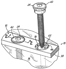
[ooll] Figure 1 is a perspective view of a section of bone plate secured to
bone by a first
bone screw, and a spacer positioned in a hole of the bone plate receiving a
corresponding second bone screw.
[0012] Figure 2 is a cross-sectional view of the bone plate of figure 1 taken
along line 2-2;
and
[0013] Figure 3 is a perspective view of the spacer of Fig. 1.
DETAILED DESCRIPTION OF EXEMPLARY EMBODIMENTS
[0ou] One or more specific embodiments of the present invention will be
described
below. In an effort to provide a concise description of these embodiments, all
features of
an actual implementation may not be described in the specification. It should
be
appreciated that in the development of any such actual implementation, as in
any
engineering or design project, numerous implementation-specific decisions must
be
made to achieve the developers' specific goals, such as compliance with system-
related
and business-related constraints, which may vary from one implementation to
another.
Moreover, it should be appreciated that such a development effort might be
complex and
time consuming, but would nevertheless be a routine undertaking of design,
fabrication,
and manufacture for those of ordinary skill having the benefit of this
disclosure.
[oois] When introducing elements of the present invention (E.G., the exemplary
embodiments(s) thereof), the articles "a", "an", "the" and "said" are intended
to mean that
there are one or more of the elements. The terms "comprising", "including" and
"having"
3
CA 02635320 2008-06-18
are intended to be inclusive and mean that there may be additional elements
other than
the listed elements.
[0016] Figs. 1-3 show an embodiment of the present invention including a
medical device
including an implant device 12, e.g., a bone plate (shown in partial), for
splinting a
fracture of a bone 14 and a spacer 16, such as a polymeric spacer, with a
coating 18
thereon used in combination with the bone plate 12 to stimulate bone growth
and/or
promote fracture healing.
(0017] With reference to Figs. 1 and 2, the bone plate 12 includes two holes
22 with each
22 hole receiving a corresponding polymeric spacer 16 and a corresponding bone
screw
24. The bone plate 12 may be composed of metals and metal alloys, such as
titanium or
titanium alloys, tantalum or tantalum alloys (e.g., Ti6A14V or ProtosulTM),
stainless steel
or alloys thereof, cobalt-based alloys, cobalt-chromium alloys, cobalt-
chromium-
molybdenum alloys, niobium alloys, zirconium alloys, as well as shape memory
alloys
such as NiTiNOL. The bone plate 12 may define, for example, a compression bone
plate (e.g. an axially compressive bone plate) or locking bone plate as are
known in the
art.
[0018] The polymeric spacer 16, as best shown in Fig. 3, includes a generally
circular-
shaped body 26 having an aperture 28 therethrough so as to receive a
correspondingly-
shaped screw 24 and further includes a protrusion 32 extending generally
perpendicularly away from the body 26 to help retain the polymeric spacer 16
within the
hole 22, as generally discussed further below. The polymeric spacer 16
functions to
improve bone fracture healing by acting as a cushion between the bone plate 12
and
bone screw 24 and by decreasing the area of contact between bone 14 and the
bone
plate 12 thereby permitting a restricted displacement in compression stressing
of the
bone 14. And, although shown as being generally circular-shaped and having the
protrusion 32 therefrom, it should be understood by one having ordinary skill
in the art
that various spacer 16 configurations may be provided for cooperation with
differently
shaped and sized holes 22 and/or screws 24.
[ooi9] The coating 18 on spacer 16 includes a therapeutic healing agent, a
polymeric
carrier, and a buffer medium. The coating 18 is applied to a bottom, or bone-
healing,
4
CA 02635320 2008-06-18
surface 34 of the spacer 16, and contacts the bone 14 (or bony tissue) when
the bone
plate 12 is implanted. Such coating 18 helps mitigate the development of
stress
shielding and further promotes bone growth and/or fracture healing. One such
suitable
bone plate 12 (with screws 24) and polymeric spacer 16, which may receive the
coating
18 in accordance with an embodiment of the present invention, are disclosed in
U.S.
Patent No. 6,540,746 to Buhler et al. entitled "Bone Plate for Splinting a
Fracture at a
Bone with a Plurality of Bone Screws", which is expressly incorporated by
reference
herein in its entirety.
[0020] As best shown in Figs. 1 and 2, the bone plate 12 is attached to the
bone 14 using
each bone screw 24. Prior to positioning the screws 24 within corresponding
holes 22, a
corresponding polymeric spacer 16 first is positioned in each hole 22. To
position the
polymeric spacer 16, the polymeric spacer 16 may be pressed into the hole 22
from the
underside 38 of the bone plate 12, which lies adjacent to the bone 14 when
implanted.
The polymeric spacer 16 is held in place within the hole 22 by a snap or
friction-type fit
and is oriented so that the coating 18 on the polymeric spacer 16 contacts
bone 14 when
the bone plate 12 is implanted. The bone screws 24 then are inserted through
the
corresponding hole 22 and spacer 16, and ultimately anchored in the bone 14
and
braced thereagainst via contact surface 40. The screw head 42, which is sunk
within the
bone plate 12, has in its upper region a shoulder 44 that lies in contact with
a ring-
shaped ledge 46 in the hole 22 of the bone plate 12 and limits the plate's
upward
movement in the direction of a screw axis 48. The contact surface 40 of the
bone screw
24 projects beyond the underside 38 of the bone plate 12, which is at least so
large that
the underside 38 does not lie in contact with the bone 14. The distance is
chosen to be
greater than about 0.2 mm in order that the underside 38 of the bone plate 12
reliably
lies spaced apart from the bone 14 between the bone screws 24.
[0021] The polymeric spacer 16 likewise projects beyond the underside 38 of
the bone
plate 12 at its bone-healing surface 34 by a distance, which can be smaller
than the
distance for the contact surface 40 of the screw 24, in order that the bone
plate 12 is
braced with only a limited force between the polymeric spacer 16 and the
shoulder 44. A
compression of the bone 14 and a moving back is possible insofar as the
polymeric
CA 02635320 2008-06-18
spacer 16 and the friction between the shoulder 44 and the ledge 46, which is
produced
by the bias force, permit. Because the material for the polymeric spacer 16
may be
bioresorbable, the deflections of micro-movements can be controlled temporally
in such
a manner that pressure peaks, which become ever greater but still remain
tractable
during backward movement, are permitted at the fracture. The bone 14 can thus
take
over its carrying function in accordance with the healing process, which has a
very
positive effect on bone forming.
[0022] The polymeric spacer 16, in accordance with embodiments of the present
invention, may be composed of a bioresorbable or biostable polymer and
includes a
desired elasticity. The bioresorbable polymer can include a poly-D, L-lactide
(PDLLA),
which may be resorbed through hydrolysis in approximately 30 weeks. A suitable
PDLLA
is Resomer R208 available from the Boehringer Company of lngelheim, Germany.
The
bioresorbable polymer can also include poly (L) lactide (PLLA), a copolymer of
PLLA and
PDLLA, polyglycolide (PGA), and copolymers of PGA and polylactide with
different
molecular weights (or inherent viscosity). Biostable polymers can include
poly(methylmethacrylate), poly (ether ether ketone), ultrahigh molecular
weight
polyethylene, and polyurethane, for example.
[0023] As best shown in Fig. 2, the bottom, or bone-healing, surface 34 of the
polymeric
spacer 16 is coated with coating 18, which is in contact with the bone 14.
That coating
18, as disclosed above, includes a therapeutic healing agent, a polymeric
carrier, and a
buffer medium. The therapeutic agent is such that it promotes bone growth
and/or
fracture healing. The coating is applied at a thickness that allows delivery
of a desired
amount of the therapeutic agent over a desired period of time.
[oo24] The therapeutic healing agent of the coating 18 can include, for
example, a drug
or biological factor, such as an osteogenic agent, an osteoinductive agent, or
mixture
thereof, which can promote bone growth and/or healing, thus, enhancing the
overall
healing characteristics of the medical device. Such osteogenic and
osteoinductive
agents can include, for example, members of the families of Bone Morphogenetic
Proteins (BMPs), Osteoprotegerin or any of the other osteoclastogenesis
inhibitors,
Connective Tissue Growth Factors (CTGFs), Vascular Endothelial Growth Factors
6
CA 02635320 2008-06-18
(VEGFs), Transforming Growth Factor-betas (TGF-(3s), Growth Differentiation
Factors
(GDFs), Cartilage Derived Morphogenic Proteins (CDMPs), and Lim Mineralization
Proteins (LMPs). Osteoconductive agents may optionally be provided in the
coating 18
along with the osteogenic and/or osteoinductive agents.
[0025] BMPs are a class of proteins thought to have osteoinductive or growth-
promoting
activities on endogenous bone tissue, or function as pro-collagen precursors.
Known
members of the BMP family that may be utilized as osteoinductive agents in
tissue
attachment formulations include BMP-1, BMP-2, BMP-3, BMP-4, BMP-5, BMP-6, BMP-
7,
BMP-8, BMP-9, BMP-10, BMP-11, BMP-12, BMP-13, BMP-15, BMP-16, BMP-17, and
BMP-18 polynucleotides and polypeptides, as well as mature polypeptides and
polynucleotides encoding the same. The BMPs may be included in the coating 18
as full
length BMPs or fragments thereof, or combinations or mixtures thereof, or as
polypeptides or polynucleotides encoding the polypeptide fragments of all of
the recited
BMPs. (Termaat et al., J Bone Joint Surg Am., 87:1367-138, 2005).
[00261 Osteoclastogenesis inhibitors inhibit bone resorption by osteoclasts of
the bone
tissue surrounding the site of implantation. Osteoclast and Osteoclastogenesis
inhibitors
include osteoprotegerin polynucleotides and polypeptides, as well as mature
Osteoprotegerin polypeptides and polynucleotides encoding the same. The
Osteoprotegerin protein specifically binds to its ligand, osteoprotegerin
ligand
(TNFSF1 1/OPGL), both of which are key extracellular regulators of osteociast
development. Osteoclastogenesis inhibitors further include chemical compounds
such
as bisphosphonates (e.g., alendronate, clodronate, etidronate, ibandronate, (3-
amino-1-
hydroxypropylidene)-1,1-bisphosphonate (APD), dichloromethylene
bisphosphonate,
aminobisphosphonatezolendronate, zoledronic acid, and pamidronate) (Morris et
al., J
Bone Joint Surf Am., 87: 1609-1618, 2005), 5-lipoxygenase inhibitors such as
those
described in U.S. Pat. Nos. 5,534,524 and 6,455,541 (herein incorporated by
reference),
heterocyclic compounds such as those described in U.S. Pat. No. 5,658,935
(herein
incorporated by reference), 2,4-dioxoimidazolidine and imidazolidine
derivative
compounds such as those described in U.S. Pat. Nos. 5,397,796 and 5,554,594
(herein
incorporated by reference), sulfonamide derivatives such as those described in
U.S. Pat.
7
CA 02635320 2008-06-18
No. 6,313,119 (herein incorporated by reference), and acyiguanidine compounds
such
as those described in U.S. Pat. No. 6,492,356 (herein incorporated by
reference).
[00271 CTGFs are a class of proteins thought to have growth-promoting
activities on
connective tissues. Known members of the CTGF family include CTGF-1, CTGF-2,
and
CTGF-4, any of which may be incorporated into the coating 18, in addition to
polypeptides and polynucleotides encoding the same.
[0028] VEGFs are a class of proteins thought to have growth-promoting
activities on
vascular tissues. Known members of the VEGF family include VEGF-A, VEGF-B,
VEGF-
C, VEGF-D and VEGF-E, any of which may be incorporated into the coating 18, in
addition to polypeptides and polynucleotides encoding the same.
[0029] TGF-(3s are a class of proteins thought to have growth-promoting
activities on a
range of tissues, including connective tissues. Known members of the TGF-R
family
include TGF-P-1, TGF-(3-2, and TGF-(3-3, any of which may be incorporated into
the
coating 18, in addition to polypeptides and polynucleotides encoding the same.
[003o] Known GDFs include GDF-1, GDF-2, GDF-3, GDF-7, GDF-10, GDF-11, and GDF-
15. GDF-1 polynucleotides and polypeptides generally correspond to GenBank
Accession Numbers M62302, AAA58501, and AAB94786; GDF-2 polynucleotides and
polypeptides correspond to GenBank Accession Numbers BC069643, BC074921,
Q9UK05, AAH69643, and AAH74921; GDF-3 polynucleotides and polypeptides
correspond to GenBank Accession Numbers AF263538, BC030959, AAF91389,
AAQ89234, and Q9NR23; GDF-7 polynucleotides and polypeptides correspond to
GenBank Accession Numbers AB158468, AF522369, AAP97720, and Q7Z4P5; GDF-10
polynucleotides and polypeptides correspond to GenBank Accession Numbers
BC028237 and AAH28237; GDF-1 1 polynucleotides and polypeptides correspond to
GenBank Accession Numbers AF100907, NP005802 and 095390; and GDF-15
polynucleotides and polypeptides correspond to GenBank Accession Numbers
BC008962, BC000529, AAH00529, and NP004855.
[0031] Known CDMPs and LMPs include CDMP-1, CDMP-2, LMP-1, LMP-2, and LMP-3.
CDMP-1 polynucleotides and polypeptides generally correspond to GenBank
Accession
Numbers NM000557, U13660, NP000548 and P43026; CDMP-2 polypeptides
8
CA 02635320 2008-06-18
correspond to GenBank Accession Numbers and P55106; LMP-1 polynucleotides and
polypeptides correspond to GenBank Accession Numbers AF345904 and AAK30567;
LMP-2 polynucleotides and polypeptides correspond to GenBank Accession Numbers
AF345905 and AAK30568; and LMP-3 polynucleotides and polypeptides correspond
to
GenBank Accession Numbers AF345906 and AAK30569.
[0032] Additional osteoinductive and osteoconductive agents, factors, and
compounds
such as hydroxyapatite (HA), tricalcium phosphate (TCP), collagen, fibronectin
(FN),
osteonectin (ON), endothelial cell growth factor (ECGF), cementum attachment
extracts
(CAE), ketanserin, human growth hormone (HGH), animal growth hormones,
parathyroid
hormone (PTH) (Aleksyniene and Hvid, Medicina (Kaunas), 40, 842-849, 2004),
epidermal growth factor (EGF), interieukin-1 (IL-1), human alpha thrombin,
insulin-like
growth factor (IGF-1), platelet derived growth factors (PDGF), fibroblast
growth factors
(FGF, RFGF, etc.), and Wnt proteins, and derivatives thereof also can be
included as
therapeutic agents.
[00331 Other examples of therapeutic healing agents can include glycogen
synthase
kinase 3 (GSK-3) inhibitors, biocidal/biostatic sugars such as dextran and
glucose,
vitamins, cartilage fragments, natural extracts, genetically engineered living
cells, or
otherwise modified living cells, permeation enhancers such as fatty acid
esters including
laureate, myristate, and stearate monoesters of polyethylene glycol, salts
such as
strontium salt, fluoride salt, magnesium salt, and sodium salt, bone marrow
aspirate,
bone marrow concentrate, and mixtures and combinations thereof.
[0034] Therapeutic agents that are full-length proteins or fragments may be
conjugated to
polyethylene glycol (PEG) moieties to increase their half-life in vivo (also
known as
pegylation). Methods of pegylating polypeptides are well known in the art. In
addition,
the biological factor(s) may be delivered by gene therapy vectors harboring
the
polynucleotides encoding the biological factor of interest. The vector may be,
for
example, a phage, plasmid, viral, or retroviral vector. Such gene therapy and
delivery
techniques are known in the art. Gene therapy vectors further comprise
suitable
adenoviral vectors. Suitable gene therapy vectors include gene therapy vectors
that do
not integrate into the host genome and gene therapy vectors that integrate
into the host
9
CA 02635320 2008-06-18
genome. A desired polynucleotide also may be delivered in plasmid
formulations.
Plasmid DNA or RNA formulations refer to polynucleotide sequences encoding
osteoinductive polypeptides that are free from any delivery vehicle that acts
to assist,
promote, or facilitate entry into the cell, including viral sequences, viral
particles,
liposome formulations, lipofectin or precipitating agents and the like.
[00351 The biological factors also may be available as heterodimers or
homodimers, as
well as multimers or combinations thereof. Recombinantly expressed proteins
may be in
native forms, truncated analogs, muteins, fusion proteins (e.g., fusion
proteins with the
FC portion of human IgG), and other constructed forms capable of inducing
bone,
cartilage, or other types of tissue formation as demonstrated by in vitro and
ex vivo
bioassays and in vivo implantation in mammals, including humans. Examples of
fusion
proteins include ligand fusions between mature osteoinductive polypeptides and
the FC
portion of human Immunoglobulin G(IgG). Methods of making fusion proteins and
constructs encoding the same are known in the art.
[00361 Examples of suitable drugs include antitumor agents and
chemotherapeutics such
as cis-platinum, ifosfamide, methotrexate, and doxorubicin hydrochloride,
immuno-
suppressants, statins, pain killers and anti-inflammatories such as non-
steroidal anti-
inflammatory drugs (NSAID) like ketorolac tromethamine, lidocaine
hydrochloride,
bipivacaine hydrochloride, and ibuprofen, antibiotics or other bactericidal
agents, and
antiretroviral drugs. Bactericidal drugs and antiretroviral drugs may be
provided to
prevent infection by pathogens that are introduced to the patient during
implant surgery.
Administration of antibiotics and antiretroviral drugs also may be useful to
account for
nosocomial infections or other factors specific to the location where implant
surgery is
conducted. Antibiotics and antiretroviral drugs include aminoglycosides such
as
tobramycin, amoxicillin, ampicillin, azactam, bacitracin, beta-lactamases,
beta-lactam
(glycopeptide), biomycin, clindamycin, chloramphenicol, chloromycetin,
cefazolin,
cephalosporins, ciprofloxacin, erythromycin, fluoroquinolones, gentamicin,
macrolides,
metronidazole, neomycin, penicillins, polymycin B, quinolones, rapamycin,
rifampin,
streptomycin, sulfonamide, tetracyclines, trimethoprim, trimethoprim-
sulfamethoxazole,
CA 02635320 2008-06-18
vancomycin, and mixtures and combinations thereof. Bactericidal agents include
the
group of metal ions such as silver and copper.
[0037] The polymeric carrier of coating 18 generally functions as a delivery
medium to
allow for regulated and sustained release of the therapeutic agent. The
polymeric carrier
can include natural or synthetic polymers such as bioresorbable or water-
soluble
polymers, hydrogel-forming polymers, polyelectrolytes, or mixtures thereof.
Examples of
suitable bioresorbable or water-soluble polymers include anionic biopolymers
such as
alginate and hyaluronic acid, cationic biopolymers such as chitin and
chitosan,
amphipathic polymers such as coliagen, gelatin and fibrin, and neutral
biopolymers such
as dextran and agarose. Examples of suitable hydrogel-forming polymers include
polyoxyethylene polyoxypropylene block copolymer (e.g. BASF Lutrol F 127),
poly
(ethylene glycol)-co-polylactide, poly (ethylene oxide), poly(amino acids),
and synthetic
polypeptides. Examples of suitable polyelectrolytes include poly(acrylic
acid), and
poly(acrylic acid) and poly(allyamine hydrochloride) such as to provide multi-
layer films
(Pavoor et al., Biomaterials, 27, 1527-1533, 2006).
[0038] The buffer medium of coating 18 can include, for example, deionized
water,
phosphate buffer saline, normal saline (e.g., 0.9% weight to volume NaCI
solution in
deionized water), serum, or whole blood, or mixtures thereof. The buffer
medium
generally is selected to provide a desirable pH environment for the
therapeutic agent. In
one embodiment, the buffer medium, in combination with the polymeric carrier,
provides
a solution for the therapeutic agent having a pH of about 4 to about 9. In
another
embodiment, the buffer medium/polymeric carrier solution has a pH of about 5
to about
8. In yet another embodiment, the buffer medium/polymeric carrier solution has
a pH of
about 5.5 to about 7.5.
[00391 Concerning the amounts of each component in the coating 18, the
therapeutic
healing agent, in one embodiment, is provided in a range of about 0.01 mg/mL
to about
50 mg/mL, expressed as weight of therapeutic healing agent(s) per volume of
polymeric
carrier(s). In another embodiment, the therapeutic healing agent is provided
in a range
of about 0.3 mg/mL to about 10 mg/mL. In yet another embodiment, the
therapeutic
healing agent is provided in a range of about 0.5 mg/mL to about 5 mg/mL.
11
CA 02635320 2008-06-18
[0040] The polymeric carrier, in one embodiment, is provided in the coating 18
in a range
of about 1% to about 90% weight per volume of buffer medium. In another
embodiment,
the polymeric carrier is provided in a range of about 5% to about 50% weight
per volume
of buffer medium. In yet another embodiment, the polymeric carrier is provided
in a
range of about 10% to about 30% weight per volume of buffer medium.
[0041] In one example, the coating 18 of the present invention includes a
growth factor, a
hydrogel-forming polymer, and a buffer medium. In another example, the coating
18
includes bone morphogenetic protein (BMP), a polyoxyethylene polyoxypropylene
block
copolymer, and deionized water. In yet another example, the coating 18
includes 1.5
mg/mL recombinant human bone morphogenetic protein 2 (rhBMP-2) and 20% wt/vol
polyoxyethylene polyoxypropylene block copolymer (i.e., BASF Lutrol F 127) in
deionized water.
[0042] The coating 18 may be coated onto the bone-healing surface 34 of the
spacer 16
at a thickness of about 10 nm to about 1000 pm. In another embodiment, the
coating 18
is coated onto the bone-healing surface 34 at a thickness of about 100 nm to
about 500
pm. In yet another embodiment, the coating 18 is coated onto the bone-healing
surface
34 at a thickness of about 300 nm to about 300 pm. While the bone- healing
surface 34
of the spacer 16 is shown as being coated, it should be understood that other
areas or
portions of the spacer 16 may be coated either alternately or in addition
thereto and that
less than or more than the entire bone-healing surface 34 may coated.
Generally
speaking, a surface (or portion) of the spacer 16 that would normally contact
bone 14 (or
bony tissue), but for the coating 18, typically is coated so as to maximize
promotion of
bone growth and/or fracture healing.
[0043] The coating 18 can be prepared by generally mixing together the
respective
components and, more specifically, can include first preparing and weighing
each of the
therapeutic agent, polymeric carrier, and buffer medium. The therapeutic agent
then
may be added to the buffer medium and the solution mixed until homogenous. The
mixing can be done by mechanical stirring, magnetic stirring, or
ultrasonically. The
polymeric carrier can be added to the homogenous solution then mixed by
mechanical
stirring, magnetic stirring, or ultrasonically until a homogenous solution is
again achieved.
12
CA 02635320 2008-06-18
The resulting homogenous solution defines the coating 18. During mixing steps,
the
solution may be subject to an elevated temperature of about 25 C to about 80
C. In
another example, the temperature is within a range of about 30 C to about 60
C. In
another example, the temperature is within a range of about 37 C to about 45
C. The
mixing process typically is carried out in a USP clean room (e.g., 10,000 or
higher).
[0044] Once mixed, the coating 18 may be sealed and packaged for sterilization
for later
coating, e.g., dip coating, of the spacer 16, such as in an operating room.
Alternatively,
the just prepared coating 18 may be subsequently applied to the spacer 16 such
as to
the bone healing surface(s) 34 thereof. Then, the spacer(s) 16 can be packaged
alone
or as a kit with the bone plate(s) 12 and corresponding bone screw(s) 24,
which may be
sterilized such as via a gas plasma process. In another embodiment, rather
than the
coating 18 being premixed or the spacers 16 pre-coated, each component of the
coating
18 may be provided separately weighed and packaged for a surgeon. Prior to
surgery,
the components, i.e., therapeutic agent, polymeric carrier, and buffer medium
can be
mixed together, as described above, then the coating can be applied, such as
via dip
coating 18, onto the surface(s) 34 of the spacer 16 that will be in contact
with bone 14 (or
bony tissue).
[0046] Dip coating of the spacer 16 may be performed in such a way that the
surface 34
that would be in contact with the bone 14 (or bony tissues), but for the
coating 18, is
immersed in the coating 18. Alternately, the entire spacer 16 may be dip
coated. In one
embodiment, the spacer 16 (or portion thereof) can be immersed in the coating
18 for
about 5 seconds to about 300 seconds. In another embodiment, the spacer 16 (or
portion thereof) can be immersed in the coating 18 for about 10 seconds to
about 180
seconds. In yet another embodiment, the spacer 16 (or portion thereof) can be
immersed in the coating 18 for about 30 seconds to about 120 seconds. After
immersion, the coating 18 is allowed to dry, e.g., air dry.
[0046) Multiple coatings 18 may be applied on the spacer 16. Subsequent
coatings may
include one or more different components. That different component, for
example, may
be different in chemistry and/or molecular weight. In one example, the
subsequent
coating(s) may define, for example, a different drug(s) with the same or
different release
13
CA 02635320 2008-06-18
profile, which may be required to act synergistically in the fracture-healing
pathway.
Multilayer coatings can modify the profiles of bone resorption and the
therapeutic agents
release to achieve desirable clinical results.
[0047] As various changes could be made in the above-described aspects and
exemplary embodiments without departing from the scope of the invention, it is
intended
that all matter contained in the above description shall be interpreted as
illustrative and
not in a limiting sense.
14