Note: Descriptions are shown in the official language in which they were submitted.
CA 02711990 2010-07-13
WO 2009/097621 PCT/US2009/032891
MEDICAL DEVICE
This application claims the priority benefit under 35 U.S.C. 119(e) of U.S.
provisional application no. 61/025,084, filed January 31, 2008; U.S.
provisional
application no. 61/025,463, filed February 1, 2008; and U.S. provisional
application
no. 61/075,710, filed June 25, 2008, the contents of each of which are
incorporated
by reference herein in their entireties.
FIELD OF THE INVENTION
[0001]The present invention is generally related to medical devices, methods
and
kits for the delivery of fluids into or through a wall of a biological space
or conduit and
optionally into the tissue adjacent to a wall of the biological space or
conduit. More
preferably, the present invention is directed to medical devices and related
methods
and kits for the delivery of fluids into or through a wall of a biological
space or conduit
and optionally into the tissue within or adjacent to a wall of the biological
space or
conduit in a controlled, uniform and minimally disruptive manner.
BACKGROUND
[0002] Numerous devices have been developed for the purpose of delivering
fluids
into and/or through blood vessel walls. For example, U.S. Pat. Nos. 5,873,852
and
6,210,392 describe a device that includes an inflatable balloon mounted on a
catheter and a plurality of injectors that extend outwardly and are deployed
in
conjunction with inflation of the balloon. U.S. Pat. Nos. 5,873,852 and
6,210,392
also disclose devices in which a grommet is used in conjunction with push-pull
wires
to forcibly insert injectors of a fixed length into the vessel wall.
Similarly, U.S. Pat.
No. 6,638,246 discloses a catheter that utilizes a balloon comprising a
plurality of
microneedles mounted on its outer surface to deliver fluids into vessel walls.
U.S.
Pat. App. Publication No. 2006/0189941A1 discloses a catheter which utilizes
numerous microneedles for distributing fluids into the adventitial tissue of a
blood
vessel. Each of these publications is incorporated herein by reference in its
entirety.
[0003] Drug delivery catheters with needles whose penetration depths into
surrounding target tissues can be modulated also have been disclosed. For
example, U.S. Patent No. 5,354,279 discloses a catheter with a plurality of
needles
that can be extended (in unison) from the catheter head and simultaneously
1
CA 02711990 2010-07-13
WO 2009/097621 PCT/US2009/032891
deflected forward and laterally for penetration to varying depths into the
wall of a
blood vessel. Similarly, U.S. Patent No. 7,141,041 discloses a catheter with a
single
needle that can be simultaneously advanced along the longitudinal axis of the
catheter and deflected perpendicularly to the longitudinal axis of the
catheter for
penetration into the tissues surrounding blood vessels or other body lumens.
[0004] In spite of these and other disclosures of devices for delivery of
fluids to walls
of biological spaces or conduits, devices that provide for more uniform,
consistent
and less disruptive and traumatic delivery of fluids into or through the wall
of a
biological space or conduit while allowing for penetration of the tissue
penetrator(s)
to a desired depth are still needed.
SUMMARY OF THE INVENTION
[0005]The present invention provides a medical device for insertion into a
biological
space or conduit, methods for using the medical device, and kits comprising
the
medical device.
[0006] In certain aspects, the present invention provides a medical device
comprising
at least one actuator having a constrained configuration, in which the at
least one
actuator is oriented substantially parallel to the longitudinal axis of the
medical
device, and an unconstrained configuration, in which at least a portion of the
at least
one actuator is oriented substantially non-parallel to the longitudinal axis
of the
medical device, and in which the at least one actuator, upon the removal of a
constraining force, adopts the unconstrained configuration without the
necessity for
the external application of a deforming force. In a preferred embodiment, the
unconstrained configuration of the at least one actuator has a predetermined
shape.
In another preferred embodiment, the unconstrained configuration is
dimensioned to
make contact with the inner surface of the wall of the biological space or
conduit into
which the medical device has been inserted. In yet another preferred
embodiment,
the transition from the constrained configuration to the unconstrained
configuration
occurs at a rate that is dependent, after removal of the constraining force,
upon the
physical properties of the resilient material from which the at least one
actuator is
constructed rather than at a rate that is dependent upon the input of external
physical force by an operator.
[0007]The present invention further provides a method for delivering a fluid
into or
through a wall of a biological conduit, the method comprising the steps of
introducing
2
CA 02711990 2010-07-13
WO 2009/097621 PCT/US2009/032891
the medical device of the present invention into the biological conduit,
advancing the
device to a target site within the biological conduit, releasing the at least
one actuator
from a constrained configuration, and delivering at least one fluid into or
through a
wall of the biological conduit, thereby delivering a fluid into or through the
wall of the
biological conduit for therapeutic, prophylactic, diagnostic or other uses. In
a
preferred embodiment, the method further comprises the steps of returning the
at
least one actuator to a constrained configuration for repositioning of the
device within
the same biological conduit for delivering additional fluid, for repositioning
of the
device in a different biological conduit for delivering additional fluid, or
for removing
the device from said biological conduit. In another preferred embodiment, the
biological conduit is a blood vessel. In yet another preferred embodiment, the
fluid to
be delivered by the medical device comprises an elastase.
[0008] In yet another aspect, the present invention provides a kit comprising
a
medical device of the present invention and at least one therapeutic agent or
at least
one diagnostic agent. In a preferred embodiment, the kit comprises an
elastase.
[0009]The present invention advantageously permits precise placement of
needles
or other similar tissue penetrators into the target delivery site in or
through the wall of
a biological space or conduit. The precise placement of the tissue penetrators
of the
device of the present invention may be achieved through a conformational
change of
one or more actuators to which the tissue penetrators are attached or within
which
the tissue penetrators are otherwise contained. Preferably, this change occurs
upon
removal by the operator of the constraining force and without the input by the
operator of any deforming forces to the device or the target tissue. This
latter feature
of the device permits the reproducible application of a known, predetermined
and
consistent force to the wall during treatment. Advantageously, the precise
placement of the tissue penetrators achieved by the present invention may
minimize
the amount of physical contact between the device and the wall, thereby
avoiding
undue compression of the wall. This feature limits the trauma to the treatment
site
and enhances delivery of fluids into the less compressed and traumatized
tissue.
The present invention also provides a medical device capable of distributing
fluids
through a plurality of tissue penetrators in a uniform manner, whereby similar
amounts of fluid are delivered through each tissue penetrator. The present
invention
advantageously permits user-controlled distribution of different amounts or
types of
fluid through each individual tissue penetrator if so desired. In a preferred
3
CA 02711990 2010-07-13
WO 2009/097621 PCT/US2009/032891
embodiment, the present invention permits repositioning or removal of the
medical
device such that no portion of the device remains at or in the target site
after
administration of the fluid.
[0010]The medical device of the present invention also advantageously permits
modulation of the depth to which a needle or other similar tissue penetrator
penetrates into a target layer of a wall of a biological space or conduit or
the tissue
beyond a wall of a biological space or conduit, and advantageously provides
for
independent control of the desired penetration depth of each of a plurality of
tissue
penetrators. Moreover, the present invention allows for the use of tissue
penetrators
with diameters much smaller than conventional needles, if desired, because the
medical device of the present invention, in certain specific embodiments,
effects wall
contact of a biological space or conduit with a larger diameter actuator
through which
a smaller diameter tissue penetrator may be advanced into or through the wall
of the
biological space or conduit.
BRIEF DESCRIPTION OF THE DRAWINGS
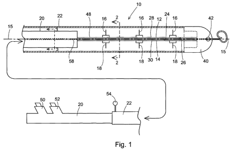
[0011] Figure 1 is a side, partially sectioned view of one embodiment of the
medical
device of the present invention.
[0012] Figure 2 is an end section view in the plane of line 2-2 in Figure 1.
[0013] Figure 3 is an end section view in the plane of line 3-3 in Figure 1.
[0014] Figure 4 is a view similar to Figure 1 that illustrates the movement of
the
actuators of the medical device.
[0015] Figure 5 is a view of the medical device shown in Figure 1, but with
the central
catheter component rotated 90 relative to its orientation in Figure 1.
[0016] Figure 6 is a partial view of the exterior of the medical device of
Figure 1 in its
constrained position.
[0017]Figure 7 is a diagram of the fluid path of the medical device of Figure
1,
extending from the Luer hubs through the fluid delivery conduits to the
reservoir and
then to the tissue penetrators.
[0018] Figure 8 is a side, partially sectioned view of a second embodiment of
the
medical device of the present invention showing the actuators in their
constrained
configurations.
4
CA 02711990 2010-07-13
WO 2009/097621 PCT/US2009/032891
[0019] Figure 9 is a view similar to Figure 8, but showing the actuators in
their
unconstrained configurations.
[0020] Figure 10 is an end perspective view of the assembly along the line 3'-
3' of
Figure 8.
[0021] Figure 11 is an end perspective view of the assembly along the line 4'-
4' of
Figure 10 showing the tissue penetrators.
[0022] Figure 12 is a side view showing the detail of the proximal end of the
device,
shown to the right in Figures 8 and 9.
[0023] Figure 13 is a three-dimensional depiction of one embodiment of a
medical
device of the invention in its deployed, or unconstrained, configuration.
[0024] Figure 14 shows the device of Figure 13 in its undeployed, or
constrained,
configuration (top panel) as well as top (middle panel) and side (bottom
panel) views
of the device in its deployed configuration.
[0025] Figure 15 shows a close up of one injection unit in a device of Figure
13 (left
panel) in the undeployed configuration, and a cross section view along the
needles
of the injection unit.
[0026] Figures 16A-16D depict the device of Figure 13 as it is deployed.
Figure 16A
shows the undeployed configuration; Figures 16B and 16C depict the device in a
partially deployed configuration, and Figure 16D shows the device in the fully
deployed configuration.
[0027] Figure 17 is a three-dimensional depiction of one embodiment of a
medical
device of the invention in its deployed, or unconstrained, configuration.
[0028] Figures 18A-18C depict the device of Figure 17 as it is deployed.
Figure 18A
shows the undeployed configuration; Figure 18B depicts the device in a
partially
deployed configuration, and Figure 18C shows the device in the fully deployed
configuration.
[0029] Figure 19 is a depiction of one embodiment of a medical device of the
invention in its deployed, or unconstrained, configuration.
CA 02711990 2010-07-13
WO 2009/097621 PCT/US2009/032891
[0030] Figures 20A-20B. Figure 20A is a close up depicting the needle
configuration
in the device. Figure 20B shows a close up of the central catheter component
and
the proximal portion of the splines and the proximal needles (left panel) and
a cross
section view along the proximal needles (right panel).
[0031] Figures 21A-21D depict the device of Figure 19 as it is deployed.
Figure 21A
shows the undeployed configuration; Figures 21 B and 21 C depict the device in
a
partially deployed configuration, and Figure 21D shows the device in the fully
deployed configuration.
[0032] Figures 22A-E are photographs of a prototype of one embodiment of a
medical device of the invention as it is deployed. Figure 22A shows the
undeployed
configuration; Figures 22B, 22C and 22D depict the device in a partially
deployed
configuration, and Figure 22E shows the device in the fully deployed
configuration.
DETAILED DESCRIPTION OF THE INVENTION
[0033]As used herein, a "wall" is any surface of any biological space or
conduit, e.g.,
an inner or outer wall of a biological conduit such as a blood vessel.
Examples of
biological spaces include, but are not limited to, the peritoneal cavity, the
epidural
space, the arachnoid and subarachnoid spaces, the subdural space, or any
potential
spaces that may be created by separating two adjacent bodily tissues. A
"biological
conduit" is any tubular structure that conveys any fluid, gas, solid, colloid,
or
combination thereof from one location to another within an organism. In a
preferred
embodiment, the organism is a mammal, most preferably a human. Examples of
biological conduits include, but are not limited to, arteries, veins, ureters,
bronchi,
bile ducts, glandular ducts, pancreatic ducts, urogenital conduits and
gastrointestinal
conduits.
[0034] In one embodiment, the medical device of the present invention has a
central
longitudinal axis, and comprises one or more actuators, wherein the one or
more
actuators can exist in a constrained configuration in which a length of said
one or
more actuators is oriented substantially parallel to the longitudinal axis of
said
medical device and an unconstrained configuration in which at least a portion
of the
length of said one or more actuators is oriented substantially non-parallel to
the
device's central longitudinal axis. After the device is positioned at a target
site
adjacent to the wall of a biological space or conduit, one or more actuators
(and if
6
CA 02711990 2010-07-13
WO 2009/097621 PCT/US2009/032891
desired, all of the actuators) may be released from a constrained
configuration and
permitted to adopt an unconstrained configuration, thereby making contact with
the
wall of the biological space or conduit. The one or more actuators may be of
any
shape, and in preferred embodiments, the movement of the one or more actuators
from the constrained configuration to the unconstrained configuration occurs
upon
release of a constraining force by the device operator but without the input
by the
operator of any deforming forces to the device or the target tissue.
[0035] In a first specific embodiment, shown in Figure 1, a device of the
present
invention is a fluid delivery catheter 10 comprising one or more actuators
that are
formed as a pair of elongate splines 12, 14, the intermediate regions of which
are
movable between a constrained configuration which is oriented substantially
parallel
to the central longitudinal axis 15 of the catheter assembly and an
unconstrained
configuration in which at least a portion of the pair of splines is oriented
substantially
non-parallel to said central longitudinal axis (see the left L and right R
portions of the
spline lengths in Figure 4). The one or more splines 12, 14 may be constructed
as
elongate bands or wires that each have opposite proximal and distal ends. In a
preferred embodiment, the splines have flat, opposing interior surfaces 24,
26, and
flat opposite facing exterior surfaces 28, 30. In this embodiment, the splines
12, 14
can translate between constrained positions and unconstrained positions, as
shown
respectively in Figures 1 and 4. In one embodiment, the pair of splines is
positioned
back-to-back in their constrained configurations as shown in Figure 1.
[0036]The catheter 10 further comprises one or more tissue penetrators 16, 18
secured to one or more surfaces of the one or more splines 12, 14, a central
catheter
component 20 having an elongate length, and an exterior catheter component 22
(sometimes referred to herein as a sheath) that can shield the tissue
penetrator or
penetrators during catheter movement within the biological space or conduit.
[0037] The tissue penetrators 16, 18 may be constructed of any suitable
material.
Preferred examples of such materials include, but are not limited to, nickel,
aluminum, steel and alloys thereof. In a specific embodiment, the tissue
penetrators
are constructed of nitinol.
[0038]The central catheter component 20 and the exterior catheter component 22
may be constructed of materials typically employed in constructing catheters.
Examples of such materials include, but are not limited to, silicone,
polyurethane,
nylon, Dacron, and PEBAXTM.
7
CA 02711990 2010-07-13
WO 2009/097621 PCT/US2009/032891
[0039]The actuators are preferably constructed of a flexible, resilient
material. In a
preferred embodiment, the flexible, resilient material is capable of being
constrained
upon the application of a constraining force, e.g., when the actuators are in
the
constrained configuration, and adopts its original unconstrained shape when
the
constraining force is removed, e.g., when the actuators are in the
unconstrained
configuration. Any such flexible, resilient material can be used, including
but not
limited to surgical steel, aluminum, polypropylene, olefinic materials,
polyurethane
and other synthetic rubber or plastic materials. The one or more actuators are
most
preferably constructed of a shape memory material. Examples of such shape
memory materials include, but are not limited to, copper-zinc-aluminum-nickel
alloys,
copper-aluminum-nickel alloys, and nickel-titanium (NiTi) alloys. In a
preferred
embodiment, the shape memory material is nitinol. In a preferred embodiment,
when the pair of splines assumes the unconstrained configuration, the shape
memory properties of the material from which each spline is formed cause the
splines, without the application of any external deforming force, to bow
radially away
from each other in a single plane as shown in Figure 4.
[0040] One or more of the splines (and preferably each of the splines) has a
flexible
fluid delivery conduit 32, 34 that extends along the length of the spline, or
within the
spline, as shown in Figure 2. As the splines 12, 14 move from their straight,
constrained configurations to their bowed, unconstrained configurations, the
fluid
delivery conduits 32, 34 also move from straight configurations to bowed
configurations. In one embodiment, the fluid delivery conduits 32, 34 are
separate
tubular conduits that are secured along the lengths of the pair of splines 12,
14. In
another embodiment, the fluid delivery conduits are conduits formed into or
within
the material of the splines.
[0041]One or more of the splines (and preferably each of the splines 12,14) is
also
formed with a zipper rail 36, 38 that extends along a length of the spline
(Figure 2).
The zipper rails 36, 38 are formed of either the same material as the splines
12, 14,
or a material that flexes with the splines 12, 14.
[0042] In certain aspects, a medical device of the invention comprises a pair
of
splines that are attached, e.g., by welding, at certain intervals along their
lengths, as
depicted in Figures 13 to 16. In this configuration, each portion of the
spline
between the two attachments, referred to herein as an "injection unit," moves
from a
straight configuration to a bowed configuration independently of other
portions of the
8
CA 02711990 2010-07-13
WO 2009/097621 PCT/US2009/032891
spline, or other injection units. See, e.g., Figure 16C showing different
injection units
at different degrees of constraint. The use of splines with multiple injection
units
minimizes wall contact. Each injection unit preferably has at least one pair
of
opposing tissue penetrators, such that at least one tissue penetrator is
secured to
the surface of the portion of each spline within the injection unit (i.e., one
above the
central longitudinal axis and one below the central longitudinal axis),
although it is
contemplated that an injection unit can have more than one tissue penetrator
attached to each spline portion within it. In certain embodiments, a device of
the
invention has a single injection unit, two injection units, three injection
units, four
injection units, five injection units or six injection units.
[0043] One or more of the tissue penetrators 16, 18 is secured to the exterior
surfaces 28, 30 of the pair of splines 12, 14 (Figure 2). The tissue
penetrators 16, 18
are connected to and communicate with the fluid delivery conduits 32, 34 that
extend
along the lengths of the splines 12, 14. The tissue penetrators 16, 18 are
positioned
to project substantially perpendicular from the exterior surfaces 28, 30 of
the splines
12, 14. The tissue penetrators 16, 18 have hollow interior bores that
communicate
with the fluid delivery conduits 32, 34 of the splines. The distal ends of the
tissue
penetrators have fluid delivery ports that communicate with the interior bores
of the
tissue penetrators.
[0044]The device permits delivery of fluids into or through one or more
distinct
layers of a wall of a biological conduit or space, for example a vascular
wall. The
vascular wall comprises numerous structures and layers, including the
endothelial
layer and basement membrane layer (collectively the intimal layer), the
internal
elastic lamina, the medial layer, and the adventitial layer. These layers are
arranged
such that the endothelium is exposed to the lumen of the vessel and the
basement
membrane, the internal elastic lamina, the media, and the adventitia are each
successively layered over the endothelium, as described in U.S. Pat. App.
Publication No. 2006/0189941A1. With the medical devices of the present
invention,
the depth to which the tissue penetrators 16, 18 can penetrate is determined
by the
length of each tissue penetrator 16, 18. For example, if the target layer is
the
adventitial layer, tissue penetrators 16, 18 having a defined length
sufficient for
penetration to the depth of the adventitial layer upon deployment of the
device are
used. Likewise, if the target layer is the medial layer, tissue penetrators
16, 18
9
CA 02711990 2010-07-13
WO 2009/097621 PCT/US2009/032891
having a defined length sufficient for penetration to the depth of the medial
layer
upon deployment of the device are used.
[0045] In specific embodiments, the length of tissue penetrators 16, 18 may
range
from about 0.3 mm to about 5 mm for vascular applications, or up to about 20
mm or
even 30 mm for applications involving other biological spaces or conduits, for
example in colonic applications. Tissue penetrators 16, 18 preferably have a
diameter of about 0.2 mm (33 gauge) to about 3.4 mm (10 gauge), more
preferably
0.2 mm to 1.3 mm (about 33 to 21 gauge). The distal tips of the tissue
penetrators
may have a standard bevel, a short bevel, or a true short bevel. In an
alternative
embodiment, the tissue penetrators attached to any one spline are not of
identical
lengths, but may be configured such that their distal ends align so as to be
equidistant from the wall of the biological space or conduit when the medical
device
is in the unconstrained position, e.g., during use. In certain embodiments,
tissue
penetrators are attached, e.g., soldered or glued, to the splines, as shown in
the
embodiment of Figure 17. In other embodiments, the tissue penetrators are
elbow
needles presented at the surface of the splines through a hole in the splines,
as
shown in the embodiment of Figure 20.
[0046]The central catheter component 20 has an elongate length with opposite
proximal and distal ends, shown to the left and right respectively in Figure
1. In one
embodiment, the central catheter component 20 has a cylindrical exterior
surface
that extends along its elongate length. The proximal ends of the splines 12,
14 are
attached e.g., soldered or glued, to the distal end of the central catheter
component
20, while the distal ends of the splines 12, 14 are attached, e.g., soldered
or glued,
to a catheter guide tip 40. The tip 40 has a smooth exterior surface that is
designed
to move easily in the biological conduit. A guide wire bore 48 extends through
the
length of the central catheter 20 and tip 40. The guide wire bore is
dimensioned to
receive a guide wire in sliding engagement through the bore.
[0047]A pair of fluid delivery lumens 44, 46 extends through the interior of
the
central catheter component 20 for the entire length of the catheter component
(Figure 3). At the distal end of the central catheter component 20 the pair of
fluid
delivery lumens 44, 46 communicates with the pair of fluid delivery conduits
32, 34
that extend along the lengths of the splines 12, 14 to the tissue penetrators
16, 18.
A guide wire bore 48 also extends through the interior of the central catheter
component 20 from the proximal end to the distal end of the central catheter
CA 02711990 2010-07-13
WO 2009/097621 PCT/US2009/032891
component (Figure 3). The proximal end of the central catheter component 20 is
provided with a pair of Luer hubs 50, 52 (Figure 1). In one embodiment, each
Luer
hub 50, 52 communicates with one of the fluid delivery lumens 44, 46 extending
through the length of the central catheter. Each Luer hub 50, 52 is designed
to be
connected with a fluid delivery source to communicate a fluid through each
Luer hub
50, 52, then through each fluid delivery lumen 44, 46 extending through the
central
catheter component 20, then through each fluid delivery conduit 32, 34
extending
along the lengths of the pair of splines 12, 14, and then through the tissue
penetrators 16, 18 secured to each of the pair of splines. In another
embodiment,
each Luer hub 50, 52 independently communicates with both of the fluid
delivery
lumens 44, 46 extending through the length of the central catheter component.
In
this configuration, a first fluid can be delivered through a first Luer hub to
both tissue
penetrators 16, 18 and a second fluid can be delivered through a second Luer
hub to
both tissue penetrators 16, 18. Delivery of fluid to both tissue penetrators
from each
Luer hub can be achieved by an independent conduit extending from each Luer
hub
to a distal common reservoir 61 as shown in Figure 7. This reservoir
communicates
with both tissue penetrators 16, 18. Alternatively, in another embodiment, the
medical device of the instant invention comprises only a single Luer hub
connected
to a single fluid delivery lumen extending through the central catheter, which
then is
attached to a distal common reservoir, permitting the delivery of a single
fluid to both
tissue penetrators 16, 18.
[0048] The exterior catheter component 22 has a tubular configuration that
surrounds
the pair of splines 12, 14 and a majority of the central catheter 20 (Figure
1). The
catheter component 22 has an elongate length that extends between opposite
proximal and distal ends of the catheter component shown to the left and
right,
respectively in Figure 1. The catheter component distal end is dimensioned to
engage in a secure engagement with the guide tip 40, where the exterior
surface of
the tip 40 merges with the exterior surface of the catheter component 22 when
the
catheter component distal end is engaged with the tip. The tubular
configuration of
the catheter component 22 is dimensioned so that an interior surface of the
catheter
component 22 is spaced outwardly of the plurality of tissue penetrators 16, 18
on the
pair of splines 12, 14 in the constrained positions of the pair of splines.
The proximal
end of the central catheter 20 extends beyond the proximal end of the catheter
11
CA 02711990 2010-07-13
WO 2009/097621 PCT/US2009/032891
component 22 when the catheter component distal end engages with the catheter
guide tip 40.
[0049]A mechanical connection 54 is provided between the exterior catheter
component 22 proximal end and the central catheter component 20 proximal end
that enables the exterior catheter component to be moved rearwardly along the
lengths of the pair of splines 12, 14 and the central catheter component 20
causing
the exterior catheter component 22 distal end to separate from the guide tip
40 and
pass over the pair of splines 12, 14, and forwardly over the length of the
central
catheter component 20 and over the lengths of the pair of splines 12, 14 to
engage
the exterior catheter component 22 distal end with the tip 40 (Figure 1). The
mechanical connection 54 could be provided by a handle or button that manually
slides the exterior catheter component 22 over the central catheter component
20.
The connection 54 could also be provided by a thumbwheel or trigger mechanism.
In addition, the connection 54 could be provided with an audible or tactile
indicator
(such as clicking) of the incremental movement of the exterior catheter
component
22 relative to the central catheter component 20.
[0050] In one embodiment, the exterior catheter component 22 is provided with
a
single zipper track 56 that extends along the entire length of one side of the
exterior
catheter component 22 on the interior surface of the exterior catheter
component
(Figure 2). The zipper track 56 in the interior of the exterior catheter
component 22
engages in a sliding engagement with the zipper rails 36, 38 at one side of
each of
the splines 12, 14. Advancing the exterior catheter component 22 forwardly
along
the lengths of the central catheter component 20 and the pair of splines 12,
14
toward the guide tip 40 of the catheter assembly causes the zipper track 56 of
the
exterior catheter component to slide along the rails 36, 38 of the pair of
splines 12,
14. This moves the pair of splines 12, 14 from their bowed, unconstrained
configuration shown in Figure 4 toward their back-to-back, constrained
configuration
shown in Figure 1. The engagement of the spline rails 36, 38 in the zipper
track 56
of the exterior catheter component 22 holds the pair of splines 12, 14 in
their back-
to-back relative positions shown in Figure 1. With the exterior catheter
component
22 pushed forward over the central catheter component 20 and the pair of
splines
12, 14 to where the distal end of the exterior catheter component 22 engages
with
the guide tip 40, the tissue penetrators 16, 18 are covered and the catheter
assembly of the present invention can be safely moved forward or backward in a
12
CA 02711990 2010-07-13
WO 2009/097621 PCT/US2009/032891
biological space or conduit. The exterior catheter component 22 covers the
tissue
penetrators 16, 18 projecting from the pair of splines 12, 14 and the
engagement of
the exterior catheter component 22 with the distal guide tip 40 provides the
catheter
assembly with a smooth exterior surface that facilitates the insertion of the
catheter
assembly into and through a biological space or conduit such as a blood
vessel. In
another embodiment, the exterior catheter component 22 is provided with two
zipper
tracks at 180 degrees from each other that extend along the entire length of
the
exterior catheter component 22 on the interior surface and the splines have
rails on
both sides.
[0051]A guide wire 58 is used with the catheter assembly (Figure 1). The guide
wire
58 extends through the central catheter component guide wire bore 48, along
the
splines 12, 14, and through the guide tip outlet 42. In certain embodiments,
the
guide wire 58 has a solid core, e.g., stainless steel or superelastic nitinol.
The guide
wire may be constructed of radiopaque material, either in its entirety or at
its distal
portions (e.g., the most distal 1 mm to 25 mm or the most distal 3 mm to 10
mm).
The guide wire 58 may optionally be coated with a medically inert coating such
as
TEFLON .
[0052] In use of this device, the guide wire 58 is positioned in the
biological space or
conduit by methods well known in the art. The guide wire 58 extends from the
biological space or conduit, through the guide wire outlet 42 in the tip 40 of
the
assembly, through the exterior shielding catheter 22 past the tissue
penetrators 16,
18, and through the guide wire bore 48 of the central catheter 20. In other
embodiments, the catheter assembly is a rapid-exchange catheter assembly,
wherein the guide wire lumen is present in the distal end of the guide tip 40
of the
catheter, but does not extend throughout the entire length of the medical
device.
[0053]After positioning of the guide wire, the device is advanced into the
biological
space or conduit along the previously positioned guide wire 58. One or more
radiopaque markers may optionally be provided on the device to monitor the
position
of the device in the biological space or conduit. Any material that prevents
passage
of electromagnetic radiation is considered radiopaque and could be used.
Preferred
radiopaque materials include, but are not limited to, platinum, gold, or
silver. The
radiopaque material can be coated on the surface of all or a part of the tip
40, on all
or part of the splines 12, 14 or other actuators, on the guide wire 58, or on
some
combination of the foregoing strucutres. Alternatively, a ring of radiopaque
material
13
CA 02711990 2010-07-13
WO 2009/097621 PCT/US2009/032891
can be attached to the tip 40. The device may optionally be provided with
onboard
imaging, such as intravascular ultrasound or optical coherence tomography. The
tip
of the device may optionally be provided with optics that are used to
determine the
position of the device or characteristics of the surrounding biological space
or
conduit.
[0054] When the device is at its desired position in the biological space or
conduit,
the operator uses mechanical connection 54 to retract the exterior catheter
component 22 rearwardly away from the guide tip 40. In a preferred embodiment,
as
the exterior catheter component 22 is withdrawn from over the tissue
penetrators 16,
18, the zipper track 56 of the exterior catheter component 22 is withdrawn
over the
rails 36, 38 of the pair of splines 12, 14. This movement releases the pair of
splines
12, 14 from their constrained, back-to-back configuration shown in Figure 1,
and
allows the shape memory material of the splines 12, 14 to adopt their
unconstrained,
bowed configurations shown in Figure 4. As the splines 12, 14 move to their
unconstrained, bowed configurations, the splines come into contact with the
inner
surface of the wall(s) of the biological space or conduit and the tissue
penetrators 16,
18 on the exterior surfaces 28, 30 of the splines 12, 14 are pressed into the
interior
surface of the biological space or conduit at the position of the device.
[0055]After the tissue penetrators 16, 18 have entered the desired layer of
the wall
of a biological space or conduit, a fluid can be delivered through the fluid
delivery
lumens 44, 46 in the central catheter component 20, through the fluid delivery
conduits 32, 34 on the pair of splines 12, 14, and through the tissue
penetrators 16,
18. When the delivery of the fluid is complete, the operator uses the
mechanical
connection 54 to move the exterior catheter component 22 (which may also be
referred to as a shielding component) forward over the central catheter
component
20 and over the pair of splines 12, 14 toward the guide tip 40. As the
exterior
catheter component 22 moves forward over the pair of splines 12, 14, the
zipper
track 56 on the interior of the exterior catheter component 22 passes over the
rails
36, 38 on the pair of splines 12, 14, causing the splines 12, 14 to move from
their
unconstrained, bowed configuration back to their constrained configuration.
When
the exterior catheter component 22 has been entirely advanced over the pair
splines
12, 14 and again engages with the guide tip 40, the zipper track 56 in the
exterior
catheter component 22 holds the splines 12, 14 in their constrained
configuration.
14
CA 02711990 2010-07-13
WO 2009/097621 PCT/US2009/032891
The device then can be repositioned for release at another location in the
biological
space or conduit or another biological space or conduit, or withdrawn from the
body.
[0056]The shape and length of the splines 12, 14 are selected such that
various
embodiments of the device can be used in biological spaces or conduits of
various
sizes or diameters. In certain embodiments, the splines may be flat or
rounded. Flat
splines preferably have a width ranging from about 0.2 mm to about 20 mm, a
height
ranging from about 0.2 mm to about 5 mm, and a length ranging from about 10 mm
to about 200 mm, depending on the particular application. Rounded splines
preferably have a diameter ranging from about 0.2 mm to about 20 mm and a
length
ranging from about 10 mm to about 200 mm, depending on the particular
application.
In specific embodiments, flat splines are 3.5 mm to 5 mm, 5 mm to 10 mm, 10 mm
to
15 mm, 15 mm to 20 mm in width, or any range therewithin (e.g., 3.5 mm to 10
mm);
3.5 mm to 5 mm, 5 mm to 10 mm. 10 mm to 15 mm, 15 mm to 20 mm in height, or
any range therewithin (e.g., 3.5 mm to 10 mm); and 10 mm to 20 mm, 20 mm to 40
mm, 40 mm to 80 mm, 80 mm to 120 mm, 120 mm to 150 mm or 150 to 200 mm in
length, or any range therewithin (e.g., 10 mm to 40 mm), or any permutation of
the
foregoing (e.g., a width of 5 mm to 10 mm, a height or 3.5 to 5 mm, and a
length of
20 to 40 mm). In other embodiments, rounded splines are 3.5 mm to 5 mm, 5 mm
to
mm, 10 mm to 15 mm, 15 mm to 20 mm in diameter, or any range therewithin
(e.g., 3.5 mm to 10 mm) and 10 mm to 20 mm, 20 mm to 40 mm, 40 mm to 80 mm,
80 mm to 120 mm, 120 mm to 150 mm or 150 to 200 mm in length, or any range
therewithin (e.g., 10 mm to 40 mm), or any permutation of the foregoing (e.g.,
a
diameter of 5 mm to 10 mm and a length of 20 to 40 mm).
[0057] In a second specific embodiment, shown in Figure 8, the device of the
present
invention is a fluid delivery catheter 110 comprising a central catheter
component
112 having an elongate length with a longitudinal axis 113, one or more (and
preferably two) flexible, resilient actuators that, in this specific
embodiment, are
formed as tissue penetrator presentation tubes 114, 116 that extend from the
distal
portion of the central catheter component 112. At least a portion of the
tissue
presentation tubes 114, 116 are movable between a constrained configuration
which
is oriented substantially parallel to the central longitudinal axis 113 of the
catheter
assembly and an unconstrained configuration which is oriented substantially
non-
parallel to the central longitudinal axis 113 of the catheter.
CA 02711990 2010-07-13
WO 2009/097621 PCT/US2009/032891
[0058]The catheter further comprises one or more (and preferably two)
flexible,
elongate tissue penetrators 118, 120 that extend through the two tissue
penetrator
presentation tubes 114, 116, and an exterior deployment tube 122 that extends
over
portions of the lengths of the central catheter component 112, the tissue
penetrator
presentation tubes 114, 116, and the middle rail 132.
[0059] The central catheter component 112 and the exterior deployment tube 122
may be constructed of any materials suitable for constructing catheters.
Examples
of such materials include, but are not limited to, silicone, polyurethane,
nylon,
Dacron, and PEBAXTM.
[0060]The tissue penetrators 118, 120 connect to respective hubs 166, 168
(Figure
12). One or more of the pair of tissue penetrators 118, 120 preferably has a
diameter of about 0.2 mm (33 gauge) to about 3.4 mm (10 gauge), more
preferably
0.8 mm to 1.3 mm (about 18 to 21 gauge). One or more of the pair of tissue
penetrators may have a standard bevel, a short bevel or a true short bevel.
The pair
of tissue penetrators 118, 120 are preferably constructed of materials that
allow the
tissue penetrators to flex along their lengths. Examples of such materials
include,
but are not limited to, nickel, aluminum, steel and alloys thereof. In a
specific
embodiment, the tissue penetrators are constructed of nitinol. The full length
of the
tissue penetrators 118, 120 can be constructed of a single material, or the
distal
ends (e.g., the distal 1 mm to the distal 20 mm), including the tips 156, 158,
of the
tissue penetrators 118, 120 may be constructed of one material and connected
to
the respective hubs 166, 168 via a tubing constructed of a different material,
e.g.,
plastic.
[0061]One or more of the pair of tissue penetrator presentation tubes 114, 116
is
preferably constructed of a flexible, resilient material. Such flexible,
resilient material
can be deformed, e.g., when the tissue penetrator presentation tubes 114, 116
are in
the straight, constrained configuration of Figure 8, but returns to its
original shape
when the deformation force is removed, e.g., when the tissue penetrator
presentation tubes 114, 116 are in the curved, unconstrained configuration
shown in
Figure 9. Any such flexible, resilient material can be used, including but not
limited
to surgical steel, aluminum, polypropylene, olefinic materials, polyurethane
and other
synthetic rubber or plastic materials. The pair of tissue penetrator
presentation tubes
114, 116 is most preferably constructed of a shape memory material. Examples
of
such shape memory materials include, but are not limited to, copper-zinc-
aluminum-
16
CA 02711990 2010-07-13
WO 2009/097621 PCT/US2009/032891
nickel alloys, copper-aluminum-nickel alloys, and nickel-titanium (NiTi)
alloys. In a
preferred embodiment, the shape memory material is nitinol.
[0062]The central catheter component 112 has a flexible elongate length with
opposite proximal 124 and distal 126 ends (Figure 8). The distal end 126 of
the
central catheter component is formed as a guide tip that has an exterior shape
configuration that will guide the distal end 126 through a biological space or
conduit.
A guide wire bore 128 within middle rail 132 extends through the center of the
central
catheter 112 from the proximal end 124 to the distal end 126. The guide wire
bore
128 receives a flexible, elongate guide wire 130 for sliding movement of the
bore 128
over the wire (Figure 10). The guide wire 130 is used to guide the catheter
assembly
through a biological space or conduit. In certain embodiments, the guide wire
130
has a solid core, e.g., stainless steel or superelastic nitinol. The guide
wire may
optionally be constructed of radiopaque material, either in its entirety or at
its distal
portions (e.g., the most distal 1 mm to 25 mm or the most distal 1 mm to 3
mm). The
guide wire 130 may optionally be coated with a medically inert coating such as
TEFLON . In other embodiments, the catheter assembly is a rapid-exchange
catheter assembly wherein a guide wire is positioned on the distal end of the
guide
tip 126 and extends therefrom.
[0063]A narrow middle rail 132 surrounding the guide wire bore 128 extends
from
the guide tip of the catheter distal end 126 toward the catheter proximal end
124.
The middle rail 132 connects the guide tip 126 to a base portion 138 of the
central
catheter component.
[0064]The central catheter component base portion 138 has a cylindrical
exterior
surface that extends along the entire length of the base portion. The base
portion
138 extends along a majority of the overall length of the central catheter
component
112. As shown in Figure 10, the guide wire bore 128 extends through the center
of
the central catheter component base portion 138. In addition, a pair of tissue
penetrator lumens 140, 142 also extend through the length of the central
catheter
component base portion 138 alongside the guide wire bore 128. At the proximal
end
124 of the central catheter component, a pair of ports 144, 146 communicate
the pair
of lumens 140, 142 with the exterior of the central catheter component 112
(Figure
8).
[0065] In an alternative embodiment, the medical device of Figure 8 also may
comprise a single flexible, resilient actuator that is formed as a tissue
penetrator
17
CA 02711990 2010-07-13
WO 2009/097621 PCT/US2009/032891
presentation tube, a single flexible, elongate tissue penetrator that extends
through
the tissue penetrator presentation tube and connects to a hub, and an exterior
deployment tube that extends over portions of the lengths of the central
catheter
component, the tissue penetrator presentation tube, and the middle rail.
[0066]The pair of first and second tissue penetrator presentation tubes 114,
116
project from the catheter central component base portion 138 toward the
catheter
distal end 126. Each of the tissue penetrator presentation tubes is formed as
a
narrow, elongate tube having a proximal end that is secured to the central
catheter
component base portion 138, and an opposite distal end 148, 150. Each of the
first
and second tissue penetrator presentation tubes 114, 116 has an interior bore
152,
154 that communicates with the respective first tissue penetrator lumen 140
and
second tissue penetrator lumen 142 in the central catheter component base
portion
138.
[0067]As shown in Figures 10 and 11, the exterior configurations of the tissue
penetrator presentation tubes 114, 116 are matched to the middle rail 132 so
that the
lengths of the tissue penetrator presentation tubes 114, 116 may be positioned
side-
by-side on opposite sides of the middle rail 132. The tissue penetrator tube
distal
ends 148, 150 can be formed as guide tip surfaces that also facilitate the
passage of
the catheter through a vascular system. The tissue penetrator tube distal ends
148,
150 are preferably larger in diameter than the tissue penetrator presentation
tubes
114, 116. In a specific embodiment, the tissue penetrator tube distal tips
148, 150
are rounded and bulbous tips. Such tips are atraumatic and the tubes will not
inadvertently puncture the wall of a biological space or conduit. The tips
148, 150
are exposed and do not extend outwardly beyond the diameter of the guide tip
126.
[0068] Each of the tissue penetrator tubes 114, 116 is preferably constructed
of a
shape memory material, such as nitinol. The tubes 114, 116 are formed with
curved,
unconstrained configurations shown in Figure 9. The tubes 114, 116 move to the
curved, unconstrained configurations shown in Figure 9 when no constraining
force
is applied against the tubes. In order for the presentation tubes 114, 116 to
lie in
straight, constrained configurations along the middle rail 132, a constraining
force
must be applied to the tubes to keep them in their straight, constrained
positions
shown in Figure 8. As each of the tubes 114, 116 moves from its straight,
constrained configuration shown in Figure 9 to its curved, unconstrained
18
CA 02711990 2010-07-13
WO 2009/097621 PCT/US2009/032891
configuration shown in Figure 9, the tissue penetrator bores 152, 154
extending
through the tubes also move from straight configurations to curved
configurations.
[0069]The pair of tissue penetrators 118, 120, from their distal tips to the
hubs 166,
168, have lengths that are slightly longer than the combined lengths of the
tissue
penetrator lumens 140, 142 extending through the central catheter base portion
138
and the tissue penetrator bores 152, 154 extending through the tissue
penetrator
presentation tubes 114, 116. The tips 156, 158 of the tissue penetrators 118,
120
are positioned adjacent to the distal ends 148, 150 of the tissue penetrator
presentation tubes 114, 116 and are positioned inside of the bores 152, 154 of
the
tubes in the constrained configuration of Figure 8. The opposite, proximal
ends of
the tissue penetrators 118, 120 project out through the side ports 144, 146 of
the
central catheter 112. The pair of tissue penetrators 118, 120 are dimensioned
to
easily slide through the tissue penetrator lumens 140, 142 of the central
catheter
component 112 and the tissue penetrator bores 152, 154 of the tissue
penetrator
presentation tubes 114, 116. The side ports 144, 146 of the central catheter
component 112 are preferably at 20 to 90 angles to the central catheter
proximal
end 124, most preferably at 30 to 60 angles to the central catheter proximal
end
124.
[0070]A pair of manual operator movement to linear movement controllers 162,
164
can be connected to the proximal ends of the tissue penetrators 118, 120 and
can be
secured to the central catheter ports 144, 146 (Figure 12). The controllers
162, 164
can be constructed to convert operator movement into controlled linear
movement of
the tissue penetrators 118, 120 through the central catheter tissue penetrator
lumens
140, 142 and through the tissue penetrator presentation tube bores 152, 154.
In one
embodiment, there are rotating controllers 162, 164 that can be manually moved
in
one direction, such that the tissue penetrator injection tips 156, 158 at the
tissue
penetrator distal ends can be adjustably positioned to extend a desired length
out
from the tissue penetrator tube bores 152, 154 at the tissue penetrator tube
distal
ends 148, 150. By rotating the controllers in the opposite direction, the
tissue
penetrators 118, 120 can be retracted back into the tissue penetrator tube
bores
152, 154. Each of the operator movement to linear movement controllers 162,
164
can be provided with a hub 166, 168 that communicates with the interior bore
extending through the tissue penetrators 118, 120 and can be used to connect a
syringe or tubing containing a solution of a diagnostic or therapeutic agent.
19
CA 02711990 2010-07-13
WO 2009/097621 PCT/US2009/032891
[0071]The exterior deployment tube 122 has a tubular length that surrounds the
central catheter 112, the tissue penetrator presentation tubes 114, 116, and
the
middle rail 132. The deployment tube 122 can be mounted on the central
catheter
component 112 and the pair of tissue penetrator presentation tubes 114, 116
for
sliding movement to a forward position of the deployment tube 122 where an
open
distal end 172 of the deployment tube is positioned adjacent the distal ends
148, 150
of the tissue penetrator presentation tubes 114, 116 as shown in Figure 8, and
a
rearward position of the deployment tube 122 where the tube distal end 172 is
positioned adjacent to the connection of the tissue penetrator presentation
tubes
114, 116 with the central catheter component 112 as shown in Figure 9. The
opposite proximal end 174 of the deployment tube 122 can be provided with a
mechanical connection 176 to the central catheter 112. The mechanical
connection
176 enables the deployment tube 122 to be moved between its forward and
rearward positions relative to the central catheter 112 and the tissue
penetrator
presentation tubes 114, 116 (Figures 8 and 9). Such a connection could be
provided
by a thumbwheel, a sliding connection, a trigger or push button or some other
connection that is manually operable to cause the deployment tube 122 to move
relative to the central catheter 112 and the presentation tubes 114, 116. When
the
deployment tube 122 is moved to its forward position shown in Figure 8, the
tube
distal end 172 passes over the lengths of the tissue penetrator presentation
tubes
114, 116 and moves the presentation tubes to their constrained positions
extending
along the opposite sides of the central catheter middle rail 132. When the
deployment tube 122 is moved to its rearward position shown in Figure 9, the
distal
end 172 of the deployment tube is retracted from over the length of the tissue
penetrator presentation tubes 114, 116 and gradually allows the presentation
tubes
114, 116 to release their constrained energy and move to their curved,
unconstrained configurations shown in Figure 9.
[0072] In use of the catheter 110, the deployment tube 122 is in the forward
position
shown in Figure 8. The guide wire 130 is positioned in a biological space or
conduit
(such as an artery or vein) in a known manner. The catheter is then advanced
into
the biological space or conduit over the guide wire. The guide wire 130
extends from
the biological space or conduit, and enters the central catheter component
distal end
126 through the guide wire lumen 128. The wire 130 passes through the length
of
the central catheter 112 and emerges at the proximal end of the central
catheter
CA 02711990 2010-07-13
WO 2009/097621 PCT/US2009/032891
component adjacent to the catheter ports 144, 146, where the guide wire 130
can be
manually manipulated.
[0073]The catheter 110 can be advanced through the biological space or conduit
and can be guided by the guide wire 130. Radiopaque markers may optionally be
provided on the assembly to monitor the position of the assembly in the
biological
space or conduit. Any material that prevents passage of electromagnetic
radiation is
considered radiopaque and may be used. Useful radiopaque materials include,
but
are not limited to, platinum, gold, or silver. The radiopaque material can be
coated
on the surface of all or a part of the tip 126, on all or part of the
presentation tubes
114, 116, on all or part of the tissue penetrators 118, 120, on the guide wire
130, or
on any combination of the foregoing structures. Alternatively, a ring of
radiopaque
material can be attached to the tip 126. The assembly may optionally be
provided
with onboard imaging, such as intravascular ultrasound or optical coherence
tomography. The tip of the assembly may optionally be provided with optics
that are
useful for determining the position of the assembly or the characteristics of
the
surrounding biological conduit. When the assembly is at a desired position,
the
exterior deployment tube 122 can be moved from its forward position shown in
Figure 8 toward its rearward position shown in Figure 9 by manual manipulation
of
the mechanical connection 176.
[0074]As the deployment tube 122 is withdrawn from over the pair of tissue
penetrator presentation tubes 114, 116, the constrained energy of the tissue
penetrator presentation tubes 114, 116 is released and the tubes move toward
their
unconstrained, curved configurations shown in Figure 9. This movement
positions
the tissue penetrator bores 152, 154 at the tissue penetrator tube distal ends
148,
150 against the interior surfaces of the biological space or conduit into
which the
assembly 110 has been inserted.
[0075]The operator movement to linear movement controllers 162, 164 then can
be
manually operated to extend the tissue penetrator distal ends 156, 158 from
the
tissue penetrator bores 152, 154 at the tissue penetrator presentation tube
distal
ends 148, 150. A gauge may be provided on each of the operator movement to
linear movement controllers 162, 164 that provides a visual indication of the
extent of
the projection of the tissue penetrator tips 156, 158 from the tissue
penetrator tube
ends 148, 150 as the controllers 162, 164 are rotated. The controllers also
could
provide an audible sound or tactile feel such as clicking to indicate
incremental
21
CA 02711990 2010-07-13
WO 2009/097621 PCT/US2009/032891
distance steps of the tissue penetrator movements. This deploys the tissue
penetrator tips 156, 158 a desired distance into the walls of the biological
space or
conduit.
[0076] In a third specific embodiment, a medical device of the instant
invention is a
fluid delivery catheter comprising one or more tissue penetrators constructed
of a
flexible, resilient material. In certain aspects, the medical device of the
present
invention has a central longitudinal axis, and comprises one or more tissue
penetrators, wherein the one or more tissue penetrators can exist in a
constrained
configuration in which a length of said one or more tissue penetrators is
oriented
substantially parallel to the longitudinal axis of said medical device and an
unconstrained configuration in which at least a portion of the length of said
one or
more tissue penetrators is oriented substantially non-parallel to the device's
central
longitudinal axis. After the device is positioned at a target site adjacent to
the wall of
a biological space or conduit, one or more tissue penetrators (and if desired,
all of
the tissue penetrators) may be released from a constrained configuration and
permitted to adopt an unconstrained configuration, thereby making contact with
the
wall of the biological space or conduit. The one or more tissue penetrators
may be
of any shape, and in preferred embodiments, the movement of the one or more
tissue penetrators from the constrained configuration to the unconstrained
configuration occurs upon release of a constraining force by the device
operator but
without the input by the operator of any deforming forces to the device or the
target
tissue.
[0077] In a preferred embodiment, tissue penetrators are constructed of
flexible,
resilient material that is capable of being constrained upon the application
of a
constraining force, e.g., when the tissue penetrators are in the constrained
configuration, and adopts its original unconstrained shape when the
constraining
force is removed, e.g., when the tissue penetrators are in the unconstrained
configuration. Any such flexible, resilient material can be used, including
but not
limited to surgical steel, aluminum, polypropylene, olefinic materials,
polyurethane
and other synthetic rubber or plastic materials. The one or more tissue
penetrators
are most preferably constructed of a shape memory material. Examples of such
shape memory materials include, but are not limited to, copper-zinc-aluminum-
nickel
alloys, copper-aluminum-nickel alloys, and nickel-titanium (NiTi) alloys. In a
preferred embodiment, the shape memory material is nitinol. In a preferred
22
CA 02711990 2010-07-13
WO 2009/097621 PCT/US2009/032891
embodiment, when the tissue penetrators assume the unconstrained
configuration,
the shape memory properties of the material from which each tissue penetrator
is
formed cause the tissue penetrators, without the application of any external
deforming force, to move from a position substantially parallel to the
longitudinal axis
of the medical device to a position substantially perpendicular to the
longitudinal axis
of the medical device.
[0078] In a preferred embodiment, the tissue penetrators are maintained in the
constrained configuration by an exterior catheter component having a tubular
configuration that surrounds the tissue penetrators. A mechanical connection
is
provided between the exterior catheter component and the central catheter
component to which the tissue penetrators are attached. The mechanical
connection enables the exterior catheter component to be moved rearwardly
along
the length of the central catheter component, thereby uncovering the
constrained
one or more tissue penetrators and permitting the one or more tissue
penetrators to
assume an unconstrained configuration wherein they make contact with the
target
delivery site. One of ordinary skill in the art would appreciate that this
specific
embodiment may be readily adapted to incorporate radiopaque markers to
facilitate
positioning of the device or rapid-exchange features to facilitate the use of
the
device.
[0079]The medical device of the present invention, in its various embodiments,
permits delivery of fluids into distinct layers of a vascular wall. The
vascular wall
consists of numerous structures and layers, structures and layers, including
the
endothelial layer and the basement membrane layer (collectively the intimal
layer),
the internal elastic lamina, the medial layer, and the adventitial layer.
These layers
are arranged such that the endothelium is exposed to the lumen of the vessel
and
the basement membrane, the intima, the internal elastic lamina, the media, and
the
adventitia are each successively layered over the endothelium as described in
U.S.
Pat. App. Publication No. 200610189941A1. With the medical devices of the
present
invention, the depth to which the tissue penetrator tips 156, 158 can
penetrate into
the target tissue can be controlled by rotating the controllers 162, 164. For
example,
if the target layer is the adventitial layer, the constrained energy of the
tubes 114,
116 is released, the tubes adopt their unconstrained, curved configurations
shown in
Figure 9, and the tissue penetrator tips 156, 158 are advanced with the
controllers to
a length sufficient for penetration to the depth of the adventitial layer.
Likewise, if the
23
CA 02711990 2010-07-13
WO 2009/097621 PCT/US2009/032891
target layer is the medial layer, the constrained energy of the tubes 114, 116
is
released, the tubes adopt their unconstrained, curved configurations shown in
Figure 9, and the tissue penetrator tips 156, 158 are advanced with the
controllers to
a length sufficient for penetration to the depth of the medial layer.
[0080] With the tissue penetrators embedded in the desired layer of the wall
of the
biological space or conduit, a fluid can then be delivered through the tissue
penetrators 118, 120. When the delivery of the fluid is complete, the
controllers 162,
164 can be operated to withdraw the tissue penetrator tips 156, 158 back into
the
interior bores 152, 154 of the tissue penetrator presentation tubes 114, 116.
The
deployment tube 122 can then be moved to its forward position where the
deployment tube distal end 172 moves the tissue penetrator presentation tubes
114,
116 back to their constrained positions shown in Figure 8. When the deployment
tube 122 has been moved to its full forward position shown in Figure 8, the
assembly
can then be repositioned or withdrawn from the body.
[0081]The medical device of the instant invention also permits delivery of
fluids to
plaque deposits on the inside of the wall of the biological conduit or within
the wall of
the biological conduit.
[0082]The medical device of the instant invention also permits delivery of
fluids to
extracellular spaces or tissues located outside of the outer wall of the
biological
space or conduit (e.g., to the exterior surface of a blood vessel or to muscle
positioned against the outer surface of vessel such as myocardium).
[0083] One advantageous feature of the devices of the present invention is
that the
actuators, by virtue of their design, make contact with less than the complete
circumference of the inner wall of a biological conduit following their
deployment
therein. In preferred embodiments, the actuators make contact with less than
100%
of the circumference of the inner wall of a biological conduit in which they
are
deployed. More preferably, the actuators make contact with less than 75%, 50%
or
25% of the circumference of the inner wall of a biological conduit in which
they are
deployed. Most preferably, the actuators make contact with less than 10%, 5%,
2.5%, 1%, 0.5% or 0.1 % of the circumference of the inner wall of a biological
conduit
in which they are deployed.
[0084]The devices can be used to deliver fluids comprising a variety of
therapeutic
and/or diagnostic agents to a wall of a biological space or conduit.
Therapeutic
agents include, but are not limited to proteins, chemicals, small molecules,
cells and
24
CA 02711990 2010-07-13
WO 2009/097621 PCT/US2009/032891
nucleic acids. A therapeutic agent delivered by the device may either comprise
a
microparticle or a nanoparticle, be complexed with a microparticle or a
nanoparticle,
or be bound to a microparticle or a nanoparticle. Protein agents include
elastases,
antiproliferative agents, and agents that inhibit vasospasm. The use of the
devices
for delivery of an elastase is specifically contemplated. Several published
patent
applications (WO 2001/21574; WO 2004/073504; and WO 2006/036804) teach that
elastase, alone and in combination with other agents, is beneficial in the
treatment of
diseases of biological conduits, including obstruction of biological conduits
and
vasospasm. Diagnostic agents include, but are not limited to, contrast,
microparticles, nanoparticles or other imaging agents.
[0085]A variety of distinct fluid delivery methods can be practiced with the
device. In
certain applications, distinct fluids can be delivered through each tissue
penetrator of
the device either simultaneously or sequentially. In other applications, the
same fluid
can be delivered through both tissue penetrators either simultaneously or
sequentially. Embodiments and/or methods where a first fluid is delivered
through
both tissue penetrators followed by delivery of a second fluid through both
tissue
penetrators are also contemplated.
[0086] Methods of using the devices to deliver fluids into or through a wall
of a
biological space or conduit are also specifically contemplated. These methods
comprise the steps of introducing the device into the biological space or
conduit,
advancing the device to a target site within the space or conduit, releasing
the
actuators from their constrained positions, optionally advancing the tissue
penetrators through lumens in the actuators to penetrate to a desired depth
into the
wall of a biological space or conduit, delivering at least one fluid into or
through the
wall, optionally returning the tissue penetrators back into the lumens of the
actuators,
retracting the actuators to their constrained position, repositioning the
device in the
same or a different space or conduit for the delivery of additional fluid if
so desired,
and removing the device from the space or conduit. Also contemplated are
methods
of manufacturing the device.
[0087] Kits that comprise the device and at least one therapeutic agent or at
least
one a diagnostic agent, and combinations thereof are also specifically
contemplated.
A kit of the invention comprises, in one or more containers, a device of the
instant
invention and one or more of the therapeutic and/or diagnostic agents. In
addition or
in the alternative, the kits of the invention may provide an instructional
material which
CA 02711990 2010-07-13
WO 2009/097621 PCT/US2009/032891
describes performance of one or more methods of the invention, or a notice in
the
form prescribed by a governmental agency regulating the manufacture, use or
sale
of the device and the therapeutic and/or diagnostic agents, which notice
reflects
approval by the agency of manufacture, use or sale for human administration.
In
one embodiment, the therapeutic agent is an elastase, such as, but not limited
to,
pancreatic type I elastase, which is preferably human or porcine. In certain
embodiments, the therapeutic agent may be pre-loaded into the medical device.
EXAMPLE
[0088]A prototype medical device of the embodiment depicted in Figure 19 was
constructed and operated.
[0089]The device has a central longitudinal axis, two splines made of a
flexible
metallic material from which project two tissue penetrators made of a more
rigid
metallic material. The splines are attached at their distal ends to a guide
tip made of
a plastic polymer. The construction was based on the following design
principles.
The construction was to include flat-wire springs to provide the outward
expansion
for the needles. A needle was to be connected at each end of each spring
plateau
so that there would be two opposing distal needles and two opposing proximal
needles. The springs were intended to be constructed of a highly elastic metal
such
as Nitinol and would be set to a shape such that in the free state the springs
are
expanded. As used, the springs would be contained until the system in moved to
the
delivery location and the sheath withdrawn. As the sheath is withdrawn, the
springs
would expand and needles attached to the springs would be forced outward into
the
vessel wall.
[0090]The springs would be constructed with holes at each end of each spring
plateau for needle connection. A needle would be connected at each hole so
that
there would be two opposing distal needles and two opposing proximal needles.
Each needle would be constructed as an L-shaped tube that would pass through a
hole in the spring. This attachment is designed to provide a secure and stable
attachment. The conduit for drug delivery to the needles would pass through
holes
in the spring and attach to the inner ends of the needle tubing. This
attachment
method is designed to provide a junction that will be secure to the needles
and easy
to seal.
26
CA 02711990 2010-07-13
WO 2009/097621 PCT/US2009/032891
[0091]A 4-to-1 scale model, shown in Figure 22, was built as a prototype of
the
design. The prototype was constructed using tempered steel for springs and
hypodermic stainless steel tubing for the springs. The springs were formed
into the
expanded shape, heat set, and then tempered. The needle tubing was attached
using adhesive. The drug delivery conduit was made of polymeric tubing that
was
attached to the ends of the needle tubing with adhesive. The polymer tip and
sheath
were made of stereolithography components.
[0092] In their constrained configuration, prior to deployment, the sheath is
holds the
springs, oriented substantially parallel to the longitudinal axis of the
prototype device,
in the compressed form (Figure 22A). As the sheath is retracted (Figures 22B-
22D),
the springs move into an unconstrained configuration such that a portion of
their
lengths is oriented substantially non-parallel to the device's central
longitudinal axis.
In Figure 22D, the first two opposing needles have exited the sheath. By the
end of
deployment, the springs have adopted an unconstrained configuration (Figure
22E).
Both pairs of opposing needles are exposed and the springs are fully expanded.
SPECIFIC EMBODIMENTS, CITATION OF REFERENCES
[0093]The present invention is not to be limited in scope by the specific
embodiments described herein. The scope of the invention contemplated herein
is
not limited by the exemplary embodiments illustrated in the schematic drawings
provided herein. Indeed, various modifications of the invention in addition to
those
described herein will become apparent to those skilled in the art from the
foregoing
description and accompanying figures, including medical devices other than
catheters in which at least one component of the device displays the ability
to adopt
an unconstrained configuration following release from a constrained
configuration,
wherein the transition between the two configurations occurs upon release of a
constraining force by the device operator but without the input by the
operator of any
deforming forces to the device or the target tissue. Such modifications are
intended
to fall within the scope of the appended claims.
[0094]Various references, including patent applications, patents, and
scientific
publications, are cited herein; the disclosure of each such reference is
hereby
incorporated herein by reference in its entirety.
27