Note: Descriptions are shown in the official language in which they were submitted.
CA 02725599 2010-11-24
WO 2009/143624 PCT/CA2009/000744
METHODS OF DIAGNOSING REJECTION OF A KIDNEY ALLOGRAFT USING
GENOMIC OR PROTEOMIC EXPRESSION PROFILING
This application claims priority benefit of U.S. Provisional application
61/129,022, filed May 30,
2008, the contents of which is herein incorporated by reference.
[0001] FIELD OF INVENTION
[0002] The present invention relates to methods of diagnosing rejection of a
kidney allograft
using genomic expression profiling or proteomic expression profiling.
BACKGROUND OF THE INVENTION
[0003] Transplantation is considered the primary therapy for patients with end-
stage vital organ
failure. While the availability of immunosuppressants such as cyclosporine and
tacrolimus has
improved allograft recipient survival and wellbeing, identification of
rejection of the allograft as
early and as accurately as possible, and effective monitoring and adjusting
immunosuppressive
medication doses is still of primary importance to the continuing survival of
the allograft
recipient.
[0004] Rejection of an allograft results from a recipient's immune response to
nonself antigens
expressed by the donor tissues, and may occur with hours or days of receiving
the allograft, or
months to years later. Renal allograft rejection is characterized by features
comprising oliguria,
rapid deterioration of renal function and mild proteinuria. Renal allograft
rejection can lead to
nephropathy and kidney failure.
[0005] At present, invasive biopsies (e.g. endomyocardial, liver core, and
renal fine-needle
aspiration) are regarded as the gold standard for the surveillance and
diagnosis of allograft
rejections, but are invasive procedures which carry risks of their own (e.g.
Mehra MR, et al. Curr.
Opin. Cardiol. 2002 Mar; 17(2):131-136.). Biopsy results may also be subject
to reproducibility
and interpretation issues due to sampling errors and inter-observer
variabilities, despite the
availability of international guidelines such as the Banff schema for grading
kidney and liver
allograft rejection (Solez et al 2008 Am J Transplant 8: 753; Table 1) An
allograft recipient may
be exposed to the biopsy procedure multiple times in the first year following
the
transplant. Noninvasive surveillance techniques are currently used (the
increase in blood
creatinine levels), however serum creatinine levels are non-specifically
reflective of kidney
-1-
CA 02725599 2010-11-24
WO 2009/143624 PCT/CA2009/000744
injury. The kidney injury can be from rejection, infection, or even recurrence
of the original
disease, thus, the test is not specific for rejection.
[0006] Indicators of allograft rejection may include a heightened and
localized immune response
as indicated by one or more of localized or systemic inflammation, tissue
injury, allograft
infiltration of immune cells, inflammatory cells which recognize donor-
specific antigens on the
graft, allospecific antibodies, cytotoxic T-cell activation, altered
composition and concentration
of tissue- and blood- derived proteins, differential oxygenation of allograft
tissue, edema,
infection, necrosis of the allograft and/or surrounding tissue, and the like.
[0007] Allograft rejection may be described as `acute' or `chronic'. Acute
rejection (also known
as acute antibody-mediated rejection, AMR or active rejection) is generally
considered to be
rejection of a tissue or organ allograft within -6-12 months of the subject
receiving the allograft.
Rejection or acute rejection may be characterized by cellular and humoral
insults on the donor
tissue, leading to rapid graft dysfunction and failure of the tissue or organ.
Rejection of a tissue
or organ allograft beyond 6-12 months is generally considered to be chronic
rejection, and may
occur several years after receiving the allograft. Such late or chronic
rejection may be the result
of sub-clinical or not fully resolved acute rejection episodes. Later-onset or
chronic rejection
may be characterized by progressive tissue remodeling triggered by the
alloimmune response
may lead to gradual neointimal formation within arteries, contributing to
obliterative
vasculopathy, parenchymal fibrosis and consequently, failure and loss of the
graft. Depending on
the nature and severity of the rejection, there may be overlap in the
indicators or clinical variables
observed in a subject undergoing, or suspected of undergoing, allograft
rejection - either chronic
or acute.
[0008] The scientific and patent literature is blessed with reports of this
marker or that being
important for identification/diagnosis/prediction/treatment of every medical
condition that can be
named. Even within the field of allograft rejection, a myriad of markers are
recited (frequently
singly), and conflicting results may be presented. This conflict in the
literature, added to the
complexity of the genome (estimates range upwards of 30,000 transcriptional
units), the variety
of cell types (estimates range upwards of 200), organs and tissues, and
expressed proteins or
polypeptides (estimates range upwards of 80,000) in the human body, renders
the number of
possible nucleic acid sequences, genes, proteins, metabolites or combinations
thereof useful for
diagnosing acute organ rejection is staggering. Variation between individuals
presents
-2-
CA 02725599 2010-11-24
WO 2009/143624 PCT/CA2009/000744
additional obstacles, as well as the dynamic range of protein concentration in
plasma (ranging
from 10-6 to 103 g/ mL) with many of the proteins of potential interest
existing at very low
concentrations) and the overwhelming quantities of the few, most abundant
plasma proteins
(constituting - 99% of the total protein mass.
[0009] PCT Publication WO 2006/125301 discloses nucleic acids that are
differentially
expressed in transplanted tissue, and methods and materials for detecting
kidney tissue rejection.
[0010] US 7235258 discloses methods of diagnosing or monitoring transplant
rejection,
including kidney transplant rejection in a subject, by detecting the
expression level of one or
more genes in the subject. Oligonucleotides useful in these methods are also
described.
[0011] Flechner et al. (Am J Transplant 2004: 4 (9) 1475-1489) identifies
several publications
that employed DNA or microarrays to identify differential expression of
various genes in subjects
receiving kidney transplants, and also describes use of microarray analysis
and RT-PCR to
examine gene expression profile of peripheral blood lymphocytes and kidney
biopsy samples
from kidney transplant subjects, and identified over 60 genes that were
differentially expressed.
[0012] Alakulppi et al, 2007 (Transplantation 83:791-798) discloses the
diagnosis of acute renal
allograft rejection using RT-PCT for eight nucleic acid markers. Further
investigations by
Alakulppi et al. (2008, Transplantation 86:1222-8) were unable to identify a
robust whole blood
gene expression nucleic acid marker for subclinical rejection.
[0013] Sarwal et al. 2003 (N. Engl. J. Med 349:125) reported that genes
associated with
apoptosis were increased in renal biopsies during acute rejection and found
transcript groups
indicating lymphocyte infiltration and activation driven by NF-kappaB and
IFNy.
[0014] Mueller et al., 2007. Am J. Transplant 7:2712 identified transcripts in
the kidney tissue
associated with cytotoxic T-lymphocytes, IFNy signaling, and epithelial cell
injury in both mouse
and human.
[0015] Mehra et al., 2008 suggests that pathways regulating T-cell homeostatis
and
corticosteroid sensitivity may be associated with future acute rejection of
cardiac transplants, but
offers no comment with respect to kidney transplantation. Expression of ITGAX
is one of the 33
genes addressed.
-3-
CA 02725599 2010-11-24
WO 2009/143624 PCT/CA2009/000744
[0016] A review by Fildes et al 2008 (Transplant Immunology 19:1-11) discusses
the role of cell
types in immune processes following lung transplantation, and discloses that
AICL (CLEC2B)
interaction with NK cell proteins may have a role in acute and chronic
rejection.
[0017] Integration of multiple platforms (proteomics, genomics) has been
suggested for
diagnosis and monitoring of various cancers, however discordance between
protein and mRNA
expression is identified in the field (Chen et al., 2002.Mol Cell
Proteomics1:304-313; Nishizuka
et al., 2003 Cancer Research 63:5243-5250). Previous studies have reported low
correlations
between genomic and proteomic data (Gygi SP et al. 1999. Mol Cell Biol.19:1720-
1730; Huber
et al., 2004 Mol Cell Proteomics 3:43-55).
[0018] Several studies have been done looking at the urine proteome of kidney
transplant
recipients (reviewed in Schaub et al., 2008. Contrib. Nephrol 160:65-75.
[0019] Bottelli et al., 2008 (J. Am Soc Nephrol 19:1904-18) teaches that
macrophage stimulating
protein (MSP) is upregulated during regeneration of injured tubule cells, and
suggests that it may
aid recovery from acute kidney injury. Gorgi et al. (2009 Transplantation
Proceedings 41:660-
662) investigated the association between acute kidney transplant rejection,
and a polymorphism
of the MBL gene, and concluded that the polymorphism could be involved in
susceptibility to
acute allograft rejection in the study population. Fiane et al., 2005 (Eur
Heart J 26:1660-5)
disclosed that a low MBL level was related to the development of acute
rejection in cardiac
transplant recipients. Fildes 2008 (J. Heart Lung Transplant 27:1353-1356)
teaches that heart
transplant recipients with MBL deficiency had fewer rejection episodes.
Neither Fiane nor Fildes
offers comment with respect to kidney transplants.
[0020] Berger et al., 2005 (Am J. Transplant 5:1361-1366) teaches that higher
MBL (Mannose-
binding lectin) may be associated with a more severe form of rejection in
kidney transplant
recipients, and suggests that pre-transplantation MBL levels may be useful for
risk stratification
prior to kidney transplantation.
[0021] Methods of assessing or diagnosing allograft rejection that are less
invasive, repeatable
and more robust (less susceptible to sampling and interpretation errors) are
greatly desirable.
-4-
CA 02725599 2010-11-24
WO 2009/143624 PCT/CA2009/000744
SUMMARY OF THE INVENTION
[0022] The present invention relates to methods of diagnosing rejection of a
kidney allograft
using genomic expression profiling or proteomic expression profiling of one or
more biological
samples obtained from a subject.
[0023] The biological sample may be a blood or a plasma sample; use of such
samples in the
methods described herein provides an advantage over biopsy-based assessment
and/or
monitoring of kidney allograft rejection (including acute rejection) as such
samples may be
obtained in a minimally invasive manner (a peripheral blood sample, for
example), with no
requirement for biopsy of the allograft. Use of a blood or plasma sample
provides a further
advantage, in that it may reduce sampling error, and detection of proteomic or
nucleic acid
markers may be less subject to interpretation - the marker is present or it is
not, or it is increased
or decreased relative to a baseline, control or the like as described herein.
[0024] Some current surveillance techinques that do employ blood sampling
(e.g. serum creatine
levels) may not be specific for rejection; the nucleic acid or proteomic
markers described herein,
when obtained from a blood or plasma sample are specific for acute kidney
allograft rejection,
thus provide a further advantage of specificity.
[0025] The complex pathobiology of acute kidney allograft rejection is
reflected in the
heterogeneity of markers identified herein. Markers identified herein
distribute over a range of
biological processes: immune signal transduction, cytoskeletal reorganization,
apoptosis, T-cell
activation and proliferation, cellular and humoral immune responses, acute
phase inflammatory
pathways, and the like.
[0026] In accordance with another aspect of the invention, there is provided a
method of
determining the acute allograft rejection status of a subject, the method
comprising the steps of:
a) determining the nucleic acid expression profile of one or more than one
nucleic acid markers
in a biological sample from the subject, the nucleic acid markers selected
from the group
comprising TncRNA, FKSG49, ZNF438, 1558448_a at, CAMKK2, LMAN2, 237442_at,
FKSG49/LOC730444, JUNB, PRO1073 and ITGAX; b) comparing the expression profile
of the
one or more than one nucleic acid markers to a control profile; and c)
determining whether the
expression level of the one or more than one nucleic acid markers is increased
relative to the
-5-
CA 02725599 2010-11-24
WO 2009/143624 PCT/CA2009/000744
control profile; wherein the increase of the one or more than one nucleic acid
markers is
indicative of the acute rejection status of the subject.
[0027] In some aspects the biological sample is blood or plasma.
[0028] In some aspects, the group of nucleic acid markers further comprises
one or more than
one of SFRS 16, NFYC, NCOA3, PGS 1, NEDD9, LIMK2, NASP, 240057_at,
LOC730399/LOC731974, FKBPIA, HLA-G, RBMS1 and SLC6A6.
[0029] In some aspects, the control profile is obtained from a non-rejecting,
allograft recipient
subject or a non-allograft recipient subject.
[0030] In some aspects, the method further comprises obtaining a value for one
or more clinical
variables.
[0031 ] In some aspects, the method further comprises at step a) determining
the expression
profile of one or more than one of the nucleic acid markers selected from
Table 2.
[0032] In some aspects, the nucleic acid expression profile of the one or more
than one nucleic
acid markers is determined by detecting an RNA sequence corresponding to one
or more than
one markers.
[0033] In some aspects, the nucleic acid expression profile of the one or more
than one nucleic
acid markers is determined by PCR.
[0034] In some aspects, the nucleic acid expression profile of the one or more
than one nucleic
acid markers is determined by hybridization. The hybridization may be to an
oligonucleotide.
[0035] In some aspects the control is an autologous control.
[0036] In accordance with another aspect of the invention, there is provided a
method of
determining acute allograft rejection status of a subject, the method
comprising the steps of a)
determining a proteomic expression profile of proteomic markers in a
biological sample from the
subject, the proteomic markers including a polypeptide encoded by one or more
than one of
KNG1, AFM, TTN, MSTP9/MST1, P116, C2, MBL2, SERPINAIO, F9 and UBR4; b)
comparing the expression profile of the proteomic markers to a control
profile; and c)
determining whether the expression level of the one or more than one
proteomics markers is
-6-
CA 02725599 2010-11-24
WO 2009/143624 PCT/CA2009/000744
increased or decreased relative to the control profile; wherein the increase
or decrease of the five
or more proteomic markers is indicative of the acute rejection status of the
subject.
[0037] In some aspects the biological sample is blood or plasma.
[0038] In some aspects,the level of polypeptides encoded by one or more than
one of KNG1 and
AFM are decreased relative to a control, and the level of polypeptides encoded
by one or more
than one of TTN, MSTP9, MST1, P116, C2, MBL2, SERPINAIO, F9 and UBR4 are
increased
relative to a control profile.
[0039] In some aspects the control profile is obtained from a non rejecting,
allograft recipient
subject or a non-allograft recipient subject.
[0040] In some aspects, the method further comprises obtaining a value for one
or more clinical
variables.
[0041 ] In some aspects, the proteomic expression profile is determined by an
immunologic
assay.
[0042] In some aspects, the proteomic expression profile is determined by
ELISA.
[0043] In some aspects the proteomic expression profile is determined by mass
spectrometry.
[0044] In some aspects the proteomic expression profile is determined by an
isobaric or isotope
tagging method.
[0045] In some aspects the proteomic markers further include a polypeptide
encoded by one or
more than one of LBP, VASN, ARNTL2, P116, SERPINA5, CFD, USHIC, C9, LCAT, B2M,
SHBG and C IS.
[0046] In some aspects the control is an autologous control.
[0047] In accordance with another aspect of the invention, there is provided a
method of
determining acute allograft rejection status of a subject, the method
comprising the steps of: a.
determining a proteomic expression profile of proteomic markers in a
biological sample from the
subject, the proteomic markers including a polypeptide included in one or more
than one of
protein group codes 111, 224, 23, 18, 100, 116, 38, 135, 125; b. comparing the
expression
profile of the proteomic markers to a control profile; and c. determining
whether the expression
-7-
CA 02725599 2010-11-24
WO 2009/143624 PCT/CA2009/000744
level of the one or more than one proteomics markers is increased or decreased
relative to the
control profile; wherein the increase or decrease of the five or more
proteomic markers is
indicative of the acute rejection status of the subject.
[0048] In some aspects the protein group codes further includes one or more
than one of groups
18, 108, 222, 97, 104, 26, 230, 103, 69 or 29.
[0049] In some aspects the biological sample is blood or plasma.
[0050] In some aspects,the level of polypeptides encoded by one or more than
one of KNG1 and
AFM are decreased relative to a control, and the level of polypeptides encoded
by one or more
than one of TTN, MSTP9, MST1, PI16, C2, MBL2, SERPINA10, F9 andUBR4 are
increased
relative to a control profile.
[0051] In some aspects the control profile is obtained from a non rejecting,
allograft recipient
subject or a non-allograft recipient subject.
[0052] In some aspects, the method further comprises obtaining a value for one
or more clinical
variables.
[0053] In some aspects, the proteomic expression profile is determined by an
immunologic
assay.
[0054] In some aspects, the proteomic expression profile is determined by
ELISA.
[0055] In some aspects the proteomic expression profile is determined by mass
spectrometry.
[0056] In some aspects the proteomic expression profile is determined by an
isobaric or isotope
tagging method.
[0057] In some aspects the proteomic markers further include a polypeptide
encoded by one or
more than one of LBP, VASN, ARNTL2, P116, SERPINA5, CFD, USH1C, C9, LCAT, B2M,
SHBG and CIS.
[0058] In some aspects the control is an autologous control.
[0059] In accordance with another aspect of the invention, there is provided
an array comprising
one or more probe sets for one or more than one of the nucleic acid markers
TncRNA, FKSG49,
-8-
CA 02725599 2010-11-24
WO 2009/143624 PCT/CA2009/000744
ZNF438, 1558448 a at, CAMKK2, LMAN2, 237442_at, FKSG49/LOC730444, JUNB,
PRO1073, ITGAX.
[0060] In some aspects, the array further comprises one or more additional
probe sets for one or
more than one of the nucleic acid markesrs , SFRS 16, NFYC, NCOA3, PGS 1,
NEDD9, LIMK2,
NASP, 240057 at, LOC730399/LOC731974, FKBPIA, HLA-G, RBMS1 and SLC6A6.
[0061 ] In some aspects, the array further comprises one or more additional
probe sets for the
nucleic acid markers of Table2.
[0062] In accordance with another aspect of the invention, there is provided
an array comprising
one or more detection reagents for one or more than one of the proteomic
markers KNG1, AFM,
1o TTN, MSTP9, MST1, PI16, C2, MBL2, SERPINA10, F9 and UBR4.
[0063] In some aspects, the array further comprises one or more additional
detection reagents for
one or more than one of LBP, VASN, ARNTL2, PI16, SERPINA5, CFD, USH1C, C9,
LCAT,
B2M, SHBG and C IS.
[0064] In accordance with another aspect of the invention, there is provided a
method of
assessing, monitoring or diagnosing kidney allograft rejection in a subject,
the method
comprising: a) determining the expression profile of at least one or more
nucleic acid markers
presented in Table 2 in a biological sample from the subject; b) comparing the
expression profile
of the at least one or more markers to a non-rejector profile; and c)
determining whether the
expression level of the at least one or more markers is up-regulated
(increased) or down-
regulated (decreased) relative to the control profile, wherein up-regulation
or down-regulation of
the at least one or more markers is indicative of the rejection status.
[0065] In some embodiments, the method further comprises obtaining a value for
one or more
clinical variables and comparing the one or more clinical variables to a
control. The control is a
non-rejection, allograft recipient subject or a non-allograft recipient
subject. In some
embodiments, the rejection is acute rejection. In some embodiments, the one or
more nucleic acid
markers includes 2, 3, 4, 5, 6, 7, 8, 9, 10, 11, 12, 13, 14, 15, 16, 17, 18,
19, 20, 21, 22, 23 or 24
nucleic acid markers selected from those presented in Table 2. In some
embodiments, the
nucleic acid markers may include one or more than one of the nucleic acid
markers presented in
Table 5.
-9-
CA 02725599 2010-11-24
WO 2009/143624 PCT/CA2009/000744
[0066] In accordance with another aspect of the invention, there is provided a
kit for assessing or
diagnosing kidney allograft rejection in a subject, the kit comprising
reagents for specific and
quantitative detection of at least one or more markers presented in Table 2,
along with
instructions for the use of such reagents and methods for analyzing the
resulting data. The kit
may further comprise one or more oligonucleotides for selective hybridization
to one or more of
a gene, transcript or sequence unit representing one or more of the markers.
Instructions or other
information useful to combine the kit results with those of other assays to
provide a non-rejection
cutoff index or control for the diagnosis of a subject's rejection status may
also be provided in
the kit.
[0067] In some embodiments, the kit may further comprise instructions or
materials for
obtaining a value for one or more clinical variables and comparing the one or
more clinical
variables to a control. The control is a non-rejection, allograft recipient
subject or a non-allograft
recipient subject. In some embodiments, the rejection is acute rejection. In
some embodiments,
the one or more nucleic acid markers includes 2, 3, 4, 5, 6, 7, 8, 9, 10, 11,
12, 13, 14, 15, 16, 17,
18, 19, 20, 21, 22, 23 or 24 nucleic acid markers selected from those
presented in Table 2. In
some embodiments, the nucleic acid markers may include one or more than one of
the nucleic
acid markers presented in Table 5.
[0068] This summary of the invention does not necessarily describe all
features of the invention.
Other aspects, features and advantages of the present invention will become
apparent to those of
ordinary skill in the art upon review of the following description of specific
embodiments of the
invention.
BRIEF DESCRIPTION OF THE DRAWINGS
[0069] These and other features of the invention will become more apparent
from the following
description in which reference is made to the appended drawings wherein:
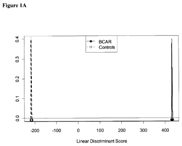
[0070] Figure 1 shows the results of a subject classification using a panel of
24 nucleic acid
biomarkers (presented in Table 5). Subjects were determined to have. A) 24-
probe-set
classifier; B) Zoomed-in view of A) to more clearly illustrate the Gaussian
peaks and samples
below. For A and B, acute rejection - solid circle; no rejection - open
circle. C) The same dataset
as in A and B, displaying the data in the same format as for Figure 2. Acute
rejection (solid
diamond) or no rejection (solid circle)
-10-
CA 02725599 2010-11-24
WO 2009/143624 PCT/CA2009/000744
[0071] Figure 2 shows the result of a subject classification using only
clinical parameters (serum
creatinine, GFR, BUN). Subjects were determined to have acute rejection (solid
diamond) or no
rejection (solid circle). .
[0072] Figure 3: Differential expression of probe-sets between subjects with
and without BCAR
detected by micro-array analysis. Points in grey indicate the probe-sets
identified by LIMMA
alone, while those in black indicate the 183 probe-sets identified by the
intersection of LIMMA,
robust LIMMA and SAM. Circles indicate the 24 probe-sets included in the
primary classifier.
[0073] Figure 4: Principal component analysis showing separation of same
subject groups,
demonstrating that the centroids of all groups are clearly separated. AR -
acute rejector; NR -
non-rejector; N - normal control (20 non-recipient subjects).The percentage
variance as
explained by the principal components are provide on the X axis (56%) and Y
axis (12%).
[0074] Figure 5. Gene ontologies and network analysis of 183 probe sets
differentially expressed
in BCAR. The x-axis shows -log10 (p-values). A Most significantly enriched
Gene ontology
categories ("Biological processes"), sorted by increasing p-value. B Most
significantly enriched
Gene ontology categories (GeneGO MetaCore Biological Categories), sorted by
increasing p-
value.
[0075] Figure 6. Performance of classifier. (A) Incremental classification
accuracy
demonstrating step-wise inclusion of 11 common most highly predictive probe-
sets. Y-axis -
classification accuracy; X-axis, biomarkers. (B) Linear discriminant analysis
showing
performance of 11 probe-set classifiers in distinguishing cases with (=, solid
line) and without ( ,
stippled line) BCAR (biopsy-confirmed acute rejection). (C) Change in
classifier score post-
transplant relative to individual pre-transplant (baseline) value. The
difference between cohorts is
significant only at the time of rejection (week 1) (p=0.0001). Y axis - change
from baseline
(mean +/- 2 se); X axis BL- baseline; WI-W12, week 1 - week 12.. "START"
indicates the
beginning of the tsep-wise analysis where there are no probe-set classifiers.
[0076] Figure 7:Volcano plot showing all 144 protein group codes that were
found in at least
two thirds of the BCAR positive samples and two thirds of the BCAR negative
samples. Circled
points indicate the 18 protein groups whose plasma concentration differed
significantly (p<0.05)
between subjects with or without BCAR.
-11-
CA 02725599 2010-11-24
WO 2009/143624 PCT/CA2009/000744
[0077] Figure 8: Linear discriminant analysis showing separation of patients
with or without
BCAR based upon plasma protein biomarkers. Solid line/ "X" - BCAR subjects;
stippled line/ "
" - control (non-rejector) subjects.
[0078] Figure 9: Estimated classification accuracy demonstrating step-wise
inclusion of protein
groups as chosen by forward-selection stepwise discriminant analysis (SDA). Y
Axis -
classification accuracy; X axis - PGC codes. "START" is as for Figure 6.
[0079] Figure 10 shows target sequences of (SEQ ID NO: 1-183) of nucleic acid
markers useful
for diagnosis of acute kidney allograft rejection, listed in Table 2.
DETAILED DESCRIPTION
[0080] The present invention provides for methods of diagnosing rejection in a
subject that has
received a tissue or organ allograft, specifically a kidney allograft.
[0081 ] The present invention provides genomic and proteomic expression
profiles related to the
assessment, prediction or diagnosis of allograft rejection in a subject. While
several of the
elements in the genomic or proteomic expression profiles may be individually
known in the
existing art, the specific combination of the altered expression levels
(increased or decreased
relative to a control) of specific sets of genomic, T-cell, proteomic or
metabolite markers
comprise a novel combination useful for assessmentf or diagnosis or allograft
rejection in a
subject.
[0082] An allograft is an organ or tissue transplanted between two genetically
different subjects
of the same species. The subject receiving the allograft is the `recipient',
while the subject
providing the allograft is the `donor'. A tissue or organ allograft may
alternately be referred to as
a `transplant', a `graft', an `allograft', a `donor tissue' or `donor organ',
or similar terms. A
transplant between two subjects of different species is a xenograft.
[0083] Subjects may present with a variety of symptoms or clinical variables
well-known in the
literature as an aid for monitoring allograft rejection. A myriad of clinical
variables may be used
in assessing a subject having, or suspected of having, allograft rejection, in
addition to biopsy of
the allograft. The information from these clinical variables is then used by a
clinician, physician,
veterinarian or other practitioner in a clinical field in attempts to
determine if rejection is
-12-
CA 02725599 2010-11-24
WO 2009/143624 PCT/CA2009/000744
occurring, and how rapidly it progresses, to allow for modification of the
immunosuppressive
drug therapy of the subject. Examples of clinical variables are presented in
Tablel.
[0084] Clinical variables (optionally accompanied by biopsy), while currently
the only practical
tools available to a clinician in mainstream medical practice, are not always
able to cleanly
differentiate between rejecting and a non-rejecting subject, as is illustrated
in Figure 2. While the
extreme left and right subjects are correctly classified as rejecting or non-
rejecting, the bulk of
the subjects are represented in the middle range and their status is unclear.
This does not negate
the value of the clinical variables in the assessment of allograft rejection,
but instead indicates
their limitation when used in the absence of other methods.
[0085] Table 1: Clinical variables for possible use in assessment of allograft
rejection.
Clinical Variable Name Renal/Heart Variable Explanation
/
Liver/ All
Primary Diagnosis All Diagnosis leading to transplant
Secondary Diagnosis All Diagnosis leading to transplant
"Transplant Procedure - Living related,
Living unrelated, or cadaveric"
Blood Type All Blood Type
Blood Rh All Blood Rh
Height (cm) All Height (cm)
Weight (kg) All Weight (kg)
BMI All Calculation: Weight/ (Height)2
Liver Ascites All
HLA Al All
HLA A2 All
HLA B1 All
HLA B2 All
HLA DR1 All
HLA DR2 All
CMV All Viral Status
CMV Date All Date of viral status
HIV All Viral Status
HBV All Viral Status
HBV Date All Date of viral status
HbsAb All Viral Status
-13-
CA 02725599 2010-11-24
WO 2009/143624 PCT/CA2009/000744
HbcAb (Total) All Viral Status
HBvDNA All Viral Status
HCV All Viral Status
HCV Genotype All Hepatitis C genotype
HCV Genotype Sub All "Hepatitis C genotype, subtype"
EBV All Viral Status
Zoster All Viral Status
Dialysis Start Date All Dialysis Start Date
Dialysis Type All Dialysis Type
Cytoxicity Current Level All
Cytoxicity Current Date All
Cytoxicity Peak Level All
Cytoxicity Peak Date All
Flush Soln All Type of Flush Solution used at transplant
Cold Time 1 All
Cold Time 2 All
Re-Warm Time 1 All
Re-Warm Time 2 All
HTLV 1 All
HTLV 2 All
HCV RNA All
24hr Urine All 24 Hour urine output
Systolic Blood Pressure All Blood Pressure reading
Diastolic Blood Pressure All Blood Pressure reading
24 Hr Urine All 24 hour urine
Sodium All Blood test
Potassium All Blood test
Chloride All Blood test
Total CO2 All Blood test
Albumin All Blood test
Protein All Blood test
Calcium All Blood test
Inorganic Phosphate All Blood test
Magnesium All Blood test
Uric Acid All Blood test
Glucose All Blood test
Hemoglobin Al C All Blood test
CPK All Blood test
Parathyroid Hormone All Blood test
-14-
CA 02725599 2010-11-24
WO 2009/143624 PCT/CA2009/000744
Homocysteine All Blood test
Urine Protein All Urine test
Creatinine All Blood test
BUN All Blood test
Hemoglobin All Blood test
Platelet Count All Blood test
WBC Count All Blood test
Prothrombin Time All Blood test
Partial Thromboplastin Time All Blood test
INR All Blood test
Gamma GT All Blood test
AST All Blood test
Alkaline Phosphatase All Blood test
Amylase All Blood test
Total Bilirubin All Blood test
Direct Bilirubin All Blood test
LDH All Blood test
ALT All Blood test
Triglycerides All Blood test
Cholesterol All Blood test
HDL Cholesterol All Blood test
LDL Cholesterol All Blood test
FEV1 All Lung function test
FVC All Lung function test
Total Ferritin All Blood test
TIBC All Blood test
Transferrin Saturated All Blood test
Ferritin All Blood test
Angiography Heart Heart function test
Intravascular ultrasound Heart Heart function test
Dobutamine Stress Echocardiography Heart Heart function test
Cyclosporine WB All Immunosuppressive levels
Cyclosporine 2 hr All Immunosuppressive levels
Tacrolimus WB All Immunosuppressive levels
Sirolimus WB All Immunosuppressive total daily dose
Solumedrol All Immunosuppressive total daily dose
Prednisone All Immunosuppressive total daily dose
Prednisone ALT All Immunosuppressive total daily dose
Tacrolimus All Immunosuppressive total daily dose
-15-
CA 02725599 2010-11-24
WO 2009/143624 PCT/CA2009/000744
Cyclosporine All Immunosuppressive total daily dose
Imuran All Immunosuppressive total daily dose
Mycophonelate Mofetil All Immunosuppressive total daily dose
Sirolimus All Immunosuppressive total daily dose
OKT3 All Immunosuppressive total daily dose
ATG All Immunosuppressive total daily dose
ALG All Immunosuppressive total daily dose
Basiliximab All Immunosuppressive total daily dose
Daclizumab All Immunosuppressive total daily dose
Ganciclovir All Anti-viral total daily dose
Lamivudine All Anti-viral total daily dose
Riboviron All Anti-viral total daily dose
Interferon All Anti-viral total daily dose
Hepatitis C Virus RNA All test for presence of HCV values Q
CMV Antigenemia All Antiviral and Virus
Valganciclovir All Anti-viral total daily dose
Neutrophil Number All Blood test
C Peptide All Blood test
Peg Interferon All Anti-viral total daily dose
GFR All Glomerular Filtration Rate
Complication Events All Complication Type
Biopsy Scores Renal Borderline 1 A, 1 B, 2A, 2B, 3
Hyperacute
Biopsy Scores Liver Portal inflammation, Bile duct
inflammation damage, Venous endothelial
inflammation, each scored from 1-2
Donor Blood Type All Donor Blood Type
Donor Blood Rh All Donor Rh
Donor HLA Al All Donor HLA Al
Donor HLA A2 All Donor HLA A2
Donor HLA B1 All Donor HLA B1
Donor HLA B2 All Donor HLA B2
Donor HLA DR1 All Donor HLA DR1
Donor HLA DR2 All Donor HLA DR2
Donor CMV All Donor CMV
Donor HIV All Donor HIV
Donor HBV All Donor HBV
Donor HbsAb All Donor HbsAb
Donor HbcAb (total) All Donor HbcAb (total)
-16-
CA 02725599 2010-11-24
WO 2009/143624 PCT/CA2009/000744
Donor Hbdna All Donor Hbdna
Donor HCV All Donor HCV
Donor EBV All Donor EBV
[0086] The multifactorial nature of allograft rejection prediction, diagnosis
and assessment is
considered in the art to exclude the possibility of a single biomarker that
meets even one of the
needs of prediction, diagnosis or assessment of allograft rejection.
Strategies involving a
plurality of markers may take into account this multifactorial nature.
Alternately, a plurality of
markers may be assessed in combination with clinical variables that are less
invasive (e.g. a
biopsy not required) to tailor the prediction, diagnosis and/or assessment of
allograft rejection in
a subject.
[0087] Regardless of the methods used for prediction, diagnosis and assessment
of allograft
rejection, earlier is better - from the viewpoint of preserving organ or
tissue function and
preventing more systemic detrimental effects. There is no `cure' for allograft
rejection, only
maintenance of the subject at a suitably immunosuppressed state, or in some
cases, replacement
of the organ if rejection has progressed too rapidly or is too severe to
correct with
immunosuppressive drug intervention therapy.
[0088] Applying a plurality of mathematical and/or statistical analytical
methods to a protein or
polypeptide dataset, metabolite concentration data set, or nucleic acid
expression dataset may
indicate varying subsets of significant markers, leading to uncertainty as to
which method is
`best' or `more accurate'. Regardless of the mathematics, the underlying
biology is the same in a
dataset. By applying a plurality of mathematical and/or statistical methods to
a microarray
dataset and assessing the statistically significant subsets of each for common
markers, uncertainty
may be reduced, and clinically relevant core group of markers may be
identified.
[0089] "Markers", "biological markers" or "biomarkers" may be used
interchangeably and refer
generally to detectable (and in some cases quantifiable) molecules or
compounds in a biological
sample. A marker may be down-regulated (decreased), up-regulated (increased)
or effectively
unchanged in a subject following transplantation of an allograft. Markers may
include nucleic
acids (DNA or RNA), a gene, or a transcript, or a portion or fragment of a
transcript in reference
to `genomic' markers (alternately referred to as "nucleic acid markers");
polypeptides, peptides,
proteins, isoforms, or fragments or portions thereof for `proteomic' markers,
or selected
-17-
CA 02725599 2010-11-24
WO 2009/143624 PCT/CA2009/000744
molecules, their precursors, intermediates or breakdown products (e.g. fatty
acid, amino acid,
sugars, hormones, or fragments or subunits thereof). In some usages, these
terms may reference
the level or quantity of a particular protein, peptide, nucleic acid or
polynucleotide, or metabolite
(in absolute terms or relative to another sample or standard value) or the
ratio between the levels
of two proteins, polynucleotides, peptides or metabolites, in a subject's
biological sample. The
level may be expressed as a concentration, for example micrograms per
milliliter; as a
colorimetric intensity, for example 0.0 being transparent and 1.0 being opaque
at a particular
wavelength of light, with the experimental sample ranked accordingly and
receiving a numerical
score based on transmission or absorption of light at a particular wavelength;
or as relevant for
other means for quantifying a marker, such as are known in the art. In some
examples, a ratio
may be expressed as a unitless value. A "marker" may also reference to a
ratio, or a net value
following subtraction of a baseline value. A marker may also be represented as
a `fold-change',
with or without an indicator of directionality (increase or decrease/ up or
down). The increase or
decrease in expression of a marker may also be referred to as `down-
regulation' or 'up-
regulation', or similar indicators of an increase or decrease in response to a
stimulus,
physiological event, or condition of the subject. A marker may be present in a
first biological
sample, and absent in a second biological sample; alternately the marker may
be present in both,
with a statistically significant difference between the two. Expression of the
presence, absence or
relative levels of a marker in a biological sample may be dependent on the
nature of the assay
used to quantify or assess the marker, and the manner of such expression will
be familiar to those
skilled in the art.
[0090] A marker may be described as being differentially expressed when the
level of expression
in a subject who is rejecting an allograft is significantly different from
that of a subject or sample
taken from a non-rejecting subject. A differentially expressed marker may be
overexpressed or
underexpressed as compared to the expression level of a normal or control
sample.
[0091 ] A "profile" is a set of one or more markers and their presence,
absence, relative level or
abundance (relative to one or more controls). For example, a metabolite
profile is a dataset of the
presence, absence, relative level or abundance of metabolic markers. A
proteomic profile is a
dataset of the presence, absence, relative level or abundance of proteomic
markers. A genomic or
nucleic acid profile a dataset of the presence, absence, relative level or
abundance of expressed
nucleic acids (e.g. transcripts, mRNA, EST or the like). A profile may
alternately be referred to
as an expression profile.
-18-
CA 02725599 2010-11-24
WO 2009/143624 PCT/CA2009/000744
[0092] The increase or decrease, or quantification of the markers in the
biological sample may be
determined by any of several methods known in the art for measuring the
presence and/or relative
abundance of a gene product or transcript, or a nucleic acid molecule
comprising a particular
sequence, polypeptide or protein, metabolite or the like. The level of the
markers may be
determined as an absolute value, or relative to a baseline value, and the
level of the subject's
markers compared to a cutoff index (e.g. a non-rejection cutoff index).
Alternately, the relative
abundance of the marker may be determined relative to a control. The control
may be a clinically
normal subject (e.g. one who has not received an allograft) or may be an
allograft recipient that
has not or is not demonstrating rejection.
[0093] In some embodiments, the control may be an autologous control, for
example a sample or
profile obtained from the subject before undergoing allograft transplantation.
In some
embodiments, the profile obtained at one time point (before, after or before
and after
transplantation) may be compared to one or more than one profiles obtained
previously from the
same subject. By repeatedly sampling the same biological sample from the same
subject over
time, a composite profile, illustrating marker level or expression over time
may be provided.
Sequential samples can also be obtained from the subject and a profile
obtained for each, to
allow the course of increase or decrease in one or more markers to be followed
over time For
example, an initial sample or samples may be taken before the transplantation,
with subsequent
samples being taken weekly, biweekly, monthly, bimonthly or at another
suitable, regular interval
and compared with profiles from samples taken previously. Samples may also be
taken before,
during and after administration of a course of a drug, for example an
immunosuppressive drug.
[0094] Techniques, methods, tools, algorithms, reagents and other necessary
aspects of assays
that may be employed to detect and/or quantify a particular marker or set of
markers are varied.
Of significance is not so much the particular method used to detect the marker
or set of markers,
but what markers to detect. As is reflected in the literature, tremendous
variation is possible.
Once the marker or set of markers to be detected or quantified is identified,
any of several
techniques may be well suited, with the provision of appropriate reagents. One
of skill in the art,
when provided with the set of markers to be identified, will be capable of
selecting the
appropriate assay (for example, a PCR based or a microarray based assay for
nucleic acid
markers, an ELISA, protein or antibody microarray or similar immunologic
assay, or in some
examples, use of an iTRAQ, iCAT or SELDI proteomic mass spectrometric based
method) for
performing the methods disclosed herein.
-19-
CA 02725599 2010-11-24
WO 2009/143624 PCT/CA2009/000744
[0095] The present invention provides nucleic acid expression profiles and
proteomic expression
profiles related to the assessment or diagnosis of allograft rejection in a
subject. While several of
the elements in the genomic or T-cell expression profiles or proteomic
expression profiles may
be individually known in the existing art, the specific combination of the
altered expression
levels (increased or decreased relative to a control) of specific sets of
genomic or proteomic
markers comprise a novel combination useful for assessment or diagnosis of
allograft rejection in
a subject.
[0096] 183 probe sets were found to specifically detect (by hybridization and
detection of a
label) and allow for quantitation of the expression level of the expressed
nucleic acids. Of this
set of 183 (listed in Table 2), representing 183 individual expressed
transcripts or nucleic acids, a
subset of 24 probe sets (Table 5) were detected, quantified and found to
demonstrate a
statistically significant fold change in the AR samples relative to non-
rejecting transplant (NR).
Figure 10 provides nucleic acid sequence information of a portion of the
nucleic acid identified
by the probe sets listed in Tables 2 and 5. Sequences in Figure 10 (SEQ ID NO:
1-183) may be
useful as probes for specific hybridization to the indicated gene (e.g. in a
microarray, blot, or
other hybridization based assay), or for the design of a primer or primers for
specific
amplification of the indicated gene (e.g. by PCR, RT-PCR or other
amplification-based assay).
[0097] 18 significant protein group codes were found to have differential
relative levels (relative
to a reference sample) in AR and NR subjects, using a multiplexed iTRAQ
methodology (Table
7). These protein group codes included proteomic markers encoded by one or
more than one of
TTN, KNG1, LBP, VASN, ARNTL2, AFM, MSTP9, MST1, P116, SERPINA5, CFD, USHIC,
C2, MBL2, SERPINAIO, C9, LCAT, B2M, SHBG, C1S, UBR4 and F9. As described
below,
accession numbers providing specific reference to the nucleic acid sequences
encoding these
polypeptides, and the amino acid sequences of these polypeptides are provided
herein. Unique
identifiers (International Protein Index accession numbers) for each member of
the indicated
protein group codes are found in Table 7. Polypeptides comprising a portion of
one or more of
these sequences may be useful for the preparation of antibodies that
specifically detect one or
more of the proteomic markers, alternately, the sequences may be used to
identify one or more
proteomic markers in a sample subjected to tryptic digest and analysis by mass
spectroscopy by
comparison of the peptide fragments generated to the sequences, or to a
database comprising
such sequences.
-20-
CA 02725599 2010-11-24
WO 2009/143624 PCT/CA2009/000744
[0098] Detection or determination, and in some cases quantification, of a
nucleic acid may be
accomplished by any one of a number methods or assays employing recombinant
DNA
technologies known in the art, including but not limited to, sequence-specific
hybridization,
polymerase chain reaction (PCR), RT-PCR, microarrays and the like. Such assays
may include
sequence-specific hybridization, primer extension, or invasive cleavage.
Furthermore, there are
numerous methods for analyzing/detecting the products of each type of reaction
(for example,
fluorescence, luminescence, mass measurement, electrophoresis, etc.).
Furthermore, reactions
can occur in solution or on a solid support such as a glass slide, a chip, a
bead, or the like.
[0099] Methods of designing and selecting probes for use in microarrays or
biochips, or for
selecting or designing primers for use in PCR-based assays are known in the
art. Once the
marker or markers are identified and the sequence of the nucleic acid
determined by, for
example, querying a database comprising such sequences, or by having an
appropriate sequence
provided (for example, a sequence listing as provided herein), one of skill in
the art will be able
to use such information to select appropriate probes or primers and perform
the selected assay.
[00100] Standard reference works setting forth the general principles of
recombinant DNA
technologies known to those of skill in the art include, for example: Ausubel
et al, Current
Protocols In Molecular Biology, John Wiley & Sons, New York (1998 and
Supplements to
2001); Sambrook et al, Molecular Cloning: A Laboratory Manual, 2d Ed., Cold
Spring Harbor
Laboratory Press, Plainview, New York (1989); Kaufman et al , Eds., Handbook
Of Molecular
And Cellular Methods In Biology And Medicine, CRC Press, Boca Raton ( 1995);
McPherson,
Ed., Directed Mutagenesis: A Practical Approach, IRL Press, Oxford (1991).
[00101] Proteins, protein complexes or proteomic markers may be specifically
identified
and/or quantified by a variety of methods known in the art and may be used
alone or in
combination. Immunologic- or antibody-based techniques include enzyme-linked
immunosorbent
assay (ELISA), radioimmunoassay (RIA), western blotting, immunofluorescence,
microarrays,
some chromatographic techniques (i.e. immunoaffinity chromatography), flow
cytometry,
immunoprecipitation and the like. Such methods are based on the specificity of
an antibody or
antibodies for a particular epitope or combination of epitopes associated with
the protein or
protein complex of interest. Non-immunologic methods include those based on
physical
characteristics of the protein or protein complex itself. Examples of such
methods include
electrophoresis, some chromatographic techniques (e.g. high performance liquid
chromatography
-21-
CA 02725599 2010-11-24
WO 2009/143624 PCT/CA2009/000744
(HPLC), fast protein liquid chromatography (FPLC), affinity chromatography,
ion exchange
chromatography, size exclusion chromatography and the like), mass
spectrometry, sequencing,
protease digests, and the like. Such methods are based on the mass, charge,
hydrophobicity or
hydrophilicity, which is derived from the amino acid complement of the protein
or protein
complex, and the specific sequence of the amino acids. Exemplary methods
include those
described in, for example, PCT Publication WO 2004/019000, WO 2000/00208, US
6670194.
Immunologic and non-immunologic methods may be combined to identify or
characterize a
protein or protein complex. Furthermore, there are numerous methods for
analyzing/detecting
the products of each type of reaction (for example, fluorescence,
luminescence, mass
measurement, electrophoresis, etc.). Furthermore, reactions can occur in
solution or on a solid
support such as a glass slide, a chip, a bead, or the like.
[00102] Methods of producing antibodies for use in protein or antibody arrays,
or other
immunology based assays are known in the art. Once the marker or markers are
identified and the
amino acid sequence of the protein or polypeptide is identified, either by
querying of a database
or by having an appropriate sequence provided (for example, a sequence listing
as provide
herein), one of skill in the art will be able to use such information to
prepare one or more
appropriate antibodies and perform the selected assay.
[00103] For preparation of monoclonal antibodies directed towards a biomarker,
any
technique that provides for the production of antibody molecules may be used.
Such techniques
include, but are not limited to, hybridomas or triomas (e.g. Kohler and
Milstein 1975, Nature
256:495-497; Gustafsson et al., 1991, Hum. Antibodies Hybridomas 2:26-32),
human B-cell
hybridoma or EBV hybridomas e.g. (Kozbor et al., 1983, Immunology Today
4:72;;Cole et al.,
1985, In: Monoclonal Antibodies and Cancer Therapy, Alan R. Liss, Inc., pp. 77-
96). Human, or
humanized antibodies may be used and can be obtained by using human hybridomas
(Cote et al.,
1983, Proc. Natl. Acad. Sci. USA 80:2026- 2030) or by transforming human B
cells with EBV
virus in vitro (Cole et al., 1985, In: Monoclonal Antibodies and Cancer
Therapy, Alan R. Liss,
Inc., pp. 77-96). Techniques developed for the production of "chimeric
antibodies" (Morrison et
al, 1984, Proc. Natl. Acad. Sci. USA 81:6851-6855; Neuberger et al, 1984,
Nature 312:604-608;
Takeda et al, 1985, Nature 314:452-454) by splicing a sequence encoding a
mouse antibody
molecule specific for a particular biomarker together with a sequence encoding
a human
antibody molecule of appropriate biological activity may be used; such
antibodies are within the
scope of this invention. Techniques described for the production of single
chain antibodies (U.S.
-22-
CA 02725599 2010-11-24
WO 2009/143624 PCT/CA2009/000744
Patent 4,946,778) may be adapted to produce a biomarker -specific antibodies.
An additional
embodiment of the invention utilizes the techniques described for the
construction of Fab
expression libraries (Huse et al, 1989, Science 246:1275-1281) to allow rapid
and easy
identification of monoclonal Fab fragments with the desired specificity for a
biomarker proteins.
Non-human antibodies can be "humanized" by known methods (e.g., U.S. Patent
No. 5,225,539).
[00104] Antibody fragments that contain an idiotype of a biomarker can be
generated by
techniques known in the art. For example, such fragments include, but are not
limited to, the
F(ab')2 fragment which can be produced by pepsin digestion of the antibody
molecule; the Fab'
fragment that can be generated by reducing the disulfide bridges of the
F(ab')2 fragment; the Fab
fragment that can be generated by treating the antibody molecular with papain
and a reducing
agent; and Fv fragments. Synthetic antibodies, e.g., antibodies produced by
chemical synthesis,
may also be useful in the present invention.
[00105] Standard reference works described herein and known to those skilled
in the
relevant art describe both immunologic and non-immunologic techniques, their
suitability for
particular sample types, antibodies, proteins or analyses. Standard reference
works setting forth
the general principles of immunology and assays employing immunologic methods
known to
those of skill in the art include, for example: Harlow and Lane, Antibodies: A
Laboratory
Manual, 2d Ed., Cold Spring Harbor Laboratory Press, Cold Spring Harbor, N. Y.
(1999);
Harlow and Lane, Using Antibodies: A Laboratory Manual. Cold Spring Harbor
Laboratory
Press, New York; Coligan et al. eds. Current Protocols in Immunology, John
Wiley & Sons, New
York, NY (1992-2006); and Roitt et al., Immunology, 3d Ed., Mosby-Year Book
Europe
Limited, London (1993). Standard reference works setting forth the general
principles of peptide
synthesis technology and methods known to those of skill in the art include,
for example: Chan et
al., Fmoc Solid Phase Peptide Synthesis, Oxford University Press, Oxford,
United Kingdom,
2005; Peptide and Protein Drug Analysis, ed. Reid, R., Marcel Dekker, Inc.,
2000; Epitope
Mapping, ed. Westwood et al., Oxford University Press, Oxford, United Kingdom,
2000;
Sambrook et al., Molecular Cloning: A Laboratory Manual, 3"d ed., Cold Spring
Harbor Press,
Cold Spring Harbor, NY 2001; and Ausubel et al., Current Protocols in
Molecular Biology,
Greene Publishing Associates and John Wiley & Sons, NY, 1994).
[00106] A subject's rejection status may be described as "rejector" (R or
"acute rejector"
or AR) or as a "non-rejector" (NR) and is determined by comparison of the
concentration of the
-23-
CA 02725599 2010-11-24
WO 2009/143624 PCT/CA2009/000744
markers to that of a non-rejector cutoff index. A "non-rejector cutoff index"
is a numerical
value or score, beyond or outside of which a subject is categorized as having
rejector status. The
non-rejector cutoff index may be alternately referred to as a `control value',
a `control index', or
simply as a `control'. A non-rejector cutoff-index may be the concentration of
individual markers
in a control subject population and considered separately for each marker
measured; alternately
the non-rejector cutoff index may be a combination of the concentration of the
markers, and
compared to a combination of the concentration of the markers in the subject's
sample provided
for diagnosing. The control subject population may be a normal or healthy
control population, or
may be an allograft recipient population that has not, or is not, rejecting
the allograft. A control,
or pool of controls, may be constant e.g. represented by a static value, or
may be cumulative, in
that the sample population used to obtain it may change from site to site, or
over time and
incorporate additional data points. For example, a central data repository,
such as a centralized
healthcare information system, may receive and store data obtained at various
sites (hospitals,
clinical laboratories or the like) and provide this cumulative data set for
use with the methods of
the invention at a single hospital, community clinic, for access by an end
user (i.e. an individual
medical practitioner, medical clinic or center, or the like). In some
embodiments the cutoff index
may be further characterized as being a genomic cutoff index (for genomic
expression profiling
of subjects), a proteomic cutoff index (for proteomic profiling of subjects),
or the like.
[00107] A "biological sample" refers generally to body fluid or tissue or
organ sample
from a subject. For example, the biological sample may be a body fluid such as
blood, serum,
plasma, lymph fluid, urine or saliva. A tissue or organ sample, such as a non-
liquid tissue
sample may be digested, extracted or otherwise rendered to a liquid form -
examples of such
tissues or organs include cultured cells, blood cells, skin, liver, heart,
kidney, pancreas, islets of
Langerhans, bone marrow, blood, blood vessels, heart valve, lung, intestine,
bowel, spleen,
bladder, penis, face, hand, bone, muscle, fat, cornea or the like. A plurality
of biological samples
may be collected at any one time. A biological sample or samples may be taken
from a subject at
any time, including before allograft transplantation, at the time of
transplantation or at anytime
following transplantation. A biological sample may comprise "nucleic acid",
such as
`deoxyribonucleic acid' (also `DNA') or `ribonucleic acid' (also `RNA' or
'mRNA'), or a
combination thereof, in either single or double-stranded form. A nucleic acid
may also be
referred to as a `transcript'.
-24-
CA 02725599 2010-11-24
WO 2009/143624 PCT/CA2009/000744
[00108] The methods described herein may be employed before a subject receives
an
allograft, or at any time following receipt of an allograft to determine
whether or not the allograft
is being rejected. For example, a sample obtained from a subject at any time
following the receipt
of the allogaft may be assessed for the presence of altered levels (increased
or decreased) of one
or more than one nucleic acid marker or proteomic marker listed in Tables 2 or
7. In some cases,
a sample can be obtained from the subject 1, 2, 3, 4, 5, 6, 7, 8, or more
hours after the allograft is
received. In some cases, a sample can be obtained from the subject one or more
days (e.g., 2, 3,
4, 5, 6, 7, 8, 9, 10, 15, 20, 25, 30, 40, or more days) after the allograft is
received. In some
examples, a sample can be obtained from 2 to 7 days (e.g., 5 to 7 days) after
receipt of the
allograft and assessed for the presence of nucleic acid markers or proteomic
markers listed in
Tables 2 or 7.
[00109] The term "subject" or "patient" generally refers to mammals and other
animals
including humans and other primates, companion animals, zoo, and farm animals,
including, but
not limited to, cats, dogs, rodents, rats, mice, hamsters, rabbits, horses,
cows, sheep, pigs, goats,
poultry, etc. A subject includes one who is to be tested, or has been tested
for prediction,
assessment or diagnosis of allograft rejection. The subject may have been
previously assessed or
diagnosed using other methods, such as those described herein or those in
current clinical
practice, or may be selected as part of a general population (a control
subject).
[00110] A fold-change of a marker in a subject, relative to a control may be
at least 0.1,
0.2, 0.3, 0.4, 0.5, 0.6, 0.7, 0.8, 0.9, 1.0, 1.1, 1.2, 1.3, 1.4, 1.5, 1.6,
1.7, 1.8, 1.9, 2.0, 2.1, 2.2, 2.3,
2.4, 2.5, 2.6, 2.7, 2.8, 2.9, 3.0, 3.1, 3.2, 3.3, 3.4, 3.5, 3.6, 3.7, 3.8,
3.9, 4.0, 4.1, 4.2, 4.3, 4.4, 4.5,
4.6, 4.7, 4.8, 4.9, 5.0 or more, or any amount there between. The fold change
may represent a
decrease, or an increase, compared to the control value. One or more than one
includes 1, 2, 3,
4, 5, 6, 7, 8, 9, 10, 11, 12, 13, 14, 15, 16, 17, 18, 19, 20, 21, 22, 23, 24
or more.
[00111] "Down-regulation" or `down-regulated' may be used interchangeably and
refer to
a decrease in the level of a marker, such as a gene, nucleic acid, metabolite,
transcript, protein or
polypeptide. "Up-regulation" or "up-regulated" may be used interchangeably and
refer to an
increase in the level of a marker, such as a gene, nucleic acid, metabolite,
transcript, protein or
polypeptide. Also, a pathway, such as a signal transduction or metabolic
pathway may be up- or
down-regulated.
-25-
CA 02725599 2010-11-24
WO 2009/143624 PCT/CA2009/000744
[00112] Once a subject is identified as an acute rejector, or at risk for
becoming an acute
rejector by any method (genomic or proteomic, or a combination thereof),
therapeutic measures
may be implemented to alter the subject's immune response to the allograft.
The subject may
undergo additional monitoring of clinical values more frequently, or using
more sensitive
monitoring methods. Additionally the subject may be administered
immunosuppressive
medicaments to decrease or increase the subject's immune response. Even though
a subject's
immune response needs to be suppressed to prevent rejection of the allograft,
a suitable level of
immune function is also needed to protect against opportunistic infection.
Various medicaments
that may be administered to a subject are known; see for example, Goodman and
Gilman's The
Pharmacological Basis of Therapeutics 11th edition. Ch 52, pp 1405-1431 and
references therein;
LL Brunton, JS Lazo, KL Parker editors. Standard reference works setting forth
the general
principles of medical physiology and pharmacology known to those of skill in
the art include:
Fauci et al., Eds., Harrison's Principles Of Internal Medicine, 14th Ed.,
McGraw-Hill
Companies, Inc. (1998). Other preventative and therapeutic strategies are
reviewed in the
medical literature- see, for example Djamali et al., 2006. Clin J Am Soc
Nephrol 1:623-630.
[00113] Genomic nucleic acid expression profiling
[00114] A method of diagnosing acute allograft rejection in a subject as
provided by the
present invention comprises 1) determining the expression profile of at least
one or more markers
in a biological sample from the subject, the markers selected from the group
presented in Table
2; 2) comparing the expression profile of the at least one or more markers to
a non-rejector
profile; and 3) determining whether the expression level of the at least one
or more markers is
up-regulated (increased) or down-regulated (decreased) relative to the control
profile, wherein
up-regulation or down-regulation of the at least one or more markers is
indicative of the rejection
status.
[00115] The invention also provides for a method of predicting, assessing or
diagnosing
kidney allograft rejection in a subject as provided by the present invention
comprising 1)
measuring the increase or decrease of at least one or more markers selected
from the group
presented in Table 2; and 2) determining the `rejection status' of the
subject, wherein the
determination of `rejection status' of the subject is based on comparison of
the subject's marker
expression profile to a control marker expression profile.
-26-
CA 02725599 2010-11-24
WO 2009/143624 PCT/CA2009/000744
[00116] The phrase "gene expression data", "gene expression profile" or
"marker
expression profile" as used herein refers to information regarding the
relative or absolute level of
expression of a gene or set of genes in a biological sample. The level of
expression of a gene may
be determined based on the level of RNA, such as mRNA, encoded by the gene.
Alternatively,
the level of expression may be determined based on the level of a polypeptide
or fragment
thereof encoded by the gene.
[00117] A `polynucleotide', `oligonucleotide', `nucleic acid' or `nucleotide
polymer' as
used herein may include synthetic or mixed polymers of nucleic acids,
including RNA, DNA or
both RNA and DNA, both sense and antisense strands, and may be chemically or
biochemically
1o modified or may contain non- natural or derivatized nucleotide bases, as
will be readily
appreciated by those skilled in the art. Such modifications include, for
example, labels,
methylation, substitution of one or more of the naturally occurring
nucleotides with an analog,
internucleotide modifications such as uncharged linkages (e.g., methyl
phosphonates,
phosphotri esters, phosphoamidates, carbamates, etc.), charged linkages (e.
g., phosphorothioates,
phosphorodithioates, etc.), pendent moieties (e.g., polypeptides), and
modified linkages (e.g.,
alpha anomeric polynucleotides, etc.). Also included are synthetic molecules
that mimic
polynucleotides in their ability to bind to a designated sequence via hydrogen
bonding and other
chemical interactions.
[00118] An oligonucleotide includes variable length nucleic acids, which may
be useful as
probes, primers and in the manufacture of microarrays (arrays) for the
detection and/or
amplification of specific nucleic acids. Oligonucleotides may comprise DNA,
RNA, PNA or
other polynucleotide moieties as described in, for example, US 5,948,902. Such
DNA or RNA
strands maybe synthesized by the sequential addition (5'-3' or 3'-5') of
activated monomers to a
growing chain which may be linked to an insoluble support. Numerous methods
are known in
the art for synthesizing oligonucleotides for subsequent individual use or as
a part of the
insoluble support, for example in arrays (Lashkari DA. et al. PNAS (1995)
92(17):7912-5;
McGall G. et al. PNAS (1996) 93(24):13555-60; Albert TJ. et al. Nucleic Acid
Res.(2003)
31(7):e35; Gao X. et al. Biopolymers (2004) 73(5):579-96; and Moorcroft MJ. et
al. Nucleic
Acid Res.(2005) 33(8):e75 and references therein). In general,
oligonucleotides are synthesized
through the stepwise addition of activated and protected monomers under a
variety of conditions
depending on the method being used. Subsequently, specific protecting groups
may be removed
to allow for further elongation and subsequently and once synthesis is
complete all the protecting
-27-
CA 02725599 2010-11-24
WO 2009/143624 PCT/CA2009/000744
groups may be removed and the oligonucleotides removed from their solid
supports for
purification of the complete chains if so desired.
[00119] A "gene" is an ordered sequence of nucleotides located in a particular
position on
a particular chromosome that encodes a specific functional product and may
include untranslated
and untranscribed sequences in proximity to the coding regions (5' and 3' to
the coding
sequence), as well as exons and/or introns. Such non-coding sequences may
contain regulatory
sequences needed for transcription and translation of the sequence or splicing
of introns, for
example, or may as yet to have any function attributed to them beyond the
occurrence of the
mutation of interest. A gene may also include one or more promoters,
enhancers, transcription
factor binding sites, termination signals or other regulatory elements.
[00120] The term "microarray," "array," or "chip" refers to a plurality of
defined nucleic
acid probes coupled to the surface of a substrate in defined locations. The
substrate may be a
solid substrate. Microarrays have been generally described in the art in, for
example, U.S. Patent
Nos. 5,143,854 (Pirrung); 5,424,186, 5,445,934, 5,744,305 and 5,800,992 to
Fodor, 5,677,195
and 6,040,193 to Winkler, and Fodor et al. 1991(Science, 251:767-777). Each of
these references
is incorporated by reference herein in their entirety.
[00121] "Hybridization" includes a reaction in which one or more
polynucleotides and/or
oligonucleotides interact in an ordered manner (sequence-specific) to form a
complex that is
stabilized by hydrogen bonding - also referred to as `Watson-Crick' base
pairing. Variant base-
pairing may also occur through non-canonical hydrogen bonding includes
Hoogsteen base
pairing. Under some thermodynamic, ionic or pH conditions, triple helices may
occur,
particularly with ribonucleic acids. These and other variant hydrogen bonding
or base-pairing are
known in the art, and may be found in, for example, Lehninger - Principles of
Biochemistry, 3`a
edition (Nelson and Cox, eds. Worth Publishers, New York.), herein
incorporated by reference.
[00122] Hybridization reactions can be performed under conditions of different
"stringency". The stringency of a hybridization reaction can determine the
ease or difficulty with
which any two nucleic acid molecules will hybridize to one another. Stringency
may be
increased, for example, by increasing the temperature at which hybridization
occurs, by
decreasing the ionic (salt) concentration at which hybridization occurs, or a
combination thereof.
Under stringent conditions, nucleic acid molecules at least 60%, 65%, 70%, 75%
or more
identical to each other remain hybridized to each other, whereas molecules
with low percent
-28-
CA 02725599 2010-11-24
WO 2009/143624 PCT/CA2009/000744
identity generally do not remain hybridized. An example of stringent
hybridization conditions are
hybridization in 6x sodium chloride/sodium citrate (SSC) at about 44-45 C,
followed by one or
more washes in 0.2xSSC, 0.1% SDS at 50 C, 55 C, 60 C, 65 C, or at a
temperature there
between.
[00123] Hybridization between two nucleic acids may occur in an antiparallel
configuration - this is referred to as `annealing', and the paired nucleic
acids are described as
complementary. A double-stranded polynucleotide may be "complementary", if
hybridization can
occur between one of the strands of the first polynucleotide and the second.
The degree of which
one polynucleotide is complementary with another is referred to as homology,
and is
quantifiable in terms of the proportion of bases in opposing strands that are
expected to hydrogen
bond with each other, according to generally accepted base-pairing rules.
[00124] In general, sequence-specific hybridization involves a hybridization
probe, which
is capable of specifically hybridizing to a defined sequence. Such probes may
be designed to
differentiate between sequences varying in only one or a few nucleotides, thus
providing a high
degree of specificity. A strategy which couples detection and sequence
discrimination is the use
of a "molecular beacon", whereby the hybridization probe (molecular beacon)
has 3' and/ or 5'
reporter and quencher molecules and 3' and 5' sequences which are
complementary such that
absent an adequate binding target for the intervening sequence the probe will
form a hairpin loop.
The hairpin loop keeps the reporter and quencher in close proximity resulting
in quenching of
the fluorophor (reporter) which reduces fluorescence emissions. However, when
the molecular
beacon hybridizes to the target the fluorophor and the quencher are
sufficiently separated to
allow fluorescence to be emitted from the fluorophor.
[00125] Probes used in hybridization may include double-stranded DNA, single-
stranded
DNA and RNA oligonucleotides, and peptide nucleic acids. Hybridization
conditions and
methods for identifying markers that hybridize to a specific probe are
described in the art - see,
for example, Brown, T. "Hybridization Analysis of DNA Blots" in Current
Protocols in
Molecular Biology. FM Ausubel et al, editors. Wiley & Sons, 2003. doi:
10.1002/0471142727.mb0210s21. Suitable hybridization probes for use in
accordance with the
invention include oligonucleotides, polynucleotides or modified nucleic acids
from about 10 to
about 400 nucleotides, alternatively from about 20 to about 200 nucleotides,
or from about 30 to
about 100 nucleotides in length.
-29-
CA 02725599 2010-11-24
WO 2009/143624 PCT/CA2009/000744
[00126] Specific sequences may be identified by hybridization with a primer or
a probe,
and this hybridization subsequently detected.
[00127] A "primer" includes a short polynucleotide, generally with a free 3'-
OH group that
binds to a target or "template" present in a sample of interest by hybridizing
with the target, and
thereafter promoting polymerization of a polynucleotide complementary to the
target. A
"polymerase chain reaction" ("PCR") is a reaction in which replicate copies
are made of a target
polynucleotide using a "pair of primers" or "set of primers" consisting of
"upstream" and a
"downstream" primer, and a catalyst of polymerization, such as a DNA
polymerase, and typically
a thermally-stable polymerase enzyme. Methods for PCR are well known in the
art, and are
taught, for example, in Beverly, SM. Enzymatic Amplification of RNA by PCR (RT-
PCR) in
Current Protocols in Molecular Biology. FM Ausubel et al, editors. Wiley &
Sons, 2003. doi:
10.1002/0471142727.mb1505s56. Synthesis of the replicate copies may include
incorporation
of a nucleotide having a label or tag, for example, a fluorescent molecule,
biotin, or a radioactive
molecule. The replicate copies may subsequently be detected via these tags,
using conventional
methods.
[00128] A primer may also be used as a probe in hybridization reactions, such
as Southern or
Northern blot analyses (see, e.g., Sambrook, J., Fritsh, E. F., and Maniatis,
T. Molecular Cloning:
A Laboratory Manual. 2nd, ed., Cold Spring Harbor Laboratory, Cold Spring
Harbor Laboratory
Press, Cold Spring Harbor, N.Y., 1989).
[00129] A "probe set" (or sometimes `primer set') as used herein refers to a
group of
oligonucleotides that may be used to detect the presence of a nucleic acid
molecule (a nucleic
acid marker) in a sample; the detection may be quantitative, or semi-
quantitative. Detection may
be, for example, through amplification as in PCR and RT-PCR, or through
hybridization, as on a
microarray, or through selective destruction and protection, as in assays
based on the selective
enzymatic degradation of single or double stranded nucleic acids. Probes in a
probe set may be
labeled with one or more fluorescent, radioactive or other detectable moieties
(including
enzymes). Probes may be any size so long as the probe is sufficiently large to
selectively detect
the desired gene - generally a size range from about 15 to about 25, or to
about 30 nucleotides is
of sufficient size. A probe set may be in solution, e.g. for use in multiplex
PCR. Alternately, a
probe set may be adhered to a solid surface, as in an array or microarray. A
probe set may detect
the expression level of a full-length gene, a splice-variant of a full-length
gene, a transcriptional
-30-
CA 02725599 2010-11-24
WO 2009/143624 PCT/CA2009/000744
unit, or a fragment of a gene or transcriptional unit. A probe set identifies
a nucleic acid marker
that is present in the sample.
[00130] In some embodiments of the invention, a probe set for detection of
nucleic acids
expressed by a set of nucleic acid markers comprising one or more than one of
TncRNA,
FKSG49, ZNF438, SFRS16, 1558448_a at, CAMKK2, NFYC, NCOA3, LMAN2, PGSI,
NEDD9, 237442 at, FKSG49/LOC730444, LIMK2, UNB, NASP, PRO1073, 240057_at,
ITGAX, LOC730399/LOC731974, FKBP 1 A, HLA-G, RBMS 1 and SLC6A6 is provided.
Such a
probe set may be useful for determining the rejection status of a subject. The
probe set may
comprise one or more pairs of primers for specific amplification (e.g. PCR, or
RT-PCR) of
nucleic acid sequences corresponding to one or more than one of TncRNA,
FKSG49, ZNF438,
SFRS16, 1558448 a at, CAMKK2, NFYC, NCOA3, LMAN2, PGSI, NEDD9, 237442_at,
FKSG49/LOC730444, LIMK2, UNB, NASP, PRO1073, 240057at, ITGAX,
LOC730399/LOC731974, FKBP 1 A, HLA-G, RBMS 1 and SLC6A6. In another embodiment
of
the invention, the probe set is part of a microarray. In another embodiment of
the invention, the
nucleic acid markers include one or more than one of TncRNA, FKSG49, ZNF438,
1558448aat, CAMKK2, LMAN2, 237442_at, FKSG49/LOC730444, JUNB, PRO1073 and
ITGAX. The markers are described in further detail below.
[00131] It will be appreciated that numerous other methods for sequence
discrimination and
detection are known in the art and some of which are described in further
detail below. It will
also be appreciated that reactions such as arrayed primer extension mini
sequencing, tag
microarrays and sequence-specific extension could be performed on a
microarray. One such
array based genotyping platform is the microsphere based tag-it high
throughput array (Bortolin
S. et al. 2004 Clinical Chemistry 50: 2028-36). This method amplifies genomic
DNA by PCR
followed by sequence-specific primer extension with universally tagged
primers. The products
are then sorted on a Tag-It array and detected using the Luminex xMAP system.
[00132] It will be appreciated by a person of skill in the art that any
numerical designations
of nucleotides within a sequence are relative to the specific sequence. Also,
the same positions
may be assigned different numerical designations depending on the way in which
the sequence is
numbered and the sequence chosen. Furthermore, sequence variations such as
insertions or
deletions, may change the relative position and subsequently the numerical
designations of
particular nucleotides at and around a mutational site. For example, the
sequences represented by
-31-
CA 02725599 2010-11-24
WO 2009/143624 PCT/CA2009/000744
accession numbers e.g. AC124566, AF211864, A1035495, A1326085, AK089167,
AK131133,
AK155816, AK170432, BC042840 and BC057200 all represent human ITGAXnucleotide
sequences, but may have some sequence differences, and numbering differences
between them.
As another example, the sequences represented by accession numbers NP_115925,
NP444509,
P20702, NP776169, NP000878, NP001706, NP04223, AAA59180, AAA51620 all
represent human ITGAX polypeptide sequences, but may have some sequence
differences, and
numbering differences between them. Other nucleic acid markers may demonstrate
variants, and
are described below.
[00133] Selection and/or design of probes, primers or probe sets for specific
detection of
expression of any gene of interest, including any of the above genes is within
the ability of one of
skill in the relevant art, when provided with one or more nucleic acid
sequences of the gene of
interest. Further, any of several probes, primers or probe sets, or a
plurality of probes, primers or
probe sets may be used to detect a gene of interest, for example, an array may
include multiple
probes for a single gene transcript - the aspects of the invention as
described herein are not
limited to any specific probes exemplified.
[00134] Sequence identity or sequence similarity may be determined using a
nucleotide
sequence comparison program (for DNA or RNA sequences, or fragments or
portions thereof) or
an amino acid sequence comparison program (for protein, polypeptide or peptide
sequences, or
fragments or portions thereof), such as that provided within DNASIS (for
example, but not
limited to, using the following parameters: GAP penalty 5, #of top diagonals
5, fixed GAP
penalty 10, k-tuple 2, floating gap 10, and window size 5). However, other
methods of alignment
of sequences for comparison are well-known in the art for example the
algorithms of Smith &
Waterman (1981, Adv. Appl. Math. 2:482), Needleman & Wunsch (J. Mol. Biol.
48:443, 1970),
Pearson & Lipman (1988, Proc. Nat'l. Acad. Sci. USA 85:2444), and by
computerized
implementations of these algorithms (e.g. GAP, BESTFIT, FASTA, and BLAST), or
by manual
alignment and visual inspection.
[00135] If a nucleic acid or gene, polypeptide or sequence of interest is
identified and a
portion or fragment of the sequence (or sequence of the gene polypeptide or
the like) is provided,
other sequences that are similar, or substantially similar may be identified
using the programs
exemplified above. For example, when constructing a microarray or probe
sequences, the
sequence and location are known, such that if a microarray experiment
identifies a `hit' (the
-32-
CA 02725599 2010-11-24
WO 2009/143624 PCT/CA2009/000744
probe at a particular location hybridizes with one or more nucleic acids in a
sample, the sequence
of the probe will be known (either by the manufacturer or producer of the
microarray, or from a
database provided by the manufacturer - for example the NetAffx databases of
Affymetrix, the
manufacturer of the Human Genome U133 Plus 2.0 Array). If the identity of the
sequence source
is not provided, it may be determined by using the sequence of the probe in a
sequence-based
search of one or more databases. For peptide or peptide fragments identified
by proteomics
assays, for example iTRAQ, the sequence of the peptide or fragment may be used
to query
databases of amino acid sequences as described above. Examples of such a
database include
those maintained by the National Centre for Biotechnology Information, or
those maintained by
the Swiss Institute of Bioinformatics, the Sanger Centre, or the European
Bioinformatics
Institute, such as the International Protein Index (IPI).
[00136] A protein or polypeptide, nucleic acid or fragment or portion thereof
may be
considered to be specifically identified when its sequence may be
differentiated from others
found in the same phylogenetic Species, Genus, Family or Order. Such
differentiation may be
identified by comparison of sequences. Comparisons of a sequence or sequences
may be done
using a BLAST algorithm (Altschul et al. 1009. J. Mol Biol 215:403-410). A
BLAST search
allows for comparison of a query sequence with a specific sequence or group of
sequences, or
with a larger library or database (e.g. GenBank or GenPept) of sequences, and
identify not only
sequences that exhibit 100% identity, but also those with lesser degrees of
identity. For
example, regarding a protein with multiple isoforms (either resulting from,
for example, separate
genes or variant splicing of the nucleic acid transcript from the gene, or
post translational
processing), an isoform may be specifically identified when it is
differentiated from other
isoforms from the same or a different species, by specific detection of a
structure, sequence or
motif that is present on one isoform and is absent, or not detectable on one
or more other
isoforms.
[00137] Access to the methods of the invention may be provided to an end user
by, for
example, a clinical laboratory or other testing facility performing the
individual marker tests -
the biological samples are provided to the facility where the individual tests
and analyses are
performed and the predictive method applied; alternately, a medical
practitioner may receive the
marker values from a clinical laboratory and use a local implementation or an
internet-based
implementation to access the predictive methods of the invention.
-33-
CA 02725599 2010-11-24
WO 2009/143624 PCT/CA2009/000744
[00138] Determination of statistical parameters such as multiples of the
median, standard
error, standard deviation and the like, as well as other statistical analyses
as described herein are
known and within the skill of one versed in the relevant art. Use of a
particular coefficient, value
or index is exemplary only and is not intended to constrain the limits of the
various aspects of the
invention as disclosed herein.
[00139] Interpretation of the large body of gene expression data obtained
from, for
example, microarray experiments, or complex RT-PCR experiments may be a
formidable task,
but is greatly facilitated through use of algorithms and statistical tools
designed to organize the
data in a way that highlights systematic features. Visualization tools are
also of value to
represent differential expression by, for example, varying intensity and hue
of colour (Eisen et al.
1998. Proc Nat! Acad Sci 95:14863-14868). The algorithm and statistical tools
available have
increased in sophistication with the increase in complexity of arrays and the
resulting datasets,
and with the increase in processing speed, computer memory, and the relative
decrease in cost of
these.
[00140] Mathematical and statistical analysis of nucleic acid or protein
expression profiles
may accomplish several things - identification of groups of genes that
demonstrate coordinate
regulation in a pathway or a domain of a biological system, identification of
similarities and
differences between two or more biological samples, identification of features
of a gene
expression profile that differentiate between specific events or processes in
a subject, or the like.
This may include assessing the efficacy of a therapeutic regimen or a change
in a therapeutic
regimen, monitoring or detecting the development of a particular pathology,
differentiating
between two otherwise clinically similar (or almost identical) pathologies, or
the like.
[00141] Clustering methods are known and have been applied to microarray
datasets, for
example, hierarchical clustering, self-organizing maps, k-means or
deterministic annealing.
(Eisen et al, 1998 Proc Natl Acad Sci USA 95:14863- 14868; Tamayo, P., et al.
1999. Proc Nat!
Acad Sci USA 96:2907-2912; Tavazoie, S., et al. 1999. Nat Genet 22:281-285;
Alon, U., et al.
1999. Proc Natl Acad Sci USA 96:6745-6750). Such methods may be useful to
identify groups
of genes in a gene expression profile that demonstrate coordinate regulation,
and also useful for
the identification of novel genes of otherwise unknown function that are
likely to participate in
the same pathway or system as the others demonstrating coordinate regulation.
-34-
CA 02725599 2010-11-24
WO 2009/143624 PCT/CA2009/000744
[00142] The pattern of nucleic acid or proteomic expression in a biological
sample may
also provide a distinctive and accessible molecular picture of its functional
state and identity.
Two different samples that have related gene expression patterns are may be
biologically and
functionally similar to one another; conversely two samples that demonstrate
significant
differences in the pattern of nucleic acid or proteomic expression may not
only be differentiated
by the complex expression pattern displayed, but may indicate a diagnostic
subset of gene
products or transcripts that are indicative of a specific pathological state
or other physiological
condition, such as allograft rejection.
[00143] Applying a plurality of mathematical and/or statistical analytical
methods to a
microarray dataset may indicate varying subsets of significant markers,
leading to uncertainty as
to which method is `best' or `more accurate'. Regardless of the mathematics,
the underlying
biology is the same in a dataset. By applying a plurality of mathematical
and/or statistical
methods to a microarray dataset and assessing the statistically significant
subsets of each for
common markers to all, the uncertainty is reduced, and clinically relevant
core group of markers
is identified.
[00144] Genomic expression profiling markers
[00145] The present invention provides for a core group of nucleic acid
markers useful for
the assessment or diagnosis of allograft rejection, including acute kidney
allograft rejection,
comprising one or more than one of the nucleic acid markers presented in Table
2, and may
include one or more than one of TncRNA, FKSG49, ZNF438, SFRS 16, 1558448_a at,
CAMKK2, NFYC, NCOA3, LMAN2, PGSl, NEDD9, 237442_at, FKSG49/LOC730444,
LIMK2, UNB, NASP, PRO1073, 240057_at, ITGAX, LOC730399/LOC731974, FKBPIA,
HLA-G, RBMSI and SLC6A6.
[00146] 183 probe sets were detected, quantified and found to demonstrate a
statistically
significant discrimination, with a false discovery rate (FDR) below I%,
comparing the rejection
(AR) samples and non-rejecting transplant (NR) controls in all of the three
moderated t-tests
applied, and may represent an increase/up-regulation or decrease/down-
regulation of the gene or
transcript in question. These probe sets specifically detect (by hybridization
and detection of a
label) and allow for quantitation of the expression level of the expressed
nucleic acids. Of this
set of 183 (listed in Table 2), representing 183 individual expressed
transcripts or nucleic acids, a
subset of 24 probe sets (Table 5) were detected, quantified and found to
demonstrate a
-35-
CA 02725599 2010-11-24
WO 2009/143624 PCT/CA2009/000744
statistically significant fold change in the AR samples relative to non-
rejecting transplant (NR)
controls in all of the three moderated t-tests applied, and may represent an
increase/up-regulation
or decrease/down-regulation of the gene or transcript in question. Of these 24
probe sets, at least
18 detect specific genes (known, or known but not described) genes or
transcripts. Figure 10
provides nucleic acid sequence information of a portion of the nucleic acid
identified by the
probe sets listed in Tables 2 and 5.
[00147] In some embodiments, the present invention provides a method for the
assessment, monitoring, prediction or diagnosis of allograft rejection,
including acute kidney
allograft rejection, comprising measuring the expression level of at least one
or more of the
markers or probe sets selected from the group listed in Table 2, and referred
to by the indicated
gene symbol. These probe sets are associated with and may specifically measure
the expression
level individual and unique genes or gene fragments referenced by the gene
symbol.
[00148] The genes or markers indicated in Tables 2 or 5 may have a biological
role in the
allograft rejection process, and represent a therapeutic target.
[00149] In another embodiment, the present invention provides for a group of
nucleic acid
markers, useful for the assessment or diagnosis of acute allograft rejection,
including kidney
allograft rejection, comprising one or more than one of TncRNA, FKSG49,
ZNF438, SFRS16,
1558448_a at, CAMKK2, NFYC, NCOA3, LMAN2, PGS1, NEDD9, 237442_at,
FKSG49/LOC730444, LIMK2, UNB, NASP, PRO1073, 240057_at, ITGAX,
LOC730399/LOC731974, FKBP 1 A, HLA-G, RBMSI and SLC6A6.
[00150] In another embodiment, the present invention provides for a subset of
markers
selected from the group of 24, that may be useful for the assessment,
monitoring, prediction or
diagnosis of allograft rejection, including acute kidney allograft rejection,
comprising one or
more than one of]FncRNA, FKSG49, ZNF438, 1558448 a at, CAMKK2, LMAN2, 237442
at,
FKSG49/LOC730444, JUNB, PRO1073 and ITGAX.
[00151] In another embodiment, the present invention provides for a subset of
markers
selected from the group of 24, that may be useful for the assessment,
monitoring, prediction or
diagnosis of allograft rejection, including acute kidney allograft rejection,
comprising TncRNA,
FKSG49, ZNF438, 1558448 a at, CAMKK2, LMAN2, 237442 at, FKSG49/LOC730444,
JUNB, PRO1073 and ITGAX and one or more than one of SFRS 16, NFYC, NCOA3, PGS
1,
-36-
CA 02725599 2010-11-24
WO 2009/143624 PCT/CA2009/000744
NEDD9, LIMK2, NASP, 240057_at, LOC730399/LOC731974, FKBPIA, HLA-G, RBMS1 and
SLC6A6. One or more than one includes 1, 2, 3, 4, 5, 6, 7, 8, 9, 10, 11, 12,
13 or more.
[00152] The results of Examples 1-3 illustrate the above embodiments - a 24
nucleic acid
classifer set (TncRNA, FKSG49, ZNF438, 1558448aat, CAMKK2, LMAN2, 237442_at,
FKSG49/LOC730444, JUNB, PRO1073, ITGAX; SFRS 16, NFYC, NCOA3, PGS1, NEDD9,
LIMK2, NASP, 240057at, LOC730399/LOC731974, FKBPIA, HLA-G, RBMS1 and SLC6A6)
are useful for discerning acute rejecting subjects from non-rejecting
subjects. Any combination
of one or more than one of the set of 24 may also be useful for discerning
acute rejecting subjects
from non-rejecting subjects. The intersecting set of 11 nucleic acid markers
(TncRNA, FKSG49,
1o ZNF438, 1558448 a at, CAMKK2, LMAN2, 237442_at, FKSG49/LOC730444, JUNB,
PRO1073 and ITGAX) may also be useful for discerning acute rejecting subjects
from non-
rejecting subjects.
-37-
CA 02725599 2010-11-24
WO 2009/143624 PCT/CA2009/000744
O
y y
s
N +'
O C y
~ p y ~ C
cC Q -y L# Z
CG ?
=.~ .~ 'S~ vi N M ~t In
y y ~ y Q
L y U /1 C 7 00
> 0 0 0 o c o 0
cz v, ',~ p ... o 0 0 0 0 0
i U y Cx. O O O O O O O
jp U C
C y
a' C y C
y O
C U ¾ y
Ia.
y crL3 v O Q C a C C C
4~r. Q.. fl.. sy. U
y fUõ
y o 00 ' N
c~ U L a s - - ^
N
O y
y n.
C U O + fJ =- M V1 N -- 00 C,
cn s=. y OU O I~ a oo
M_ C U o 0 0 0 0
y O N
cC U N C
N M Q C'
O C H f.~ n
In It C M M O 00 C1 M C' C' O
Q. Z rt In ^- '-- M I 'c M M N O N O
Vy] r~= '~ C N C' -- 00 n
-v'i N O O M N N O t`
C p fl- =~ o vii cOn~ nI n O ~n ~n N O O o f C'
Q p bA y C~- O'--~.-- --I OI--I^I- O~ OI OI Ir
C) U C E x Q Z Z Z Z Z Z Z Z Z Z Z Z Z Z Z
s- O
v p U 73 u r y
U Ln l/~ 'r~+ U =M U'O L y
CA cu ob
a. CO U ' fl. vyi _J p E ,.., C
s 1 C O a C
U U C H O C cC ,=yn .^- C O.. C i, y- .L-y y 6/ O O =v y n C7 Q
C- O = CO ~: C ~-J v v M ate. aC CUx
O -
cC c3
y i -a y ` ~, a Z a
N U O cz'. ~4 L)
y C C
- `n Q O
I C3 I C13 I
C 4- y ~I III 'I ~I ^I _I NI
C/] ' 7 0o C C' N
CC +. S-. y N V~ N 'O -t "t
L y N N rn `7 In In
y ~,? C n n n n n n n
38
CA 02725599 2010-11-24
WO 2009/143624 PCT/CA2009/000744
y 0
>
c y
y :J
u
n 7 z
y y 0 O N rn n 'C N- 00
CC rn 00 C' -'
L
00
N C n = 'C C n C
N v1 [- rn N- N-
0 0 O O O C O O
Q O O O O O O O O O C C
Ca.
~ d a a a a ~C n a n o
"O C r- C - M C~ ~n C 'C M
N- 00 00 N l- N M 'd' O 00 Q1
Ci U N N
Cf
N ~C O ~n r- C' d' N C, n ~n
O w t C' 00 N N 00 rh O 00 O1
U C C O C C C O C
o r
N
00 00
c r 00 00 N r `7 C C C M 00
N- N- N 00 00 00 --C N C M
ct N N N N C - L C'
-t rl-
6. =N C O O O N C O O 0 C. C r-l C14
¾ z z z zzzz I zz z>CX z
N y y y rn ilh11 U L^ o c ^ x .0 . bD b
a ,~ J _ S~. =~ ,n N d N rn O > ca "~ a. _ r- IN
In d n d N rcu v c = s c
a p n y a o
z c z w o y o c ^i o' ri n U c
c1l
C7 c y -- L a y U U D U y i- C a U n uc. d t ..C _7
N
~ DD
oll L c'3 :3i c3 4 N
oc all
~n n ^t C C1 Y
00
J d U d m w w - aI ~I ~I ~I ~I ~I ~I NI NI ~I
00 C\ 7 C` N- ON a) V] C n N - 0 00 C, o C
E N- 00 C, a d= ~n -S In C- 'C C
v-, N- 00 fl) n W'~ n o0 C'
C n In n 'C 'C 'C
d y v n n n n n In 39
CA 02725599 2010-11-24
WO 2009/143624 PCT/CA2009/000744
yd
y p a o ^ N - ~n .o ^ 00 a
CG N N N N N N N N N N N M M
7
a N N N- 00 '.O 00 V1
I~ = . cC N- ', I~ S. Vl 00 ~O N M ~O
N- l- N 00 N 00 N- N 00 C N
rL C C O C C O C O O O O O O
~ C O O O O C O O O O O C O
^. C O C O C O C O O O O O C
a a a a C. a a a C. a n.
7 C 7 7 7 7 3 O 7 C 7
0A
b O O O N 7 00 N kn vl C' -
n n vl n n O N- v 'O 00
01
N a C' N C N M ' -t 10
0q C ~n ~n M n `O r 00 I'D W') N 00
O U C O O O C O O O O C C O
C
00
O ^ ~O ^ M M M ~O O O v'i N 1=
z O- M D r- O - r- oo - O
= oo O 4A a1 01 [~ oo N V) O M O oo N 7
O _r 1. N N V' vl vl ^ C `7 N M
n O N oc 00 O O O O O O C
y~ C C O O- O- O O O OI O C O O O
z z z z z z z z z z z z z 7-17- z z
.s E
o E 8 c Q - y `^ 7n
fl oM - -
> a y c3 p i
_C y O y CG C~ O y u cC N O '~ C
U ca = :a rn ^ .~ a O a
-0 u y Q U U U y = Q, L ti G C ) G, c3 Q k 2 y v
y CO N M M '~ c v 'j 'G y y _ 'y U L U =3 y Q 4=~. N
_G x
~' 1 ? y' v v 5 v v __ o " c U c N W E
~.i i-z. - C~ .-+ :) C rn i_ (n - - N 0f) .D a J E U .-+ - c3 = M Q Q N N J
E M N fn
z N W M Q.' N
U v
C7 =n Z CC s d H Q -~ N
0 cJ c5 ='3 c3
.L :3 V1~ V1~ u:~ :C tI ^J c3 rn~ '/,' :7 7 c3
y I I I I I I I I
d) J] C' C' 'C n N C rrl O C M
U C M a C' C Ul ~n C' 7 n d
o N O
N N N N N N O N C N O N N ^ C
N N N ri N
CA 02725599 2010-11-24
WO 2009/143624 PCT/CA2009/000744
1
L =z
y N M r ~n ~O t~ 00 r O - N
S~ ~ a M M M M M M M M t ~' ~
cC - - N N - - N M N C'
00 N N 00 N G'000 N N N 7
Q O 0 0 C O O O C O O O
C C O C C O C O C O O
Li, O O O C C O C O C O C
Lx
1
TS ^ - 'C N O M C N 'C rr~
LL L) N
N 00 CT O 00 N -t N 'n N 00
04 O f- ~G ~n - l~ ~C ~n 00 'n n o0
2 ~" U o O C O C O C C C C
N r1
~
z O (n O ^ - N 00 -t M IO 00 M N IC r- i-fl O c3l o of f f
flfl
cG~ z zz Z z>C zzzzz zzz zzZ
Z
Qj M - M N
y C y C .~-~ N V V C .~~' c: ^
r-A
v~ C1. C C y y, C C~ CG /~ Q _ p ~ ,ll :n 1 v
v .~ i =.. O Q 1 - cCC .C i =~ is ran = ,`~' J =W W
'n N .~ R O 6 .~ J `) 7 y C C 'w O U
C 7 C ~C y L CC C C CC C ~+ M i. c0 r- C s-.
C C^ v -~ a v" C- C C^ o =. y x ci v C ,cs v
'C
C O
z C. r 0.
0 v ¾00 z z z
Q cs cs cad ='~~ :a~ csl csl ~~ ~o~
y
~I ~I - ~I ~I ~I nI = ~I ~I
y J] - O rt C O C 'O M O
y ~n N 'C V1 'n N 00 - N -
- - - N N N N N
O O O o C O C C C
Q d N N N N N N N N N N N
41
CA 02725599 2010-11-24
WO 2009/143624 PCT/CA2009/000744
y d
C v
v -
V i O
L
[] M ~n C [- oo C` O -- N c~7 ~n C
y
oo C, O C' N 7 C' N 0o N C' 7 N
C' N t- 00 C` oo 'S oo C C C1 N M n
Q c c 0 0 0 0 0 0 o c 0 c 0 0
c o ~ o o c o o c 0 0 0 0
L o 0 c o c o 0 0 0 0 0 0 0 0
=~ c3s C~ O ^ ^ 00 1 O v1 ~n 7 O
N c3 n C1 M 00 N C' M O 00 00 N 0 0 C
31) C oc C' C' C, C 00 00 'C) 00 N 00 00 N
C ~" ~.J O O O O O C O O O O O O O
M C' N
N N .O C1 r'1
z 00 O C N ~n ^ 00 00 C Cn 7 In O oo CT rn 00 l- C C' C r~ C
C1 17, N 00 00 t' - C -~ 00 C O C M M^ 00 C\ M M O C n
C ^ O O O 4 N 7 O ~n N C t-- O c cn O o0 00 00 0
p N 00 N O^ 7 C N M O C C N F- N C C^ F-
U" C O 7S oc CI C O CIO O C O O O O O O^ O 0 0 0 0 0 0
rte: ¾ z z z z z z z z z z z z 7-1 z z z z z z z z z z z z
c n o y
wol
cl. C C N C s.. Q v ,~ O
u C =1 H C 'C 3 'C C "O C
L C cn y '' `v _C cn v
y .~ '""' :~ [\ ~ M 7 ~ =~ .--. .1 C ~ ,a C C ~ ..vim u ~ E ,_
S C O' C
C7 c, 'f U asp
0
E
cn N
[/J 00 H z
ci n v U F- a. V:
Q cS c3 c3 cs cs ^~I
L cS a cs c3 n I n n cs n I cs /, 3 a
_I I I I I I I I
J V) F- O M C~. 7 ^ C '7 00 C 00
ti M Q ^ ~n M M v N o o N C
v1 00 C7~ T N N N 7 v1 v1 C ^ C'
;~' p N N N N rte, M M M M r) 7 ^t
O O O O C C C C C C O C O
Q 0 N N N N N N N N (V N N (V f4 fV
42
CA 02725599 2010-11-24
WO 2009/143624 PCT/CA2009/000744
yam'
7) J
CL
'C 'C O
y - l~ oo O. O ^ N M 'C 'C 'C 'C
V
C
N N r- C' N a N O 00 --+ ^ T O
> Vl 1- 00 ^ N -t N n 00 (~ rn V1 Ch C-
v'i oo O [~ C' N- N 7 N C' C`. M r- 00
Q _O O C O O O C O O O C O C O
O O O C C O O C O O C O C O
w O O O C C O O O O O O O O O
CG
yam,
a a a C C a C C a a C a
y
0A
b N M C, n 00 00 N^ M M l- N C,
N o0 V1 C- V1 00 r- l` 00 t C- 'J
w U N
N ;y r N N In M 1` It N 00 1- O 1- 'C 00 10
bA O '-D C~ oo =n 'C C' r- s r- oc ~n C- l~
~" U^ O 0 o O 0 0 O O o O 0 0 O O
n fn
t` o fn
cn o0 C
00 `~ ' N M 00 N M `7' [` 1 C` C, 00 'C In c N C M r N N
M M
Z -- ' t l~ 00 --+ 7 0 't C0 M M 10 N N 't
N N - o0 00 O O C ^ -- - -D
O ^ - en 'C O - a, C1 x
p IO ^ M ^ C O C N N N O N^ O O^
C O O O O O 00 00 00 C --~ N O 0 0 O C O O O O C
v cl o of o of of of of 0 0 0l of CI C Col of c of of of o of
ILI
C Q Z X Z Z Z z z z z z z z z z z z z z z 7-1 X Z Z X z
N
y p
T ycn ^ T c~ v
c c o 1 y a^ o 0
a O..O % C ^ ) p ¾ G fn C tij
o y E a c o o o o a vU = o
U O C Ir p J C N a) 'O <~C U V=i ~. y ~J N y O i
f-. L LCC L , L ,~ i-. L OJ L-. L N ^ ;~ fn C` -] L ,~ p
~, y O G c) O f, bfl O S.' oq U u
_ A U ti u C t 0 .b i
v ~_ 07 ar a s o a o c o~ Q _ ? s o asf a~ a p ~. a o
> p C .- y y p oD y ,.., y C V1 C Cn y0 S', _ >y
1- .4
V v w a _ E o s x .7, .c o 2 E ') p M x
C. sw "mss
0
C-
Q O
V ~. z a" ' LY. c N
C/ `/ w C, /l Q C~ F ,` rS w 7 L)
c3 c1 cS h cn ='3 c3 c3 cC c3
x I I I i I I I I I
.~ y csl rnl :=s yl ~I / nl I.I il l v~l liii'
l M r- N N M N~O 00 Y ^ N Q1 N 1\ i. ^ C^ C C Q 2 N N N Cl N N N N N Cl N N N
N N
43
CA 02725599 2010-11-24
WO 2009/143624 PCT/CA2009/000744
v d
C v
v v .
L
~z
'll N M '7 ~n ~O r x O - N f 0 00
n 00 x x 00 00
r- F- N (V M N C N r N 00 N N
> 1~ N - t O 10 N N M Gv x r- 'C
C' C' C' x r- C' C' N M M C' C' C'
O O O 0 0 O O OC O O 0 0 O
LL O O O C O O C C O O C O O
o~
'3 7 7 '3 '3 7 7 C C C
1
- " O C N N 00 00 n C' 00
75 o r- W) r-- r- 00 00 In 00 In
til
N ~n fV N- - ~t M N - x ~n C C C'
an o x x x C x x c x o o c n
'~ U c o 0 0 0 o c o 0 0 - 0 0
a`
a 'C
r 00 C 00 'S 'C 00 x- a't P l- 'C C1 N- N 00
z -t r` C' J' C' 7 G\ - 00 M In r- -t C' M M 00 N 'C O
C N- C1 'C ''O Y N N C C In 'C O -t 00 00 x 00 - N-
- t- 'C N 'C 'C C - M 'O
C ^t l no N M C 'C x
N N ^ ~n C N O O O O O-- C O C O
v C C O C O O O C C O C- C C O O O C O O O
I I I I I I I I I I I I I
zzzz z z z z z z z z z z z z z z >C z z
1~ It
v
cU = ^ O
to 7D
C M W) , y p Oy' e... C cn = y ><' G "'3 G G
czs ") -0 v
"J r
y 1 Q c It c c p C) z c y u o '6 ~ c o C7
z q +. C U U U .~ Chi m U m ~ 0. .-. s
:n
'C Q W C'
y z~ z 4 x C o m x
v V] N - ~n C' N O M 10 00 ~t M
y r- 00 N 'C x 00 'O 00 x 70 r- 00 C, C) rA oc
C C C C C O C O p O O C C
Q 0.. N N N N N N N N N N N N N
44
CA 02725599 2010-11-24
WO 2009/143624 PCT/CA2009/000744
'1) d
C v
7) U
y C Q
L ~z
a
v v C 00
00 00 00 a O N r1
o0 00 00 00 00 a a a a
3
N N- N 00 C- 00 O N '-
> N 00 N 00 Ic N O
O O O O O O O O
0 C O O O O O O
6z, C C O O O O O O
Q ~ a a a C. a a a C
a4
n o 00 a N- -t N- 00
CC N ~n 'C N -t
u U
ti,
NOA 00 0 0
oa0 00 'C N 'C 00 n
O ~' U o 0 0 0 o O O O o
O - N
., a N- O a a O '7 n n V'1 M N
z 7 N 7 O n 7 a 0 -N O O O'C o0
00 00 } N M n 7 n N N N N 0 _ O
2 M N M "S N N 'C rr) rry CA N N N- O 0 0
=;~ O O O ~n O O Vl V1 N- [~ N- N- O O O N O
I u cl Ol of ._,I "'I OI OI - -I i -I -I _- of I o
GGQ z z z z z z zzzzzzzzzzz z
r'l
y ~ ^ 71 '~ v v O
2 r4 7:
CLO
O. J :o n co 'J L a . y -p v C ^ .L C1. cN3 C -
.~_ U ~ L R = j ~ Gl. .J cC3 = '7 C C C _~-. bvA ~ > U U ~ C v ~ (G
F"' C v C .C t M U O C C v U U C i '6 O1J N C
00
a . n, v ~' ,^ M
v n o rs U
tij
v H CL Q o v o v v 'v o `~ - p
~. L C
C7 y 7 L s U a 'v 1 L Ca => n O 1 i
FH
N- 'C
N
v ~/ Q ,~ N CC Q
C7 CL x U V V] - L) L H
O s1 al ~l of ~~ c3 a ~l ro
L y
~I ~I ,=I ~I =I NI ~I xl Y
cJ :/] -r "f M a 'C 7 N- N 00
o0 o0 00 a In
;.' rt O O O o O O O
Q ~.. N N N N N N N N N
CA 02725599 2010-11-24
WO 2009/143624 PCT/CA2009/000744
c v,_
v ~ .
00 r- -t
00 v1 O N ==3= ac N- n S f` rrr
CG C O C 0 0 O C C O O O O C
C C O O O C C C O O O O O
w c c o 0 0 0 0 0 0 0 0 0 0
01
'O - O N CT _ ^ N O rn M N- t10 M
Y J N N
L
0
N r3 C C' ~n CT I- oC C' N v1 O
aA ~O S oo t` C ~n [~ O 00 'C [~
O O O C O - C C O O O
C`1 M n in n r', ,n ~n <}' V1 Vl V1 ~n M CT C`I CT M
00 N O O'C 00 oc C- 'C NO 'C M N Vl M 7 N- 00 00
C O N O M NO [- = rh M't m N CC M 00 Cl N
p 'C `S r^ O O O M -t Cl CO N - ~n '10 M M
r~1 O C G~ O CT - O C O -+ O O
,.`~ O O O O~ O~ N O C O O O- O ^- O O O C C N (4
¾ zz zzzz;z zzz 77777 z<zz
to o E ^ a
th Q. , o o v aCO ,o e
- =~ E o - co ao '~ r
vJi cam. O m u bA a4 ~' m C O 2
c cs cv 3 n cs Es c E a c cn 2
ca r .~ ~ '~ ~ = ~ w ~ Cq J G = ~ C 3 C
_ E=
C7 c uJ a`~ a:n -o
E u a U
c m >- C7 U a] 07 >. z
L r z w w i > H w z cx
O ~ csl cad a :al ca cs csl cnl ~sl cal ~I
/ Y Y' /I / nl n e nl ~~ ~I ~I ~I YI
L y
C) /] N 11 N- `t In N- NO t
l~ 'n a v1 N 1~ V1 00 C' r C' N N
E r- 00
Q LL. N N N N N N N N N N N N N
46
CA 02725599 2010-11-24
WO 2009/143624 PCT/CA2009/000744
J J
M d'
(~ O C O
y
3
.- 00 C O\ O\ 7 00
U
CG O O O O C C C O
C O O O O O O
w c C 0 C C 0 0
aQ a in. C C C
n
00 n S 5 00 7
U - N
C
G
00 O S 0O0 m 'C .C t- 00 C' 'n
O ~'.~' u O O 0 0 C C O
N
C'
N M
O N 00 00 l~ N- C "Y C\ [~ 00 N C' N ~1 N- N- Vl N [~ N
Z M M O O^^ N M-t M 'O 00 00 00 7 00 00 00 C\ O c1 C n 00
00 00 00 00 00 00 00 00 00 00 'C C 'C C N C ^ 'C r- a, C oc
c
O N N 5 r) m m M ^ m ") .-. fn M M in N 00 7S N N f1 M
01 ^
y
fn M M M M- C M O M M 7 7
O^
= C CCI~~C' C' C' C' C' C' C' C' ClC C OI C OHO IC' C' C' ~ OI C
rLQ ZZXXXXXXXXXXXXZ Z ZZ ZXXXXXZ ZZ
Cl, L
V3 s
cs n' Q O O p 0] ' f~ v
O v 7 p y Q _
= 04
~" = y ., O ~" . y y 'cam., O ro G. '""' N 0,0 C y
av~; y y c o c? _ 3 a~ Z :~ c
y O> C C 2 1) oL = v ."1 ;- 0.1 3% y j `'S O ::] La
M LJ
N- -- n O
00 ) f^ -~
8 U M M
.~- ~ I~ a /l a .L ~ Vl
c3 cC
f7 c3 :7
L y
I ~I I ti kl ~I ~I rI
v .C .C C C' C 00 '1- ~n
E y O\ M n '1 00 C 1. O
Ic 10
',~~' p N N N N N N fN
'-
Q N N N N N N N
47
CA 02725599 2010-11-24
WO 2009/143624 PCT/CA2009/000744
v a
olw
N =o
L 'll
r- 00
n 'S O N N N N
vn
C
> C'
00 M C
\C I a ^
Q, O N '0 '0 M 00 M
C, -1 N 00 00 f^I t- f'1 00 "f
~ O C O O O O C O C O
0 C O O O O O O O O O
^., O O O O O C C O O O
CC
yx
0 0. a
oQ C n. a a 0.
CIO
'17 'n Q, a, - t~ ^ M M
O S C 'n 'n M M 'C 'C l: ,C --
U N N N
2 r- oc C,
:w 'C `7 N r- M 00 r- ,C
U^ O O O O O
Or OC r N
IC) r-
O N 00 00 wl
C1 M 00 Vi kn C ' I oo `C .C
1 C. C7 00 C, C' N^ M O 00 O 00 O `C M
N c'1 M C. C 00 00 C1 C '~ '"" O M T O l~
p ^ fry M 'C n M O ~} ^ t` 00
,S =-~ O ff1 m C. O n O O O O C O O O C. M
v y ... CIO C C^ CI C ..I C C CI C rr~
Q z z z z z 'Z z z z z z z z z z z z z z z
N C
7z cO
.C L y .~ COO y .a N v C U L `n rn C Q.
eq .2 v cs m c C C v ~_ 2 a c
Z- 72
2 7 U 9 CO OO v v v o> CO UM r,
CLO
c3 v¾ c. ^ "' IdifI11IflJ1iFtII
O , i CC a -0 00 "m s c- CIO a CO ._ C. > c L n o7
o.
r
N
T x Q N
>~ a m
Q _ s a a as s O cs at
v
~I ~I ~I ~I ~I CI > CI
N :/] C .C O n 'C C N C
M
8 y C' 't 'C C^ fr -' "t Ic C,
õp v1 ,C M N N ,C 00 C`
M M 7 n ~ n n '(C '(C
L
d+ N N N N N N N N N rl
48
CA 02725599 2010-11-24
WO 2009/143624 PCT/CA2009/000744
Y U ^"1
~.z~ ~ roo a c -- N M ~
(~ N N N N N m cn M M cn
a cn C r 00 'C ' a m
n a r o 0 N 00 a M
x o 0 0 0 0 0 0 0 0 0
p o 0 0 0 0 0 0 0 ~
n o 0 0 0 0 0 0 0 0 0
cG
yx
O Q a c n a c C 3. C.
v)
s 00 aao ~n r r~ In Lt U N - - N
N C N C 00 a 00 r N N-
mo 'J a 7 ^. ~O r O o0 r r r
" U o 0 0 0 0 0 0 0
r cn
0 o n
00 r o
- r ~o moo a 00C en -t'CN
z M 7 'C r 7 M n m r N N M c,~ cn a O O O
C a 7 ~n -- ~~ O r^ 'C cn C C C .~ C O O
C 'C M C n - C C rY N _ cn M M
C /~ O O O C N O M M M
O' .. O C C C O C C O O O C C C C O O O
x x G G G G G L G L
xd zz z z ~G>C z z z z z z zzzzzzz
N L N L .C N cC - G
T ,ll T yLJ, L C C y T i_ ti .. ; ,~
-0 = to
y ,.,p %. y oA i C L c3
i L .3 L C a', 11
[-.= c3 C C G E C-1
Y ._ M cn C C; .C y u y N ,~C C
c 'v c c c _ O a a 2 L v y c
C7 ti " Y [i rRS ~ v t' ~ ~' ~ ¾ c cr rL
N
77,
wll L
E It
- Q C Q VI
v Q ttii M r m 'It r CA
C: v: M L Cr r f w Q z
c~ i7 cS ~ c3 c3 !'~
i y
~I ZI ~I ~I ~I ~I cI ~I cl
n) C/1 'C O 'C .n r 00 N r C
y M V1 00 M r O N a 00
E
;~"~' 'C ~C r r r r r 00 00
Q Cl. N N N N N N N N N N
49
CA 02725599 2010-11-24
WO 2009/143624 PCT/CA2009/000744
y~^
n o Z n 'C oo C' - N M v, .o
p M M M M M d' 7 ~F Z
9)
C
00 M C' C' r - ~n 00 O
N 731 C r C1 --i Vl O
N C' 'Cr C' N 'O 00 N
O O C O O O O C O O O C
[~ C C C O O O O C O O O O
G.. O O C O O C O O C O O O
IS" c a n a a a n C a C.
E Q C C C C C C C C C C C
y
t1b
'O = N 7 M o0 'C 00 N n o0 'O o0 kn
C 'C N C C C1 00 't 00 1 C W
Li CJ - N N
y
N rj C' O N ~n N -- O a N V M
O [C = c 00 C O C' a n o0 'n a 'C O
c O O -~ O O O O C O O
r M
00
O oo ~n C' N O N a` 0000 N C' O N
N 00 O M N O N M N N O M C
C' M rt M C C N 'C -- M O '7 C- C1
O N M N -r 'T CN N o0 M O C N - 09 C 0 n
C" N C C O O O C C C C CO O C N O C
¾ z z z zz z z zx z z zz>G
y z v
CC .^J. > - fl. C = T 31 ol) U U S. U .O C
oll
v g 3) on ~~ a a > y a) u "' u
C cn ao . C
C13 =y
'j N C ca C.~ p, N t 7 .C 7 .9 >_ E
y r Oy -? ,~ C U '^
d o .~
c
LL~
'^" '^' .~ v~ c7 ran r N N L y >; '1 N
r:: 7D y C3 SC f3 y 'JA a C C C C Ez a.f C~ j L ~' C
c y s G1u n o y o^ s .F = s
V u 3) a. v) v C. CC- .C C ~, m C a -- C u w '7 n ti C u
d 00
N a M .,f
Ci C^ S V U z / L H U
c3 c3 c3 :'7 c1 c7 c3
r 7 NI cl c 3I , I n1 NI f1 nl a
v _I I I I I I I I I I
a) V) IC .o N 0n
oc v3 n C'
y O o0 C' It C N M C~ `-t r 3 3 C
M C M M 'O N ^t C' C
;i/ C C' C' C O C N N N rr1
N N N N N N N N N
d d. N N N N N N N N N N N N
CA 02725599 2010-11-24
WO 2009/143624 PCT/CA2009/000744
y
v C ::
v
O. ~. Z r 00 C1 O ^ N M ' n 00 C' C
yu t.n n n n n n n n C
V
C
^ a C' 'C C' 00 ^ N 00 C' N V1 C
> a, r- C1 M 'C `7 'n r "f 'C r-a, oc -t In n It CO 00 M N -
Q C C C O C C O O C O C C C C
C C O O C O O O O O O O O
LL C C O C O O O O O O C C O
a C C C C C C C C C C
a)
'O Q` 7 N 1~ -- ~t 00 ^ 'C N M R 00
o 00 'n 'C C' 00 00 o N 00 - N --
Li. U N N N N
y
N fj Y N OC M It 00
Cp O S 00 N 00 M N 0 00 00 30 ~n
C ~' U C O C- O C C C C O C
0C N N M
00 00 00
N C' ~1 O O C C M C' N v1 M 00 fr1 O n
z N M N C' N N N N N C` C' M M C' n n
M a 00 00 00 C .O. O C oc .7 M ~C `+t C N
n O
M C n O
C.. =-
0\ O N CN 0 0 0 0M= N M
j C1 ^ OI CI 01 CI I CI I CI ^I CI , CI CI
aCQ I z z z z z zzzzzz z zz zz z z
C ^
M j O. '- N r~'i, w C
, N
c ,c o o v O v N X? fl ~e
=Y =, Q L' v L O C4 C Q V Lt C .~ T
v M 1l ;~ C .a Q' h 'i ^y3 > u O L' U 'roll J c) 5
t In E C)
C, 0 fl.
C7 Y c C7 C. r l;, 8) E C c. Q Q L_ V a. M. 3 E
cs Q
M
f C M ~ Q .-.d N N C
n z 2 > r. M
V G N H `i 6. Q CG 0. - 0 V]
ro ro ro c'J c3
. % cS 7 r3 c3 ca rn I c7 7 cC c
v 1 I 1 I
'5 00 ^ -V '0 C C., M 'C 7 N C' O 0
~n Ic r- C,
n n N "n 00 G1 N M 00 M N 'n N
' C f^ f~
L N N N N N N N N N N N N N N
N N N N N N N N N N N N
51
CA 02725599 2010-11-24
WO 2009/143624 PCT/CA2009/000744
U d
:3 v
D. z N M d' Ic tc 00 Ic C N M ~t Vl
U
CO
N- O N N N N ~n 'C M l- N- r) l-
oc C 1` V' 'C C- Cl N N N- 1 N
Q` ' 00 M 'C to 00 '7 r~) 00 'C 'n [~ O
Q C C ^ O O O O O O C O O O O O
C C C C C C O O O O O O C O O
LL. C C O O C O O C O O O O O O
C
0¾ CO CO CO CO CO CO CO CO C C CO CO CO 7 CO
U
'O oc C o0 C O '-h rt= O O O "t
O ,~ OG M 7 00 C' 00 M '.c 01 --~ N C " h
LL U -- Cl N N M N N N Cl N N
Of
N C - N D1 O 'C 'C N M 00
D s D` N M C' C' oc N 00
M ~n O =--~ O O cM t`
- U O O O O
t~
O
O t-
z Vr 00 M `v N 00 7 00 l- r O N N
r}' N- M N M C C N M' -C
c N l~ O O 00 00 OO 00 00 00 00 'C 00
O N 7 N
C'y O M O N
C 01 M M M M M M M N O
y O~ O O~ 7~ O C' C C' C C 01 O~ C
IJ,
cGd ZZZZ XXXXXXXXZ
o c
o _ o Y _
c Ic
rn (n ,, v rn .~- O '? y U " rn rn rn
U U C Y U rf J 00 Y U C U U 't=. Q Y
c o U - - No IC U c o
v Y Y o Y
It v o n
v
7E- .2 72 't 5
It t
n rn 00 O C7 L N ti O rUn
ILI
v ~'' o O o o p o T O c rs ? o gcs
Ci F- F- ._ U U H U I _. c U v s N -o H{ H c
r
x
N-
cO CO CO CO X I CO CO CO CO
vll
;C 4714 rIA eq -t
U Cl O0 U JM 't '? z N
In U n n to .c [i n n fY~ C-
00 OC ~~~' Q C N r') 't U 'C t- 1\ U 00
u N N -~i U Cl Cl Cl N O N N N N N N
= L 'S Y X c1 v) :S c3 X cS C3 cY c1 ca :G cC
y V1 'C; N M I O I V) n' M O I r V') oc Cl 'T O I N
2 y - 00 C` N r7 n C 1 10 In Cl =7 -t Cl oc n 00 f- M 'C -^t ~n M N
00 00 00 C N M
7 ~n Vr J (~ t- 00 00
z. Cl Cl Cl N K; M to M M M to to r^ M r7
Q 0.. N Cl N N Cl Cl N N N N Cl Cl Cl Cl N
52
CA 02725599 2010-11-24
WO 2009/143624 PCT/CA2009/000744
o -a
I U V
O
y y .~ U C 3
Ion
'CJ" Z .p f~ 00 "S O- N M V
cG vv p 00 00 00 00
4) -o
on ~
Ic r-
o o o c o o 0 o t
p o 0 0 0 0 0 c o
rJ. 0 00000 00 ~
U
p d _ c c/a
v p z Q
'~ 00 M 01 M ~n M C r1 mfr" W
O N 7 N N N o0 v'? 7 CC cl
C7.., U N ^- N N N -O O
U Q U
U N
y
O~. o .D t ono n O V
z p n oM 6 Q C
C in l~ M U N
o vl N !t `n > >>
C'
y : G G
~Q z zz cl
Y (w
f 00 72
C 'J = cC r =c ^' U
C '.- F r N r'j C/1 N cz
vi -- 'y - 7!3 -5 rn 5 0
cs
v u :v
y ~n ~n G m v] (y ' y +. C C~ Yr VU]
y o n ~n~ n L c 5 0 u~ v
C7 E- 8 X >< N a '
0
V U C
C6 d)
Q U V
f -~ .o c~ c~ --
N In N In N .* '3
CJ N N N N N v N 0. 6
N
rn 4y
N yV,~
p > O V
a n a n <s cs V C
y +'
v n -~ r- In N RI aj
8 y n, n t- ^ n In n n 00 ~,
M In N N V V
U sY- U
H S3 O V
53
CA 02725599 2010-11-24
WO 2009/143624 PCT/CA2009/000744
[00154] Biological pathways associated with genomic biomarkers of the
invention
[00155] Large scale gene expression analysis methods, such as microarrays have
indicated
that groups of genes that have an interaction (often with two or more degrees
of separation) are
expressed together and may have common regulatory elements. Other examples of
such
coordinate regulation are known in the art, see, for example, the diauxic
shift of yeast (DiRisi et
al 1997 Science 278:680-686; Eisen et al. 1998. Proc Natl Acad Sci 95:14863-
14868).
[00156] Microarray analysis using peripheral blood samples may be used to
document the
biological processes invoked during graft rejection; identification of nucleic
acid markers of
BCAR has also been demonstrated in the preceding examples. These markers have
been
demonstrated to correctly classify samples with high cross-validation
specificity. The biological
functions of the genes differentially expressed during rejection (Table 2)
encompass three major
biological categories of processes related to immune signal transduction,
cytoskeletal
reorganization, and apoptosis, and emphasize the participation of the cytokine-
activated Jak-Stat
pathway, interferon signaling, and lymphocyte activation, proliferation,
chemotaxis and
adhesion.
[00157] Upregulation of 4 mammalian Jak family kinases was identified in the
rejecting
subjects, as well as STAT3, STATS and STAT6 in patients with BCAR - the Jak
tyrosine kinase-
Stat transcription factor pathway is known to be involved in immune cell
development,
proliferation and function While acute rejection may be classically ascribed
to cytotoxic T cell
mediated events, these data demonstrate that Th2/STAT6 processes are also
important. Genes
involved in interferon (IFN) signaling are also upregulated in BCAR, including
interferon-
inducible guanylate-binding protein (GBP), the interferon-response factor 1
(IRF1) and STAT1.
Two MHC class I genes, HLA-E and HLA-G are known to have immunomodulatory
functions
and are increased in AR subjects.
[00158] T cell activation and proliferation are known to involve actin
remodeling. On
MHC-peptide/TCR engagement, the actin cytoskeleton is bundled at the site of
engagement and
is essential to forming the immune synapse; this bundling is known to be
mediated by structural
proteins like SLP-76, and ADAP, CDC42EP, and the actin bundling protein LCP-2.
The actin
cytoskeleton is remodeled to link to the integrin-receptor complex through
proteins like talin and
paxillin. The genes encoding these proteins are upregulated in AR subjects.
AVIL (Advillin) was
-54-
CA 02725599 2010-11-24
WO 2009/143624 PCT/CA2009/000744
one of the most highly differentially expressed genes, and codes for known to
be a Ca2+ regulated
actin-binding protein and a member of the gelsolin/villin family of actin
regulatory proteins.
Apoptotic cell death, another central theme detected in this dataset, was
represented by caspase 4,
presenilin1, NACHT leucine rich repeat and PYD containing 1 (NLRP1), and tumor
necrosis
factor receptor 1 (TNF-R1). ANP32A (Acidic nuclearphosphoprotein 32 family,
member a), was
a highly differentially expressed nucleic acid marker and this gene encodes a
protein known to
have pro-apoptotic function and as illustrated in this dataset, is linked to
acute rejection in AR
subjects. The apoptotic signature detected in peripheral blood samples of AR
subjects may thus
represent a combination of T cell activation (TNF-R1 is a T cell co-receptor)
and activation
induced cell death (AICD) of cells which have transited from the organ.
Interestingly, SIGLEC-9
(Sialic-acid binding Ig-like lectin 9), another of the most highly
differentially-expressed genes,
encodes a cell-adhesion molecule expressed on blood leukocytes which is
upregulated during
inflammation and is known to negatively regulate T cell and other leukocytes
through induction
of apoptosis.
[00159] A product of the CAMKK2 (calcium/calmodulin-dependent protein kinase
kinase
2, beta) gene encodes a protein which belongs to the Serine/Threonine protein
kinase family, and
plays a role in calcium-mediated signaling. Seven transcript variants encoding
six distinct
isoforms have been identified for this gene. CAMKK2 beta is ubiquitously
expressed and known
to regulate activation of the transcription factor NfkappaB. Additional splice
variants have been
described but their full-length nature has not been determined. The identified
isoforms undergo
autophosphorylation and also phosphorylate other kinases. Nucleotide sequences
of human
CAMKK2 are known (e.g. GenBank Accession No. AB018081, CH473973).
[00160] A product of the FKBP 1 A (FK506 binding protein 1 A, l2kDa) gene
encodes a
protein which is a member of the immunophilin protein family, which play a
role in
immunoregulation and basic cellular processes involving protein folding and
trafficking.
Nucleotide sequences of human FKBP 1 A are known (e.g. AB241120, AB241121,
AB241122,
AF483488, AF483489, A1847849, AK002777, AK010693, AK019362, AK085599,
AK141261,
AK145400, AK145986, AK151047, AK154751, AK168333, AK169186, AK169242,
AL928719, B0004671, BG074872, BY065108, CH466551, U65098, U65099, U65100,
X60203).
- 55 -
CA 02725599 2010-11-24
WO 2009/143624 PCT/CA2009/000744
[00161] A product of the HLA-G (HLA-G histocompatibility antigen, class I, G)
gene
encodes a protein which belongs to the HLA class I heavy chain paralogues and
is a heterodimer
consisting of a heavy chain and a light chain. Nucleotide sequences of human
HLA-G are known
(e.g. AB088083, AB103589).
[00162] A product of the ITGAX (integrin, alpha X (complement component 3
receptor 4
subunit) gene encodes a heterodimeric integral membrane protein composed of an
alpha chain
and a beta chain. Nucleotide sequences of human ITGAX are known (e.g. AC
124566,
AF211864, A1035495, A1326085, AK089167, AK131133, AK155816, AK170432,
BC042840,
BC057200).
[00163] A product of the JUNB (jun B proto-oncogene) gene encodes a.
Nucleotide
sequences of human JUNB are known (e.g. BC053234, BX548032, EC268690).
[00164] A product of the LIMK2 (LIM domain kinase 2) gene encodes a protein
which
belongs to the LIM-domain containing family of proteins. LIMK2 is involved in
regulation of
actin cytoskeleton. Nucleotide sequences of human LIMK2 are known (e.g.
NC_000022.9
NT_011520.11).
[00165] A product of the LMAN2 (lectin, mannose-binding 2) gene encodes an
intracellular lectin which is known to function as a chaperone protein and
transmembrane cargo
receptor in the endoplasmic reticulum and golgi apparatus. Nucleotide
sequences of human
LMAN2 are known (e.g. X76392).
[00166] A product of the NASP (nuclear autoantigenic sperm protein (histone-
binding))
gene encodes a protein which is involved in transporting histories into the
nucleus of dividing
cells. Multiple isoforms are encoded by transcript variants of this genes. The
nucleotide sequence
of the human NASP are known (e.g. BC081913, CH474008).
[00167] A product of the NCOA3 (nuclear receptor coactivator 3) gene encodes a
nuclear
receptor coactivator that interacts with nuclear hormone receptors to enhance
their transcriptional
activator functions. Nucleotide sequences of the human NCOA3 are known (e.g.
AF322224,
BC088343, CH474005)
[00168] A product of the NEDD9 (neural precursor cell expressed,
developmentally down-
regulated 9) gene encodes a docking protein which plays a central coordinating
role for tyrosine-
-56-
CA 02725599 2010-11-24
WO 2009/143624 PCT/CA2009/000744
kinase-based signaling related to cell adhesion. Nucleotide sequences of the
human NEDD9 are
known (e.g. AC167669, AF009366, AK030985, AK033729, AK046357, AK054179,
AK083374, BB458177, B0004696, BC053713, CH466546, CT025639, D10919).
[00169] A product of the NFYC (nuclear transcription factor Y, gamma) gene
encodes one
subunit of a trimeric complex, forming a highly conserved transcription factor
that binds with
high specificity to CCAAT motifs in the promoter regions in a variety of
genes. Nucleotide
sequences of human MFYC are known (e.g. BC045364, BC065645, BC155102,
CR388024,
CT027763).
[00170] A product of the PGS 1 (phosphatidylglycerophosphate synthase 1) gene
encodes a
protein which is a phosphatidyltransferase and participates in metabolic
pathways. Nucleotide
sequences of human PGS I are known (e.g. AC061992, AK024529, AK225030,
AL359590,
B0008903, BC015570, BC025951, BC035662, BC108732, CH471099, CR594011,
CR749720,
DQ892813, DQ896059).
[00171] A product of the RBMS 1 (RNA binding motif, single stranded
interacting protein
1) gene encodes a protein which is a member of a small family of proteins
which bind single
stranded DNA/RNA. Nucleotide sequences of human RBMS1 are known (e.g.
AB009975).
[00172] A product of the SFRS 16 (splicing factor, arginine/serine-rich 16)
gene encodes a
protein which may participate in processes such as mRNA processing or RNA
splicing..
Nucleotide sequences for human SFRS16 are known (e.g. AC011489, AF042800,
AF042802,
AF042803, AF042804, AF042805, AF042806, AF042807, AF042808, AF042809,
AF042810,
AK074590, AK094681, AL080189, AY358944, BC013178, BC080554, BC131496,
CH471126,
CR604154).
[00173] A product of the SLC6A6 (solute carrier family 8 (neurotransmitter
transporter,
taurine) member 6) gene encodes a protein which may have a role in amino acid
transport or
neurotransmitter transport. Nucleotide sequences of human SLC6A6 are known
(e.g.
NC_006602, NW876271).
[00174] A short noncoding RNA, designated TncRNA (trophoblast-derived ncRNA),
originates from the 3-prime end of NEAT1 and is expressed exclusively in
trophoblasts.
TncRNA is known to suppress MHC class II expression in mice through inhibition
of CIITApI1I
activity, and may be a target for TP53 (p53), suggesting involvement in
apoptosis or cell cycle
-57-
CA 02725599 2010-11-24
WO 2009/143624 PCT/CA2009/000744
control Nucleotide sequences of human TncRNA are known (e.g. AF001892,
AF001893,
AF080092, AF508303, AK027191, AP000769, AP000944, CR611820, CR618687, U60873).
[00175] A product of the ZNF438 (zinc finger protein 438) gene encodes a
protein which
belongs to the family of zinc-finger motif containing proteins and may play a
role in regulation of
DNA-dependent transcription of immunoglobulins. Nucleotide sequences of human
ZNF438 are
known (e.g. AF428258, AF440405, AK057323, AK131357, AK292730, AL359532,
AL591707,
AL596113, AL833056, BC101622, BC104757, CH471072, DQ356011, DQ356012).
[00176] A product of the PRO 1073 gene (MALAT 1, metastasis associated lung
adenocarcinoma transcript 1) encodes a protein which may be involved in cell
cycle progression.
Nucleotide sequences of human PRO 1073 are known (e.g. AEO17126, NP_875465).
[00177] Probe set 1558448_a at is unannotated in the AffymetrixTM NetAffxTM
Annotation database, but the target sequence is part of the IMAGE clone
5215251, according to
NCBI Blast. IMAGE clone 5215251 is uncharacterized. A nucleotide sequence of
IMAGE clone
5215251 is known (e.g. GenBank Accession No. BC0324515.1).
[00178] Probe set 208120 x_at is unannotated in the AffymetrixTM NetAffxTM
Annotation
database, but the target sequence is part of the gene FKSG63, according to
NCBI Blast. FKSG63
is uncharacterized. A nucleotide sequence of FKSG63 is known (e.g. GenBank
Accession No.
AF338192).
[00179] Probe set 237442_is unannotated in the AffymetrixTM NetAffxTM
Annotation
database identifies a nucleic acid marker that includes sequences on
chromosome 10 and may be
part of the gene APBB 1 IP (amyloid beta (A4) precursor protein-binding family
B member I
interacting protein).Nucleotide sequence of APBB I IP is known (e.g. GenBank
Accession No.
A160287.18).
[00180] Probe set 240057_at is unannotated in the AffymetrixTM NetAffxTM
Annotation
database, and is part of an EST, according to NCBI Blast. Nucleotide sequence
of the human
EST is known (e.g. GenBank Accession No. AP000763.5).
[00181] Probe set 217436_x_at is annotated as coding for a "hypothetical
protein" in the
AffymetrixTM NetAffxTM Annotation database, but was found to be part of Homo
sapiens major
histocompatibility complex, class I, G, mRNA (cDNA clone IMAGE:4694038),
partial cds in
-58-
CA 02725599 2010-11-24
WO 2009/143624 PCT/CA2009/000744
NCBI Blast. Nucleotide sequences of human HLA-I, G, are known (e.g. GenBank
Accession No.
BC020891.1)
[00182] FKSG49 is unannotated in the AffymetrixTM NetAffxTM Annotation
database.
Nucleotide sequence of the human FKSG49 is known (e.g. GenBank Accession No.
AC113404.3).
[00183] While the specific biological roles of FKSG49, FKSG49/LOC730444, and
1558448_a at are as yet unknown, their identification and upregulation in AR
samples is
indicative of their suitability as nucleic acid markers of acute rejection.
[00184] Proteomic profiling for diagnosing allograft rejection
[00185] Proteomic profiling may also be used for diagnosing allograft
rejection. Proteomic
profiling may be used alone, or in combination with genomic expression
profiling or metabolite
profiling.
[00186] In some embodiments, the invention provides for a method of assessing
or
diagnosing allograft rejection, including acute kidney allograft rejection in
a subject comprising
1) determining the expression profile of one or more than one proteomic
markers in a biological
sample from the subject, the proteomic markers selected from the group
comprising a
polypeptide encoded by TTN, KNG1, LBP, VASN, ARNTL2, AFM, MSTP9, MST1, PI16,
SERPINA5, CFD, USH1C, C2, MBL2, SERPINA10, C9, LCAT, B2M, SHBG, C1S, UBR4 and
F9; 2) comparing the expression profile of the one or more than one proteomic
markers to a non-
rejector profile; and 3) determining whether the expression level of the one
or more than one
proteomic markers is increased or decreased relative to the control profile,
wherein increase or
decrease of the one or more than one proteomic markers is indicative of the
acute rejection status.
These markers are described in further detail below.
[00187] The invention also provides for a method of assessing or diagnosing
allograft
rejection, including acute kidney allograft rejection, in a subject as
provided by the present
invention comprises 1) measuring the increase or decrease of one or more than
one proteomic
markers selected from the group comprising a polypeptide encoded by TTN, KNG1,
LBP,
VASN, ARNTL2, AFM, MSTP9, MST1, P116, SERPINA5, CFD, USH1C, C2, MBL2,
SERPINAIO, C9, LCAT, B2M, SHBG, C1S, UBR4, and F9; and 2) determining the
`rejection
status' of the subject, wherein the determination of `rejection status' of the
subject is based on
-59-
CA 02725599 2010-11-24
WO 2009/143624 PCT/CA2009/000744
comparison of the subject's proteomic marker expression profile to a control
proteomic marker
expression profile.
[00188] In some embodiments, the one or more than one proteomic markers are
KNGI,
AFM, TTN, MSTP9/MST1, P116, C2, MBL2, SERPINAIO and UBR4.
[00189] A myriad of methods for protein identification and quantitation are
currently
available, such as glycopeptide capture (Zhang et al., 2005. Mol Cell
Proteomics 4:144-155),
multidimensional protein identification technology (Mud-PIT)Washburn et al.,
2001 Nature
Biotechnology (19:242-247), and surface-enhanced laser desorption ionization
(SELDI-TOF)
(Hutches et al., 1993. Rapid Commun Mass Spec 7:576-580). In addition, several
isotope
labelling methods which allow quantification of multiple protein samples, such
as isobaric tags
for relative and absolute protein quantification (iTRAQ) (Ross et al, 2004 Mol
Cell Proteomics
3:1154-1169); isotope coded affinity tags (ICAT) (Gygi et al., 1999 Nature
Biotechnology
17:994-999), isotope coded protein labelling (ICPL) (Schmidt et al., 2004.
Proteomics 5:4-15),
and N-terminal isotope tagging (NIT) (Fedjaev et al., 2007 Rapid Commun Mass
Spectrom
21:2671-2679; Nam et al., 2005. J Chromatogr B Analyt Technol Biomed Life Sci.
826:91-107),
provide a format suitable for high-throughput performance, a trait
particularly useful in
biomarker screening/identification studies.
[00190] A multiplexed iTRAQ methodology was employed for identification of
plasma
proteomic markers in allograft recipients. iTRAQ was first described by Ross
et al, 2004 (Mol
Cell Proteomics 3:1154-1169). Briefly, subject plasma samples (control and
allograft recipient)
were depleted of the 14 most abundant proteins and quantitatively analyzed by
iTRAQ-MALDI-
TOF/TOF, resulting in the identification of 460 protein group codes in at
least one BCAR
positive and BCAR negative sample. 144 protein group codes were detected in at
least 8 out of
I 1 BCAR positive samples, and in at least 14 of 21 controls. Table 7 presents
the 18 significant
protein group codes identified.
[00191] Thus, while a single candidate biomarkers may not clearly
differentiate AR and
NR subjects, together, a set of proteomic markers comprising KNG1, AFM, TTN,
MSTP9/MST1, P116, C2, MBL2, SERPINAIO and UBR4 achieved a satisfactory
classification
(63% sensitivity and 86% specificity). As described below and in the
accompanying examples,
amino acids sequences of the isoforms of the proteomic markers identified as
members of the
-60-
CA 02725599 2010-11-24
WO 2009/143624 PCT/CA2009/000744
protein group codes are known, and may be specifically identified by the
accession numbers
described herein (e.g. GenBank, GenPept, IPI or the like).
[00192] While iTRAQ was one exemplary method used to detect the peptides,
other
methods described herein, for example immunological based methods such as
ELISA may also
be useful. Alternately, specific antibodies may be raised against the one or
more proteins,
isoforms, precursors, polypeptides, peptides, or portions or fragments
thereof, and the specific
antibody used to detect the presence of the one or more proteomic marker in
the sample.
Methods of selecting suitable peptides, immunizing animals (e.g. mice, rabbits
or the like) for the
production of antisera and/or production and screening of hybridomas for
production of
monoclonal antibodies are known in the art, and described in the references
disclosed herein.
[00193] Proteomic expression profiling markers ("proteomic markers")
[00194] One or more precursors, splice variants, isoforms may be encoded by a
single
gene Examples of genes and the isoforms, precursors and variants encoded are
provided in Table
7, under the respective Protein Group Code (PGC).
[00195] A polypeptide encoded by TTN (Titin, Connectin,TMD, CMH9, CMD1G,
CMPD4, EOMFC, HMERF, LGMD2J, FLJ26020, FLJ26409, FLJ32040, FLJ34413, FLJ39564,
FLJ43066, DKFZp451N061) is a muscle protein expressed in regions of cardiac
and skeletal
muscle. Nucleotide sequences encoding TTN are known (e.g. GenBank Accession
Nos.
A0009948.3, AF321609.2, NM_133437.2, NM_133432.2, NM_003319.3, NM_133378.3,
NM133379.2,). Amino acid sequences for TTN are known (e.g. GenPept Accession
Nos.
NP_597676.2, NP596870.2, NP_597681.2NP003310.3, NP_596869.3, Q4ZG20, Q8WZ50,
Q6ZP81, Q8WZ42.2).
[00196] A polypeptide encoded by KNG1 (Kininogen 1, BDK) may have a role in
assembly of plasma kallikrein, and has high and low molecular weight isoforms,
generated by
alternate splicing. Nucleotide sequences encoding KNG1 are known (e.g. GenBank
Accession
Nos. NM 000893.2, NM001102416.1, AC109780.7, A1133186.1, BC060039.1, ). Amino
acid
sequences for KNGI are known (e.g. GenPept Accession Nos. NP_000884.1,
NP001095886.1,
AAH600396.1, P01042.2, Q05CF8).
[00197] A polypeptide encoded by LBP(lipopolysaccharide binding protein) may
have a
role in an acute-phase immunologic response to a bacterial infection.
Nucleotide sequences
-61-
CA 02725599 2010-11-24
WO 2009/143624 PCT/CA2009/000744
encoding LBP are known (e.g. GenBank Accession Nos. NM004139.2, AF013512.1,
AF106067/1, M35533.1, DQ891394.2). Amino acid sequences for LBP are known
(e.g. GenPept
Accession Nos. NP004130.2, AAC39547.1, AAD21962.1, AAA59493.1, ABM85360.1,
P 18428.3, Q8TCFO).
[00198] A polypeptide encoded by VASN (vasorin) is a TGF-beta binding protein
found
in vascular smooth muscle cells. Nucleotide sequences encoding VASN are known
(e.g.
GenBank Accession Nos. NM_138440.2, CH471112.2, AY166584.1). Amino acid
sequences for
VASN are known (e.g. GenPept Accession Nos. NP_612449.2, EAW85311.1, Q6EMK4.
1,
AA027704. 1).
[00199] A polypeptide encoded by ARNTL2 (aryl hydrocarbon receptor nuclear
translocator-like-2, BMAL2, MOP9) is a member of the basic helix-loop-helix
family of
transcription factors, which may have roles in various physiological processes
including
circadian rhythms. Nucleotide sequences encoding ARNTL2 are known (e.g.
GenBank
Accession Nos. NM_020183.3, AC068794.25, AB03992.1). Amino acid sequences for
ARNTL2
are known (e.g. GenPept Accession Nos. NP064568.3, Q8WYA1.2, BAB01485.4).
[00200] A polypeptide encoded by AFM (afamin, ALB2, ALBA, ALF, MGC125338,
MGC125339, AFM) is a serum transport protein of the albumin gene family.
Nucleotide
sequences encoding AFM are known (e.g. GenBank Accession Nos. NM 001133.2,
AC 108157.3, AK290556. 1). Amino acid sequences for AFM are known (e.g.
GenPept Accession
Nos. NP_001124.1, BAF83245.1, P43652.1, Q4W5C5).
[00201] A polypeptide encoded by MSTP9 is a putative macrophage-stimulating
protein
(brain rescue factor 1), and a homolog of hepatocyte growth factor-like
protein. Nucleotide
sequences encoding MSTP9 are known (e.g. GenBank Accession Nos. AF083416.1,
AF116647.1, AY192149.1, U28055. 1). Amino acid sequences for MSTP9 are known
(e.g.
GenPept Accession Nos. Q2TV78.2, AAP20103.12, AAC35412.1).
[00202] A polypeptide encoded by MST1 (macrophage stimulating 1, MSP, HGFL,
NF15S2, D3Fl5S2) may have a role in inflammatory bowel disease. Nucleotide
sequences
encoding MSTI are known (e.g. GenBank Accession Nos. NM020998.3, AC099668.2,
AK222893.1, M74178.1). Amino acid sequences for MST1 are known (e.g. GenPept
Accession
Nos. NP066278.3, P26928.2, Q13208, Q49A61, Q53GN8, BAD96613.1, AAA50165.1).
-62-
CA 02725599 2010-11-24
WO 2009/143624 PCT/CA2009/000744
[00203] A polypeptide encoded by PI16 (Peptidase inhibitor 16, PSPBP, CRISP9,
MSMBBP, MGC45378, DKFZp586B1817) is a blood protein that may interact with
prostate
secretory proteins. Nucleotide sequences encoding P116 are known (e.g. GenBank
Accession
Nos. NM 153370.2, AL122034.29, AK075470.1, AK124589.1, AK302193.1, AK312785.1,
BC022399.1). Amino acid sequences for PI16 are known (e.g. GenPept Accession
Nos.
NP 699201.2, Q6UXB8. 1, BAC11640.1, BAG35648. 1, AAH22399.2).
[00204] A polypeptide encoded by SERPINA5 (serpin peptidase inhibitor, Glade A
member 5, PAI3, PCI, PROCI, protein C inhibitor) is a plasma protein inhibitor
of activated
protein C. Nucleotide sequences encoding SERPINA5 are known (e.g. GenBank
Accession Nos.
NM 000624.4, AF361796.1, AK096131.1, BC018915.2, U35464.1). Amino acid
sequences for
SERPINA5 are known (e.g. GenPept Accession Nos.NP_000615.3,
P05154.2AAB60386.1,
AAH08915.1, BAG53218.1).
[00205] A polypeptide encoded by CFD (complement factor D, adipsin) is a
member of
the trypsin factor of peptidases. Nucleotide sequences encoding CFD are known
(e.g. GenBank
Accession Nos. NM-001 928.2, AC112706.2,AJ313463.1, BC034529. 1, BC057807. 1,
M84526. 1). Amino acid sequences for CFD are known (e.g. GenPept Accession
Nos.
NP 001919.2, P00746.5, Q6FHW3, AAA35527.1, AAH570807.1, CAC48304.1).
[00206] A polypeptide encoded by USH 1 C is a scaffold protein that functions
in the
assembly of Usher protein complexes. Nucleotide sequences encoding USH 1 C are
known (e.g.
GenBank Accession Nos. NM 005709.3, NM 153676.3, kAC124799.5, AB006955.1,
AF039699.1, AK000936.1, BK000147.1). Amino acid sequences for USH1C are known
(e.g.
GenPept Accession Nos. NP_005700.2, NP_710142.1,AAC18049.1, BAG62565.1,
DAA00086.1, Q7RTU8, Q9H758, Q9Y6N9.3 ).
[00207] A polypeptide encoded by C2 (complement component 2, C02,
DKFZp779MO311) is a serum glycoprotein having a role in the classical
complement pathway.
Nucleotide sequences encoding C2 are known (e.g. GenBank Accession Nos.
NM000063.4,
NM-001 145903. 1, AF019413.1, AK096258.1, BC029781.1, BX537504.1, M26301.1,
X04481.1). Amino acid sequences for C2 are known (e.g. GenPept Accession Nos.
NP 000054.2, NP 001139375.1, AAA35604.1, CAA28169.1, CAD97767. 1).
-63-
CA 02725599 2010-11-24
WO 2009/143624 PCT/CA2009/000744
[00208] A polypeptide encoded by MBL2 (mannose binding lectin 2, MBL, MBP,
MBP1,
COLECI, HSMBPC, MGC116832, MGC116833) is a soluble mannose-binding lectin
found in
serum. Nucleotide sequences encoding MBL2 are known (e.g. GenBank Accession
Nos.
NM000242.2, AB025350.1, AF360991.1, BC096181.2). Amino acid sequences for MBL2
are
known (e.g. GenPept Accession Nos. NP000233.1, BAB17020.1, AAK52907.1,
AAH96182.3,
P11226.2, Q5SQS3, Q9HCS8).
[00209] A polypeptide encoded by SERPINA 10 (serpin peptidase inhibitor Glade
A ember
10, ZPI, PDI) is a serpin that inhibits the activated coagulation factors X
and XI. Nucleotide
sequences encoding SERPINAI O are known (e.g. GenBank Accession Nos.
NM_001100607.1,
NM016186.2, CH471061.1, AF181467.1, BC022261.1, CR606434.1). Amino acid
sequences
for SERPINA 10 are known (e.g. GenPept Accession Nos. NP_001094077.1,
NP_057270.1,
EAW81564.1, AAD53962. 1, CAD62339. 1, Q9UK55. 1).
[00210] A polypeptide encoded by LCAT (lecithin-cholesterol acetyltransferase)
is an
extracellular cholesterol esterifying enzyme, affecting cholesterol transport.
Nucleotide
sequences encoding LCAT are known (e.g. GenBank Accession Nos. NM_000229.1,
AC040162.5, BC014781.1, X06537.1). Amino acid sequences for LCAT are known
(e.g.
GenPept Accession Nos. NP_000299.1, P04180.1, Q53XQ3, Q9Y5N3, AAH14781.1,
CAB56610.1).
[00211] A polypeptide encoded by B2M (Beta-2-Microglobulin) is a serum protein
found
in association with the major histocompatibility complex (MHC) class I heavy
chain on the
surface of most nucleated cells. Nucleotide sequences encoding B2M are known
(e.g. GenBank
Accession No. NM_004048, BU658737.1, BC032589.1 and A1686916.1). Amino acid
sequences
for B2M are known (e.g. GenPept Accession No. P61769, AAA51811, CAA23830).
[00212] A polypeptide encoded by SHBG (Sex-hormone binding globulin, androgen-
binding protein, ABP, testosterone-binding beta-globulin, TEBG) is a plasma
glycoprotein that
binds sex steroids. Nucleotide sequences encoding SHBG are known (e.g. GenBank
Accession
No. AK302603.1, NM_001040.2). Amino acid sequences for SHBG are known (e.g.
GenPept
Accession No. P04728.2, CAA34400.1, NP001031.2).
[00213] A polypeptide encoded by CIS (complement component 1, S subcomponent)
is a
serine protease and a component of the human complement Cl. Nucleotide
sequences encoding
-64-
CA 02725599 2010-11-24
WO 2009/143624 PCT/CA2009/000744
C1S are known (e.g. GenBank Accession Nos. NM_001734.3, NM201442.2,
AB009076.1,
AK025309.1, J04080.1, M18767.1). Amino acid sequences for CIS are known (e.g.
GenPept
Accession Nos. NP 001725.1, NP 958850.1, BAA86864.1, AAA51852.1, AAA51853.1).
[00214] A polypeptide encoded by UBR4 (ubiquitin protein ligase D3 component n-
recognin 4, p600; ZUBR1; RBAF600; FLJ41863; KIAA0462; KIAA1307; RP5-1126H10.1)
may
have a role in regulation of anchorage-independent growth associated with some
oncogenic
viruses. Nucleotide sequences encoding UBR4 are known (e.g. GenBank Accession
Nos.
NM 020765.2, AL137127.7, AA748129.1, AB007931.1, BC096758.1). Amino acid
sequences
for UBR4 are known (e.g. GenPept Accession Nos. NP_065816.2, CA119268.1,
BAA32307.1,
io AAH96758.1, Q5T4S7.1, Q6ZUC7, Q96HY5).
[00215] A polypeptide encoded by F9 (coagulation factor XI) is a vitamin K-
dependent
coagulation factor found in the blood as an active zymogen. Nucleotide
sequences encoding F9
are known (e.g. GenBank Accession Nos. NM_000133.3, A01819.1, AB186358.1,
A13997.1,
M11390.1). Amino acid sequences for F9 are known (e.g. GenPept Accession Nos.
NP 1000124.1, CAA00205.1, BAD89383.1, P00740.2, Q14316, CAA01140.1,
AAA52023.1).
[00216] Table 7 and the IPI accession numbers provided therein further
indicate database
records where the amino acid sequence information of specific isoforms of the
indicated protein
group code members may be obtained.
[00217] Interpretation of the large body of expression data obtained from, for
example,
iTRAQ protein or proteomic experiments, but is greatly facilitated through use
of algorithms and
statistical tools designed to organize the data in a way that highlights
systematic features.
Visualization tools are also of value to represent differential expression by,
for example, varying
intensity and hue of colour. The algorithm and statistical tools available
have increased in
sophistication with the increase in complexity of arrays and the resulting
datasets, and with the
increase in processing speed, computer memory, and the relative decrease in
cost of these.
[00218] Mathematical and statistical analysis of protein or polypeptide
expression profiles
may accomplish several things - identification of groups of genes that
demonstrate coordinate
regulation in a pathway or a domain of a biological system, identification of
similarities and
differences between two or more biological samples, identification of features
of a gene
expression profile that differentiate between specific events or processes in
a subject, or the like.
-65-
CA 02725599 2010-11-24
WO 2009/143624 PCT/CA2009/000744
This may include assessing the efficacy of a therapeutic regimen or a change
in a therapeutic
regimen, monitoring or detecting the development of a particular pathology,
differentiating
between two otherwise clinically similar (or almost identical) pathologies, or
the like.
[00219] Methods for selecting and manufacturing such antibodies, as well as
their
inclusion on a `chip' or an array, or in an assay, and methods of using such
chips, arrays or assays
are referenced or described herein.
[00220] Other embodiments
[00221] Nucleic acid profiling may also be used in combination with metabolite
("metabolomics") or proteomic profiling. Minor alterations in a subject's
genome, such as a
single nucleotide change or polymorphism, or expression of the genome (e.g.
differential gene
expression) may result in rapid response in the subject's small molecule
metabolite profile.
Small molecule metabolites may also be rapidly responsive to environmental
alterations, with
significant metabolite changes becoming evident within seconds to minutes of
the environmental
alteration - in contrast, protein or gene expression alterations may take
hours or days to become
evident. The list of clinical variables includes, for example, cholesterol,
homocysteine, glucose,
uric acid, malondialdehyde and ketone bodies. Other non-limiting examples of
small molecule
metabolites are listed in Table 3.
[00222] Table 3: Metabolites identified and quantified in NMR spectra of serum
samples
obtained from subject population.
Compound Name
Glucose Lactate
Glutamine Alanine
Glycine Proline
Glycerol Valine
Taurine Lysine
Citrate Serine
Leucine Ornithine
Creatinine Tyrosine
Phenylalanine Pyruvate
Histidine Carnitine
Glutamate Acetate
Isoleucine As ara ine
Betaine 3 -H drox but ate
Creatine Propylene glycol
2-H drox but ate Formate
-66-
CA 02725599 2010-11-24
WO 2009/143624 PCT/CA2009/000744
Methionine Choline
Acetone
[00223] Various techniques and methods may be used for obtaining a metabolite
profile of
a subject. The particulars of sample preparation may vary with the method
used, and also on the
metabolites of interest - for example, to obtain a metabolite profile of amino
acids and small,
generally water soluble molecules in the sample may involve filtration of the
sample with a low
molecular weight cutoff of 2-10 kDa, while obtaining a metabolite profile of
lipids, fatty acids
and other generally poorly-water soluble molecules may involve one or more
steps of extraction
with an organic solvent and/or drying and resolubilization of the residues.
While some
exemplary methods of detecting and/or quantifying markers have been indicated
herein, others
will be known to those skilled in the art and readily usable in the methods
and uses described in
this application.
[00224] Some examples of techniques and methods that may be used (either
singly or in
combination) to obtain a metabolite profile of a subject include, but are not
limited to, nuclear
magnetic resonance (NMR), gas chromatography (GC), gas chromatography in
combination with
mass spectroscopy (GC-MS), mass spectroscopy, Fourier transform MS (FT-MS),
high
performance liquid chromatography or the like. Exemplary methods for sample
preparation and
techniques for obtaining a metabolite profile may be found at, for example,
the Human
Metabolome Project website (Wishart DS et al., 2007. Nucleic Acids Research
35:D521-6).
[00225] Standard reference works setting forth the general principles of such
methods
useful in metabolite profiling as would be known to those of skill in the art
include, for example,
Handbook of Pharmaceutical Biotechnology, (ed. SC Gad) John Wiley & Sons,
Inc., Hoboken,
NJ, (2007), Chromatographic Methods in Clinical Chemistry and Toxicology (R
Bertholf and R.
Winecker, eds.) John Wiley & Sons, Inc., Hoboken, NJ, (2007), Basic One- and
Two-
Dimensional NMR Spectroscopy by H., Friebolin. Wiley-VCH 4th Edition (2005).
[00226] Access to the methods of the invention may be provided to an end user
by, for
example, a clinical laboratory or other testing facility performing the
individual marker tests -
the biological samples are provided to the facility where the individual tests
and analyses are
performed and the predictive method applied; alternately, a medical
practitioner may receive the
marker values from a clinical laboratory and use a local implementation or an
internet-based
implementation to access the predictive methods of the invention.
-67-
CA 02725599 2010-11-24
WO 2009/143624 PCT/CA2009/000744
[00227] Kits
[00228] The invention also provides for a kit for use in assessing or
diagnosing a subject's
rejection status. The kit may comprise reagents for specific and quantitative
detection of one or
more nucleic acid markers, selected from the group comprising TncRNA, FKSG49,
ZNF438,
SFRS16, 1558448_a at, CAMKK2, NFYC, NCOA3, LMAN2, PGS1, NEDD9, 237442_at,
FKSG49/LOC730444, LIMK2, UNB, NASP, PRO 1073, 24005 7-at, ITGAX,
LOC730399/LOC731974, FKBPIA, HLA-G, RBMS1 and SLC6A6, along with instructions
for
the use of such reagents and methods for analyzing the resulting data. In some
embodiments, the
nucleic acid markers are TncRNA, FKSG49, ZNF438, 1558448_a_at, CAMKK2, LMAN2,
237442 at, FKSG49/LOC730444, JUNB, PRO1073 and ITGAX. The kit maybe used alone
for
predicting or diagnosing a subject's rejection status, or it may be used in
conjunction with other
methods for determining clinical variables, or other assays that may be deemed
appropriate. The
kit may include, for example, a labelled oligonucleotide capable of
selectively hybridizing to the
marker. The kit may further include, for example, an oligonucleotide operable
to amplify a region
of the marker (e.g. by PCR). Instructions or other information useful to
combine the kit results
with those of other assays to provide a non-rejection cutoff index for the
prediction or diagnosis
of a subject's rejection status may also be provided.
[00229] The invention also provides for a nucleic acid array. The array may be
a two-
dimensional array, and may contain at least 10 different nucleic acid
molecules (e.g., at least 20,
at least 30, at least 50, at least 100, or at least 200 different nucleic acid
molecules). Each nucleic
acid molecule may have any length sufficient to specifically identify a
nucleic acid marker by
hybridization. For example, each nucleic acid molecule may be between 10 and
250 nucleotides
(e.g., between 12 and 200, 14 and 175, 15 and 150, 16 and 125, 18 and 100, 20
and 75, or 25 and
50 nucleotides, or any amount therebetween) in length. For example, the
nucleic acid molecules
of the arrays provided herein may comprise sequences that hybridize with and
specifically
identify one or more than one of the nucleic acid markers presented in Table
2. Exaples of such
sequences include SEQ ID NO: 1-183.
[00230] The invention also provides for a kit for use in assessing or
diagnosing a subject's
rejection status. The kit may comprise reagents for specific and quantitative
detection of one or
more than one proteomic markers selected from the group comprising TTN, KNG1,
LBP,
VASN, ARNTL2, AFM, MSTP9, MST1, P116, SERPINAS, CFD, USH1C, C2, MBL2,
-68-
CA 02725599 2010-11-24
WO 2009/143624 PCT/CA2009/000744
SERPINA10, C9, LCAT, B2M, SHBG, Cl S, UBR4 and F9, along with instructions for
the use
of such reagents and methods for analyzing the resulting data. In some
embodiments, the one or
more than one proteomic markers are KNG1, AFM, TTN, MSTP9, MST1, PI16, C2,
MBL2,
SERPINAIO, F9 and UBR4. For example, the kit may comprise antibodies or
fragments
thereof, specific for the proteomic markers (primary antibodies), along with
one or more
secondary antibodies that may incorporate a detectable label; such antibodies
may be used in an
assay such as an ELISA. Alternately, the antibodies or fragments thereof may
be fixed to a solid
surface, e.g. an antibody array. The kit may be used alone for predicting or
diagnosing a
subject's rejection status, or it may be used in conjunction with other
methods for determining
clinical variables, or other assays that may be deemed appropriate.
Instructions or other
information useful to combine the kit results with those of other assays to
provide a non-rejection
cutoff index for the prediction or diagnosis of a subject's rejection status
may also be provided.
[00231] The invention also provides for computer-readable storage medium
configured
with instructions for causing a programmable processor to determine whether an
allograft is
being rejected. Methods for determining whether an allograft is being rejected
(rejection status
of the subject) are described herein, and the processor comprises instructions
to receive a signal
(e.g. light emission, a change in intensity or frequence of fluorescence, or
the like, representative
of the relative quantity of the nucleic acid or proteomic marker present in
the sample) and assess
the level of a nucleic acid or proteomic marker relative to a control and
determine if the level is
increased or decreased. The processor may be further provided with
instructions to interpret the
pattern of increase and/or decrease of the indicated nucleic acid or proteomic
marker, and
provide information to a user (for example a physician) on the rejection
status of the subject.
Instruction and information for removal of baseline noise or other aberrant
signals from the
detected signals may also be included. The instructions may be provided on a
computer-readable
storage medium and may be implemented in a high level procedural or object
oriented
programming language to communicate with a computer system. Alternatively,
such instructions
can be implemented in assembly or machine language. The language further can
be compiled or
interpreted language.
[00232] The nucleic acid detection signals can be obtained using an apparatus
(e.g., a chip
or an array reader) and a determination of tissue rejection can be generated
using a separate
processor (e.g., a computer). Alternatively, a single apparatus having a
programmable processor
may combine these and/or other functions and obtain the detection signals and
process the
-69-
CA 02725599 2010-11-24
WO 2009/143624 PCT/CA2009/000744
signals to generate a determination of the rejection status of the subject.
The processing step may
be performed simultaneously with the step of collecting the detection signals
(e.g., "real- time").
[00233] Methods for selecting and manufacturing such antibodies, as well as
their
inclusion on a `chip' or an array, or in an assay, and methods of using such
chips, arrays or assays
are referenced or described herein.
[00234] Methods
Subjects and Specimens
[00235] All subjects in this study received a renal transplant between 2005
and 2007 at St.
Paul's Hospital or Vancouver General Hospital, Vancouver, UBC, Canada, and
appropriate
consent was obtained. Immunosuppression was mainly based on Mycophenolate
Mofetil (MMF)
in combination with Tacrolimus and/or Prednisolone. Age, gender, ethnicity and
primary disease
of the subjects are summarized in Table 4, below. Whole blood was drawn using
PAXgeneTM
tubes pre-transplant (baseline) and post-transplant at 0.5, 1, 2, 3, 4, 8, 12,
and 26 weeks, every 6
months through year 3, and at the time of suspected rejection. Urine samples
were obtained for
the same time points. PAXgeneTM whole blood samples were also taken from a
cohort of
control subjects with no disease using representative ages and sexes from the
transplant patients.
All samples were stored at -80 C until selection for analysis. 33 subjects
were included in the
genomic marker study, and 32 of these 33 were included in the proteomic marker
study.
[00236] Table 4: Kidney transplant subject demographics.
Subjects Subjects
with AR without AR
(n=11) (n=22)
Mean Age (standard deviation) 41.85 (11.98) 48.97 (10.57)
Gender (n, % male) 8 (72.73%) 14 (63.64%)
Ethnicity (n,%)
Caucasian 9 (81.82%) 15 (68.18%)
North American Indian 1 (9.09%) 2 (9.09%)
Asian 0(0%) 2 (9.09%)
Indian Sub-continent 1 (9.09%) 2 (9.09%)
Other 0 (0%) 1 (4.55%)
Primary Disease (n,
Chronic renal failure, aetiology uncertain 4 (36.36%) 2 (9.09%)
Cortical or tubular necrosis 1 (9.09%) 1 (4.55%)
Diabetic nephropathy associated with Type II 1 (9.09%) 1 (4.55%)
Focal glomerulosclerosis - adults 3 (27.27%) 4 (18.18%)
-70-
CA 02725599 2010-11-24
WO 2009/143624 PCT/CA2009/000744
Pol c stic kidneys, adult type (dominant) 0 (0%) 5 (22.73%)
IgA Ne hro ath (proven by immunofluorescence) 2(18.18%) 1 (4.55%)
Other 0 (0%) 8 36.36%)
Donor
Living 3 (27.3%) 8 (38.1%)
Deceased 8 (72.7%) 13 (61-9%)
[00237] All kidney transplant subject clinical data was reviewed. Samples were
selected
from subjects with acute rejection, borderline rejection or no rejection who
had no significant co-
morbidities (infections, disease recurrence, or other co-morbid events). To
ensure homogeneous
phenotypes and to minimize biological variability for this analysis, patients
were considered
eligible if they were less than 75 years of age; were not receiving
immunosuppression prior to
transplantation; had not received pre-transplant immunological
desensitization; had received a
kidney transplant from a deceased or non-HLA-identical living donor; had a
negative AHG-CDC
anti-donor T-cell cross-match; had not received depleting antibody induction
therapy with ATG
or OKT3; were able to receive oral medication, had immediate graft function,
and had no clinical
or laboratory evidence of infections, disease recurrence, and other major co-
morbid events.
Biopsies were diagnosed and recorded using the Banff criteria (Solez et al
2008 Am J Transplant
8: 753; Table 1). The cohort for this study consisted of 11 acute rejection
(AR) subjects within
the first week, and 22 non-rejection (NR) subjects within the first week
(biopsy-confirmed acute
rejection, BCAR). For all NR subjects data was available at weeks 1, 2, 3, 4
and baseline (BL).
One AR subject did not have a baseline sample, and three subjects did not have
a week 1, week 2
and week 4 sample, respectively. Several subjects had data for additional time
points at weeks 8
and 12. Two AR patients had their rejection at day 3. For the analysis, these
rejections were
considered in the week 1 group. 20 normal samples from 20 healthy individuals
are included to
calculate results relative-to-normal. Thus, the analysis includes samples from
53 individuals, 33
of which were patients who provided samples at different time points during
the 3-month post-
transplant period
[00238] The study employed a closed cohort case-control design to compare
differential
gene expression in subjects with or without BCAR during the first 3 months
post-transplant.
Patients with BCAR (cases) diagnosed during the first 12 weeks post-transplant
were matched
1:2 with those who did not have evidence of clinical or BCAR (controls) during
the same period
of observation. All rejection episodes were diagnosed by conventional clinical
and laboratory
parameters, were confirmed by biopsy, and graded according to the Banff
criteria for working
-71-
CA 02725599 2010-11-24
WO 2009/143624 PCT/CA2009/000744
classification of renal allograft pathology. Banff categories 2 and 4
(antibody-mediated or
acute/active cellular rejection) were considered significant. Subjects with
borderline changes
(Category 3) were analyzed separately. All baseline demographic and follow-up
data were
recorded in the transplant program electronic database and there was no loss
to follow-up during
the period of study.
[00239] Immunosuppression: Immunosuppression consisted of basiliximab at 20 mg
i.v.
on days 0 and 4, with tacrolimus 0.075 mg/kg b.i.d and mycophenolate 1000 mg
b.i.d. Drug
concentrations were measured by tandem mass spectrometry; the tacrolimus dose
was adjusted to
achieve 12-hour trough levels of 8-12 ng/mL for the first month post-
transplant, 6-9 ng/ml for the
second month, then 4-8 ng/ml thereafter. First graft and non-sensitized
subjects received
methylprednisolone 125 mg iv on the day of transplantation, and oral
prednisone of 1 mg/kg on
day 1, declining to zero by day 3 post-transplant. For recipients of a second
or subsequent graft,
the prednisone dose was reduced slowly and in a stepwise fashion to a
maintenance dose of 10
mg on alternate days after three months. Rejection episodes were treated with
methylprednisolone 500 mg i.v. daily for 3-5 days. Steroid resistant
rejections were treated with
OKT3 5 mg i.v. or ALG 15mg/kg i.v daily for 7-10 days.
[00240] Plasma collection and depletion: Whole blood samples from transplant
recipients, taken at the scheduled time-points and at the time of suspected
rejection, and similar
blood samples from normal disease-free controls of comparable ages and sexes,
were drawn into
EDTA tubes, stored on ice before processing. Plasma was separated and stored
at -80 C within 2
hours then transferred to liquid nitrogen until selected for analysis. Plasma
samples were then
thawed to room temperature, diluted 5 times with 10 mM phosphate buffered
saline (PBS) at pH
7.6, and filtered with spin-X centrifuge tube filters. Diluted plasma was
injected via a 325 L
sample loop onto a 5 mL avian antibody affinity column (Genway Biotech; San
Diego, CA)
capable of removing the 14 most abundant plasma proteins: HAS, IgG,
fibrinogen, transferring,
IgA, IgM, haptoglobin, a2-macroglobulin, al-acid glycoprotein, al-antitrypsin,
Apoliprotein-I,
Apoliprotein-II, Complement C3 and low density lipoproteins (mainly
Apoliprotein B). Flow-
through fractions were collected and precipitated by adding TCA to a final
concentration of 10%
and incubated at 4 C for 16-18 hours. The protein precipitate was recovered
by centrifugation at
3200g at 4 C for 1 hour, washed three times with ice cold acetone (EMD;
Gibbstown, NJ) and
re-hydrated with 200-300 L iTRAQ buffer consisting of 45:45:10 saturated urea
(J.T. Baker;
-72-
CA 02725599 2010-11-24
WO 2009/143624 PCT/CA2009/000744
Phillipsburg, NJ), 0.05 M TEAB buffer (Sigma-Aldrich; St Louis, MO), and 0.2%
SDS (Sigma-
Aldrich; St Louis, MO). Each sample was then stored at -80 C.
RNA Extraction and Microarray Analysis
[00241] RNA extraction was performed on thawed samples using the PAXgeneTM
Blood
RNA Kit [Cat #762134] to isolate total RNA. Between 4 and 10 g of RNA was
routinely
isolated from 2.5 ml whole blood and the RNA quality confirmed using the
Agilent BioAnalyzer.
Samples with 1.5 g of RNA, an RIN (RNA integrity number) >5, and A240/A280
>1.9 were
packaged on dry ice and shipped by overnight courier to the Microarray Core
(MAC) Laboratory,
Children's Hospital, Los Angeles, CA for Affymetrix microarray analysis. The
microarray
analysis was performed by a single technician at the CAP/CLIA accredited MAC
laboratory.
Nascent RNA was used for double stranded cDNA synthesis. The cDNA was then
labeled using
the Affymetrix cDNA Synthesis Kit (Affymetrix Inc., Santa Clara, CA),
fragmented, mixed with
hybridization cocktail and hybridized onto GeneChip Human Genome U133 Plus 2.0
Arrays. The
arrays were scanned with the Affymetrix System in batches of 48 with an
internal RNA control
made from pooled normal whole blood. Microarrays were checked for quality
issues using
Affymetrix version 1.16.0 and affyPLM version 1.14.0 BioConductor packages
(Bolstad, B., Low
Level Analysis of High-density Oligonucleotide Array Data: Background,
Normalization and
Summarization. 2004, University of California, Berkeley; Irizarry et al. 2003.
Biostatistics 4(2):
249-64). The arrays with lower quality were repeated with a different RNA
aliquot from the same
time point. The AffymetrixTM NetAffxTM Annotation database Update Release 25
(March 2008)
was used for identification and analysis of microarray results.
Gene expression analysis
[00242] The microarray analysis produced one Cel file per sample with 54,000
probe sets
that analyzes over 47,000 transcripts and variants from over 38,500 well-
substantiated human
genes. All Cel files were pre-processed before the final analysis. The pre-
processing steps were:
(1) quality control of gene chip results, (2) adjustment of background
intensities, (3)
normalization of all data together, (4) summarization of probe-level data into
probe-set intensity
values, and (5) filtering of probe-sets to removed probe-sets that did not
show a high enough
intensity across samples.
[00243] Quality control was performed using issues using Affy version 1.16.0
and
affyPLM version 1.14.0 BioConductor packages. Samples with low quality were
repeated. Cel
-73-
CA 02725599 2010-11-24
WO 2009/143624 PCT/CA2009/000744
files were RMA normalized (Bolstad, et al. Bioinformatics, 2003. 19(2): p. 185-
93) and log2-
transformed with the Affy BioConductor package version 1.16.0 (Bolstad, 2004,
supra). A raw
expression filter left 21,771 probe sets with a signal intensity of 26=64 in
at least 3 of 416
samples. The filtering step was then used to include probe-sets with a log2-
expression value of at
least 6 in at least 3 samples over all 416 samples that were used in the
normalization. The overall
number of samples included in the pre-processing steps was 416; 33 of these
were from
transplant subject samples were used in the final analysis.
[00244] Trypsin Digest and iTRAQ labeling: Total protein concentration was
determined using
the bicinchoninic acid assay (BCA) (Sigma-Aldrich, St Louis, MO USA) were used
to obtain 100
pg of total protein from each sample. Each sample was then precipitated by the
addition of 10
volumes of HPLC grade acetone at -20 C (Sigma-Aldrich, Seelze, Germany) and
incubated for
16-18 hours at -20 C. The protein precipitate was recovered by centrifugation
at 16,11 Og for 10
min and dissolved in 50 mM TEAB buffer (Sigma-Aldrich; St Louis, MO) and 0.2%
electrophoresis grade SDS (Fisher Scientific; Fair Lawn, NJ). Proteins in each
sample were
reduced with TCEP (Sigma-Aldrich; St Louis, MO) at 3.3 mM and incubated at 60
C for 60
min. Cysteines were blocked with methyl methane thiosulfonate at a final
concentration of 6.7
mM and incubated at room temperature for 10 min.
[00245] Reduced and blocked samples were then digested with sequencing grade
modified trypsin
(Promega; Madison, WI) and incubated at 37 C for 16-18 hours. Trypsin
digested peptide
samples were then dried in a speed vacuum (Thermo Savant; Holbrook, NY) and
labeled with
iTRAQ reagent according to the manufacturer's protocol (Applied Biosystems;
Foster City, CA).
Labeled samples were pooled and acidified to pH 2.5-3.0 with concentrated
phosphoric acid
(ACP Chemicals Inc; Montreal, QC, Canada).
[00246] 2D-LC Chromatography: iTRAQ labeled peptides were separated by strong
cation
exchange chromatography (SCX) using a 4.6 mm internal diameter (ID) and 100 mm
in length
polysulphoethyl A column packed with 5 gm beads with 300 A pores (Po1yLC Inc.,
Columbia,
MD USA) on a VISION workstation (Applied Biosystems; Foster City, CA). Mobile
phases
used were Buffer A composed of 10 mM monobasic potassium phosphate (Sigma-
Aldrich; St
Louis, MO) and 25% acetonitrile (EMD Chemicals; Gibbstown, NJ) pH 2.7, and
Buffer B that
was the same as A except for the addition of 0.5 M potassium chloride (Sigma-
Aldrich St Louis,
MO, USA). Fractions of 500 L were collected over an 80 minute gradient
divided into two
-74-
CA 02725599 2010-11-24
WO 2009/143624 PCT/CA2009/000744
linear profiles: 1) 0-30 min with 5% to 35% of Buffer B, and 2) 30-80 min with
35% to 100% of
Buffer B. The 20 to 30 fractions with the most peptides detected by UV trace
were selected and
their volumes were reduced to 150 L in preparation for nano reverse phase
chromatography.
Peptides were desalted by loading fractions onto a C18 PepMap guard column
(300 pm ID x 5
mm, 5 m, 100 A, LC Packings, Amsterdam) and washing for 15 min at 50 L/min
with mobile
phase A consisting of water/acetonitrile/TFA 98:2:0.1 (v/v). The trapping
column was then
switched into the nano flow stream at 200 nL/min where peptides were loaded
onto a Magic C 18
nano LC column (15 cm, 5 gm pore size, 100 A, Michrom Bioresources Inc.,
Auburn CA, USA)
for high resolution chromatography. Peptides were eluted by the following
gradient: 0-45 min
with 5% to 15% B (acetonitrile/water/TFA 98:2:0.1, v/v); 45-100 min with 15%
to 40% B, and
100-105 min with 40% to 75% B. The eluent was spotted directly onto MALDI ABI
4800 plates
using a Probot microfraction collector (LC Packings, Amsterdam, Netherlands).
Matrix solution,
3 mg/mL a-cyano-4-hydroxycinnamic acid (Sigma-Aldrich, St Louis, MO USA) in
50% ACN,
0.1 % TFA, was then added at 0.75 4L per spot.
[00247] Proteomic Methodology: Proteomic analysis was performed using iTRAQ-
MALDI-
TOF/TOF methodology. The multiplexing capability of iTRAQ technology allows
simultaneous
processing of four samples per experimental run. To ensure interpretable
results across different
experimental runs, a reference sample was processed together with 3 patient
samples in all
iTRAQ runs. The reference sample consisted of a pool of plasma from 16 healthy
individuals and
was consistently labeled with iTRAQ reagent 114. Patient samples were randomly
labeled
between reagents 115, 116 and 117. Each iTRAQ run enabled the identification
and quantitation
of proteins of 3 patient samples relative to the reference sample.
[00248] Mass Spectrometry and Data Processing: For each experiment, peptides
spotted
on MALDI plates and analyzed using the 4800 MALDI TOF/TOF analyzer (Applied
Biosystems;
Foster City, CA) controlled using 4000 series Explorer version 3.5 software.
The mass
spectrometer was set in the positive ion mode with an MS/MS collision energy
of 1 keV. A
maximum of 1400 shots/spectrum were collected for each MS/MS run causing the
total mass
time to range from 35 to 40 hours. Peptide identification and quantitation was
carried out by
ProteinPilotTM Software v2.0 (Applied Biosystems/MDS Sciex, Foster City, CA
USA) with the
integrated ParagonTM Search Algorithm (Applied Biosystems) and Pro GroupTM
Algorithm.
Database searching was performed against the International protein index (IPI
HUMAN v3.39)
(Kersey et al., 2004. Proteomics 4:1985-8) to identify the polypeptides
present in the samples.
-75-
CA 02725599 2010-11-24
WO 2009/143624 PCT/CA2009/000744
The precursor tolerance was set to 150 ppm and the iTRAQ fragment tolerance
was set to 0.2 Da.
Identification parameters were set for trypsin cleavages, cysteine alkylation
by MMTS, with
special factors set at urea denaturation and an ID focus on biological
modifications. The detected
protein threshold was set at the 85% confidence interval.
[00249] Pro GroupTM Algorithm (Applied Biosystems) assembled the peptide
evidence
from the ParagonTM Algorithm into a comprehensive summary of proteins in the
sample. The set
of identified proteins from each iTRAQ run were organized into protein groups
to avoid
redundancies. Relative protein levels (levels of labels 115, 116 and 117
relative to 114,
respectively) were estimated for each protein group by Protein Pilot based on
a weighted average
of the log ratios of the individual peptides for each protein. The weight of
each log ratio is the
inverse of the Error Factor, an estimate of the error in the quantitation,
calculated by Pro Group
Algorithm. These weighted averages were then converted back into the linear
space and
corrected for experimental bias using the Auto Bias correction option in Pro
Group Algorithm.
Peptide ratios coming from the following cases are excluded from the
calculation of the
corresponding average protein ratios: shared peptides (i.e., the same peptide
sequence is claimed
by more than one protein), peptides with a precursor overlap (i.e., the
spectrum yielding the
identified peptide is also claimed by a different protein but with an
unrelated peptide sequence),
peptides with a low confidence (i.e., peptide ID confidence < 1.0%), peptides
that do not have an
iTRAQ modification, peptides with only one member of the reagent pair
identified, and peptide
ratios where the sum of the signal-to-noise ratio for all of the peak pairs is
less than 9. When all
(non-blank) peptide ratios are 0 or 9999 (indicating that only one member of
the reagent pair was
identified), the average ratio for the corresponding protein is shown as 0 or
9999. Further
information on these and other quantitative measures assigned to each protein
and on the bias
correction are given in ProteinPilot Software documentation.
[00250] Although each protein group in an iTRAQ experiment may consist of more
than
one identified protein, a single set of three iTRAQ ratios was assigned for
the entire group based
on its corresponding list of identified peptides. An in-house algorithm,
called the Protein Group
Code Algorithm (PGCA) was employed to link protein groups across all iTRAQ
experiments.
PGCA assigns an identification code to all the protein groups within each
iTRAQ run and a
common code to similar protein groups across runs. The latter code, also
referred to as the
protein group code (PGC), was then used to match proteins across different
iTRAQ runs. This
-76-
CA 02725599 2010-11-24
WO 2009/143624 PCT/CA2009/000744
process ensures common identifier nomenclature for related proteins and
protein families across
all experimental runs.
[00251] Statistical Analysis
[00252] The statistical analysis for the microarray experiments was performed
using SAS
version 9.1, R version 2.6.1 and BioConductor version 2.1 (Gentleman, R., et
al., Genome
Biology, 2004. 5: p. R80). Robust Multi-array Average (RMA) (Bolstad, 2003,
supra) technique
was used for background correction, normalization and summarization as
available in the Affy
BioConductor package. A noise minimization was then performed; probe sets with
expression
values consistently lower than 50 across at least 3 samples were considered as
noise and
1o eliminated from further analysis. The remaining probe sets were analyzed
using three different
moderated T-tests. Two of the methods are available in the Linear Models for
Microarray data
(limma) BioConductor package - robust fit combined with eBayes and least
square fit combined
with eBayes. The third statistical analysis method, Statistical Analysis of
Microarrays (SAM), is
available in the same BioConductor package. A gene was considered
statistically significant if it
had a false discovery rate (FDR) <0.01 in all three methods (Smyth, G., Limma:
linear models
for microarray data, in Bioinformatics and Computational Biology Solutions
using R and
Bioconductor, R. Gentleman, et al., Editors. 2005, Springer: New York). The
fold-change and
maximum FDR value [the highest FDR from the 3 methods] are presented in Table
2.
[00253] The nucleic acid markers were identified by applying Stepwise
Discriminant
Analysis (SDA) with forward selection on the statistically significant probe
sets. Linear
Discriminant Analysis (LDA) was used to train and test the biomarker panel as
a `classifier
marker' to generate a minimal or small subset of markers with optimal
diagnostic qualities. An
11-fold cross-validation of the entire process of classifier construction was
used to evaluate the
performance of the principal classifier based on the biomarker panel. Samples
were randomly
divided into 11 disjoint sets, each consisting of one sample from subjects
with and two without
BCAR, mirroring the one-to-two distribution in the overall study cohort. For
each of the 11
disjoint sets, a new classifier was constructed in the same manner as the
principal classifier:
identification of a list of differentially expressed probe sets based on 3
moderated t-tests,
followed by forward selection discriminant analysis. The classification
accuracy (sensitivity and
specificity) of each of the 11 classifiers was then determined based on the 3
samples left out at
-77-
CA 02725599 2010-11-24
WO 2009/143624 PCT/CA2009/000744
each fold. Sensitivity and specificity for the principal classifier were
estimated by averaging the
performance across the 11-fold cross-validation samples.
[00254] Statistical analysis for proteomics: A one-protein at a time
evaluation of differential
relative levels was performed using a robust moderated t-test (empirical
Bayes, eBayes; Smyth et
al., 2004 Stat Appl Genet Mol Biol 3: Article 3) on a set of proteins that
have been detected,
using the assigned protein group code by PGCA, in at least two thirds within
each analyzed
group). Using the eBayes approach decreases the number of false positives
caused by artificially
low sample variance estimates when the sample size in the study is small. In
addition, its robust
version assigns less analytical weight to protein levels that are statistical
outliers. This makes the
procedure less sensitive to observations deviating from the bulk of the data
than classical, non-
robust tests. Protein group codes with mean relative concentrations (relative
to pool control's
level) differing significantly between BCAR positive and negative (i.e., with
p-value < 0.05)
were identified as potential markers.
[00255] The proteomic biomarker panel proteins were then determined using a
forward selection
stepwise discriminant analysis (SDA) based on the identified list of potential
markers. The SDA
algorithm incorporates one protein group code at a time from the list of
potential markers. In the
first step it identifies the protein group code that best classifies samples
based on leave-one-out
cross validation. In the second step it identifies the second protein group
code that, together with
the previously identified code, best classify samples in a leave-one-out cross
validation. This
procedure is repeated until all protein group codes are sequentially
incorporated or until (n-2)
steps are performed, where n is the number of available samples. The proteomic
biomarker panel
is defined by the first k protein group codes selected by the SDA algorithm,
where k=kõ+k,,, is the
step at which the maximum cross-validation accuracy is reached for the first
time (k0~) and
maintained for k,,, additional steps. In each cross-validation, sample
classification is performed
using a linear discriminant analysis (LDA) with prior probabilities for each
group set to 0.5. In
LDA, the relative concentration for each protein undetected in patient
sample(s) and/or pooled
control was imputed using the average relative concentration calculated from
remaining training
samples in each group (BCAR positive and negative).
[00256] Internal validation (proteomics data): Statistical validation was
performed by a
leave-one-out cross-validation of the entire process of biomarker panel
selection. More
specifically, at each step of the leave-one-out cross-validation one sample is
left out for
-78-
CA 02725599 2010-11-24
WO 2009/143624 PCT/CA2009/000744
classification (test set) and the remaining samples are used to build a
classifier (training set). The
entire biomarker selection process is then performed on the training set,
i.e., from the selection of
protein group codes detected in at least 2/3 of the samples in each group
through the biomarker
panel selection by SDA. A classifier based on the resulting proteomic
biomarker panel is built
using LDA and tested on the test set (priors and missing values have been
treated as explained
above). This process is repeated until all samples are used as test set once.
The overall specificity
and sensitivity are estimated based on the classification accuracy of each
run. All statistical
analyses were implemented using R version 2.7.0 (The R Project for Statistical
Computing).
[00257] Technical validation: 2 proteins from a panel of 9 proteomic
biomarkers were
selected for validation by Enzyme-Linked ImmunoSorbent Assay (ELISA) using
commercially
available kits, following manufacturer's directions: Hepatocyte growth factor-
like protein
homolog (R&D DHGOO) and E3 ubiquitin-protein ligase UBR4 (DiaPharma -
DPGR032A).
[00258] The present invention will be further illustrated in the following
examples.
However it is to be understood that these examples are for illustrative
purposes only, and should
not be used to limit the scope of the present invention in any manner.
[00259] Example 1:Comparison of biomarkers with clinical diagnosis
[00260] A total of 33 subjects were included in the study, comprising 11
patients with an
acute rejection within the first week of transplantation, and 22 patients who
were free of rejection
for at least 6 months following transplantation. The 33 transplanted patients
were clinically stable
3 months following renal transplantation. A total of 183 probe sets
representing 160 genes were
found to be statistically significantly and consistently differentially
expressed between AR and
NR subjects (Table 2). The sequences that the probe sets represent are
presented in Figure 10.
Samples from subjects with acute rejection within the first week after
transplantation clustered
together, separately from samples from non-rejection patients.
[00261] Classifying the test subjects using the panel of nucleic acid markers
listed in Table
5 divided the subjects into rejectors (AR) or non-rejectors (NR) (Figure lA-
C).
[00262] As a comparison, an independent classification of a set of subjects
using only
clinical parameters did allow for separation of AR and NR subject, however the
boundary
between the two groups was not as clear as demonstrated for the set of
subjects illustrated in
Figures 1 A-C, as some overlap of AR subjects and NR subjects was observed
(Figure 2).
-79-
CA 02725599 2010-11-24
WO 2009/143624 PCT/CA2009/000744
[00263] Table 5: Primary classifier (24 nucleic acid markers) associated with
acute graph
rejection.
Affymetrix Gene Symbol Gene Title Iog2 Fold Direction SEQ ID
Probe Set (Fold Change (AR NO:
ID Change) versus
NR)
238320_at ++TncRNA trophoblast-derived 1.34 2.54 up 150
noncoding RNA
211454_x ++FKSG49 FKSG49 0.75 1.69 up 69
at
244752_at ++ZNF438 zinc finger protein 438 0.67 1.59 up 182
204978_at SFRS16 splicing factor, 0.76 1.70 up 56
arginine/serine-rich 16
1558448_ ++1558448a CDNA FLJ35687 fis, 0.79 1.73 up 12
a at at clone SPLEN2019349
210787_s ++CAMKK2 calcium/calmodulin- 0.62 1.54 up 91
_at dependent protein
kinase kinase 2, beta
211251_x NFYC nuclear transcription 0.49 1.40 up 40
at factor Y, gamma
209060_x NCOA3 nuclear receptor 0.83 1.77 up 77
at coactivator 3
200805_at ++LMAN2 lectin, mannose-binding 0.72 1.64 up 23
2
226266_at PGS1 phosphatidylglyceropho 0.91 1.88 up 137
sphate synthase 1
202150_s NEDD9 neural precursor cell 0.49 1.40 up 38
_at expressed,
developmentally down-
regulated 9
237442_at ++237442_at --- 1.03 2.05 up 172
208120_x ++FKSG49/LO FKSG49 hypothetical 0.56 1.47 up 69
at C730444 protein LOC730444
217475_s LIMK2 LIM domain kinase 2 0.78 1.71 up 129
at
201473_at ++JUNB jun B proto-oncogene 0.76 1.69 up 30
201970_s NASP nuclear autoantigenic 0.52 1.43 up 37
_at sperm protein (histone-
bindin
227510_x ++PRO1073 PRO1073 protein 1.16 2.24 up 147
at
240057_at 240057_at Transcribed locus 0.52 1.43 up 177
210184_at ++ITGAX integrin, alpha X 0.65 1.57 up 81
(complement
component 3 receptor 4
subunit)
217436_x LOC730399/L hypothetical protein 0.60 1.51 up 128
_at OC731974 LOC730399
hypothetical protein
LOC731974
200709_at FKBP1A FK506 binding protein 0.59 1.50 up 19
1A, 12kDa
-80-
CA 02725599 2010-11-24
WO 2009/143624 PCT/CA2009/000744
210514x HLA-G HLA-G 0.58 1.50 up 86
_at histocompatibility
antigen, class I, G
203748_x RBMS1 RNA binding motif, 0.80 1.74 up 54
_at single stranded
interacting protein 1
205921_s SLC6A6 solute carrier family 6 0.65 1.57 up 60
_at (neurotransmitter
transporter, taurine),
member 6
++ intersection of the 11 probe sets identified in the cross-validation
process to estimate out-of-
sample performance
[00264] Example 2:
[00265] Subjects: Of the 305 subjects who received a renal transplant during
the period of
observation, 27 (8.9%) developed BCAR with a Banff grade of > 1 a during the
first 3 months
post-transplant, while a further 24 (7.9%) had only borderline changes. A
total of 11/27 (40.74%)
subjects with grade> la rejection on biopsy (range: 3-10 days, mean: 6 days)
fulfilled the case
selection criteria with immediate graft function, and absence of infection or
other confounding
co-morbid events, as did 5/24 (20.83%) subjects with borderline changes on
biopsy (range: 5-7
days, mean: 6 days). A further 22 subjects who had immediate graft function,
with no clinical or
BCAR for at least 6 months following transplantation, and no confounding
clinical co-morbid
events, were selected as matched controls, and 20 normal control subjects
served as a comparator
group. Demographic details are shown in Table 4. Graft function was
significantly inferior in
cases with BCAR at the first week post-transplant (27+10 vs. 42 13
ml/min/1.73M2, P = 0.004),
but was comparable in both cases and controls by month 3 (48 11 vs. 51 8
ml/min/1.73M2, P =
0.359) and remained clinically stable with good allograft function throughout
the 12 months
period of observation (54 13 vs. 53 15 ml/min/1.73M2 at month 12, P = 0.859).
[00266] Micro-array expression: Peripheral blood samples were selected from
each of the cases
with BCAR at the time of biopsy for acute rejection, and from the respective
controls without
BCAR at a time-point identical to the respective case, and were compared with
samples from
normal comparators. Microarray analysis of the samples from patients with or
without BCAR at
an FDR < 0.01 identified a total of 239 probe-sets that were differentially
expressed using
LIMMA, 575 probe-sets with robust LIMMA and 2677 probe-sets using SAM. The
intersection
of the three methods found a more restricted set of 183 probe sets which were
differentially
expressed between cases (BCAR) and controls (no BCAR) for all three analytical
methods. Of
the 183 significantly differentially expressed probe sets, 182 were over-
expressed in subjects
-81-
CA 02725599 2010-11-24
WO 2009/143624 PCT/CA2009/000744
with BCAR while one (1565484_x_at coding for the epidermal-growth factor
receptor; EGFR)
was under-expressed (Figure 3).
[00267] Unsupervised two-way hierarchical clustering and principal component
analysis based
upon these probe-sets showed discrete separation between normal subjects,
patients with BCAR
and those without BCAR. A principle component analysis (Figure 4) illustrates
the separation of
the subject groups (AR, NR and N), demonstraing that the centroids of all
groups are clearly
separated. When samples from subjects with borderline changes were introduced,
they were
distributed heterogeneously among the cases and controls with and without BCAR
. The
biological processes encompassed by the 183 differentially expressed probe
sets, representing
approximately 160 genes, are shown in Figure 5. Combination of overlapping
networks in which
probe-sets were shared identified three major biological categories implying
involvement of
processes related to immune responses, signal transduction, and cytoskeletal
reorganization.
Analysis of gene-gene and protein-protein networks (Ekins et al., 2007.
Methods Mol Biol
356:319-50) revealed that the cytokine-activated Jak-Stat pathway, interferon
signaling,
lymphocyte activation, proliferation, chemotaxis, and apoptosis were
prominently represented
among the 183 differentially expressed probe-sets.
[00268] Classifier selection: Although many genes were highly associated with
BCAR, co-
linearity implied that not all were necessary to develop a classifier for this
event. Forward
selection discriminant analysis was therefore employed to identify a linear
discriminant function
consisting of a more parsimonious classifier from among the 183 differentially
expressed probe-
sets initially documented. The principal 24 probe-sets identified within this
classifier, and their
respective genes, are shown in Table 5.
[00269] Example 3: Cross validation of nucleic acid biomarkers
[00270] Cross-validation of the entire gene set using the same reductive
process was employed to
enhance the robustness of this classifier and to estimate the out-of-sample
performance. An 11
nucleic acid marker set lists produced by this process contained a mean of 103
probe-sets, and
the six most significantly differentially expressed of the original 183 probe-
sets (TncRNA,
FKSG49, AVIL, SIGLEC9, ANP32A, SLC25A16) were present in each list. Forward
selection
discriminant analysis identified a group of 11 classifiers with a union of 87
probe-sets. Eleven of
these probe-sets, depicted in Table 5 , were contained within the original 24
probe-set classifier.
-82-
CA 02725599 2010-11-24
WO 2009/143624 PCT/CA2009/000744
Cross-validation yielded an overall mean sensitivity of 73% and specificity of
91% for the
identification of samples with or without BCAR.
[00271] Performance of the final 11 probe-set (nucleic acid marker) classifier
is shown in Figure
6. The set of 11 nucleic acid markers included TncRNA, FKSG49, ZNF438,
1558448_a at,
CAMKK2, LMAN2, 237442 at, FKSG49/LOC730444, JUNB, PRO1073 and ITGAX.
[00272] Diagnostic accuracy improved rapidly with addition of sequential probe-
sets (Figure 6A),
and the linear discriminant scores for the full 11 probe-set classifier showed
clear separation of
the samples with and without BCAR (Figure 6B). Finally, longitudinal
monitoring over the first
3 months post-transplant showed a significant increase in classifier score at
the time of BCAR
(p=0.001), with a subsequent return to the baseline value following treatment
and resolution of
the rejection episode. No comparable increase occurred in subjects who did not
experience
BCAR and there was no significant difference between these curves at any other
time post-
transplant (Figure 6C).
[00273] An 11 cross-validation analysis demonstrated an average prediction
accuracy of
72.7% (sensitivity) for AR and 90.9% (specificity) for NR (Table 6) and is an
estimate of the
prediction accuracy of the panel of 24 biomarkers listed presented in Table 5.
The "++"
designation in Table 5 indicates the nucleic acid markers in the intersecting
set of the 11 probe
sets identified in the cross-validation process to estimate out-of-sample
performance.
[00274] Table 6: Sensitivity and specificity outcome of cross-validation
analysis of nucleic
acid markers.
Sensitivity Specificity
Fold 1 100% 100%
Fold 2 0% 100%
Fold 3 100% 100%
Fold 4 100% 100%
Fold 5 100% 100%
Fold 6 100% 100%
Fold 7 100% 100%
Fold 8 100% 100%
Fold 9 0% 100%
- 83 -
CA 02725599 2010-11-24
WO 2009/143624 PCT/CA2009/000744
Fold 10 100% 50%
Fold 11 0% 50%
[00275] Example 4: Proteomics biomarker identification and validation
[00276] A total of 305 subjects received a renal transplant during the period
of observation, of
whom 27 (8.8%) developed BCAR > 1 a during the first 3 months post-transplant.
Eleven of
these fulfilled the case selection criteria, with immediate graft function,
BCAR > 1 a within the
first 4 weeks post transplant (range: 3-10 days, mean: 6 days), and no
infection or other
confounding co-morbid events. A further 21 subjects who had immediate graft
function, with no
clinical or BCAR for at least 6 months following transplantation, and no
confounding clinical co-
morbid events, were selected as controls; for a total of 32 transplanted
subjects. Except for the
incidence of BCAR, all patients were otherwise clinically stable, with good
allograft function
throughout the 12-month period of observation. Six additional BCAR negative
samples were
selected for an internal validation, one each from three patients without BCAR
included in the
discovery study, and three from new patients.
[00277] After depletion of the 14 most abundant proteins (albumin, fibrinogen,
transferin, IgG,
IgA, IgM, haptoglobin, a2-macroglobulin, al-acid glycoprotein, al-antitrypsin,
Apoliprotein-I,
Apoliprotein-II, complement C3 and Apoliprotein B) by immuno-affinity
chromatography
(Genway Biotech; San Diego, CA), less than 5% of the total protein mass
remained. The
remaining protein was trypsin digested with sequencing grade modified trypsin
(Promega;
Madison, WI) and labelled with iTRAQ reagents according to manufacturer's
(Applied
Biosystems; Foster City, CA) protocol and was examined to identify plasma
proteomic markers
of renal acute rejection. A total of 460 protein group codes were identified
in at least one BCAR
positive sample and one BCAR negative sample, among which 144 protein group
codes were
detected in at least 8 out of 11 BCAR positive samples and in at least 14 out
of 21 controls,
passing the two-thirds selection criteria per group. Analysis of the 144
protein group codes with
the robust eBayes identified a total of 18 protein group codes whose
concentrations differed
significantly (p<0.05) between the two groups (Figure 7). The results for the
18 significant
protein group codes are shown in Table 7.
[00278] Forward selection stepwise discriminant analysis (SDA) identified a
subset of 9
protein group codes that constitutes the proteomic biomarker panel (blue bold
font in Table 7).
-84-
CA 02725599 2010-11-24
WO 2009/143624 PCT/CA2009/000744
Seven of the biomarker panel PGCs were up-regulated (TTN, MSTP9, PI16, C2,
MBL2,
SERPINA10, UBR4) and two were down-regulated (KNG1 and AFM) in patients with
compared
to those without BCAR. Figure 8 illustrates the marginal classification
performance achieved by
protein group codes at each step of the forward selection. The x-axis shows
the protein group
code selected at each step to join the protein group codes selected in
previous steps. The y-axis
shows the classification accuracy achieved by each successively larger panel.
The marginal gain
in prediction accuracy quickly stabilized as protein group codes were added
into the panel, and
even three proteins were sufficient to achieve maximum accuracy (Figure 8).
[00279] Table 7: Plasma proteins with differential relative concentrations at
p-value<0.05.
Protein group identified in the column AR vs NR constitute the plasma
proteomic biomarker
panel. The column "PGC" contains the code assigned by the PGCA. Accession
numbers and
protein names of all proteins in each group, corresponding genes, p-values
calculated by the
robust-eBayes test, fold changes and their directions (up- or down-regulated)
in BCAR positive
relative to negative are given in the remaining columns.
Adj. AR
PGC Gene P- P- Fold vs
Accession # Symbol Protein Name Value Value Change NR
IPI00759754.1 TTN Isoform I of Titin
IPI00749039.2 TTN titin isoform N2-A
IPI00179357.2 TTN Isoform 7 of Titin
IPI00023283.3 TTN Isoform 2 of Titin
IPI00759542.1 TTN Isoform 8 of Titin
IPI00759637.1 TTN Isoform 4 of Titin
IP100759613.1 TTN Isoform 5 of Titin
IPI00375499.2 **111 TTN titin isoform novex-2 0.00003 0.0045 1.21 up
IPI00375498.2 TTN titin isoform novex-1
IPI00455173.4 TTN Isoform 3 of Titin
IPI00412307.8 TTN 2268 kDa protein
IPI00436021.3 TTN Titin (Fragment)
Cellular titin isoform
PEVK variant 3
IPI00884109.1 - (Fragment)
IPI00789376.1 KNG1 KNG1 protein
IPI00797833.3 KNG1 Kininogen 1
**18 Isoform HMW of 0.00149 0.1108 1.18 down
IP100032328.2 KNG1 Kininogen-1 precursor
Isoform LMW of
IP100215894.1 KNG 1 Kinino en-1 precursor
108 Lipopolysaccharide- 0.00641 0.2024 1.22 up
1PI00032311.4 LBP binding protein precursor
-85-
CA 02725599 2010-11-24
WO 2009/143624 PCT/CA2009/000744
IP100395488.2 VASN Vasorin precursor
Isoform 7 of Aryl
hydrocarbon receptor
nuclear translocator-like
IP100827866.1 ARNTL2 protein 2
Isoform 1 of Aryl
hydrocarbon receptor
nuclear translocator-like
IPI00142781.3 ARNTL2 protein 2
Isoform 2 of Aryl
hydrocarbon receptor
nuclear translocator-like
IP100163662.3 ARNTL2 protein 2
Isoform 5 of Aryl
hydrocarbon receptor
nuclear translocator-like
IP100465306.3 222 ARNTL2 protein 2 0.00666 0.2024 1.14 up
Isoform 6 of Aryl
hydrocarbon receptor
nuclear translocator-like
IP100788724.2 ARNTL2 protein 2
Isoform 3 of Aryl
hydrocarbon receptor
nuclear translocator-like
IP100789255.2 ARNTL2 protein 2
Isoform 4 of Aryl
hydrocarbon receptor
nuclear translocator-like
IP100795339.2 ARNTL2 protein 2
Isoform 8 of Aryl
hydrocarbon receptor
nuclear translocator-like
IP100827897.1 ARNTL2 protein 2
IP100019943.1 **23 AFM Afamin precursor 0.00679 0.2024 1.29 down
IP100873854.1 MSTP9 64 kDa protein
Hepatocyte growth factor-
IPI00292218.4 MST1 like protein precursor
**224 Hepatocyte growth factor- 0.00863 0.2143 1.09 up
IPI00384647.1 MST1 like protein homolog
IP100718805.1 MSTP9 Brain-rescue-factor-1
IP100816378.1 - 21 kDa protein
IP100847702.2 MST1 14 kDa protein
Isoform 1 of Peptidase 0.01286 0.2738 1.25 up
IPI00301143.5 **135 P116 inhibitor 16 precursor
Isoform 2 of Peptidase
IPI00845506.1 P116 inhibitor 16 precursor
97 SERPIN Plasma serine protease 0.01925 0.2870 1.22 down
IP100007221.1 AS inhibitor precursor
-86-
CA 02725599 2010-11-24
WO 2009/143624 PCT/CA2009/000744
Complement factor D
IPI00165972.3 CFD preproprotein
Harmonin (Usher
syndrome type-IC
protein) (Autoimmune
enteropathy- related
antigen AIE-75) (Antigen
NY-CO-38/NY-CO-37)
104 (PDZ-73 protein) (Renal 0.02044 0.2870 1.43 up
carcinoma antigen NY-
IPI00218195.3 USHIC REN-3). Isoform 3
IPI00412105.2 USHIC harmonin isoform b3
IPI00478105.4 USHIC Isoform 4 of Harmonin
IPI00478519.3 USHIC Isoform 3 of Harmonin
1PI00790818.1 USHIC 29 kDa protein
IPI00872537.1 USHIC 60 kDa protein
Complement C2 precursor
1P100303963.1 **38 C2 (Fragment) 0.01939 0.2870 1.09 up
IPI00643506.3 C2 Complement component 2
**116 Mannose-binding protein 0.02119 0.2870 1.3678 up
IPI00004373.1 MBL2 C precursor
Protein Z-dependent
**125 SERPIN protease inhibitor 0.02335 0.2899 1.2317 up
IPIOOO07199.4 A10 precursor
26 Complement component 0.02917 0.2962 1.1321 up
IPI00022395.1 C9 C9 precursor
Phosphatidylcholine-
230 sterol acyltransferase 0.03031 0.2962 1.1822 down
IPI00022331.1 LCAT precursor
IPI00868938.1 - Beta-2-microglobulin
1P100796379.1 103 132M 132M protein 0.03179 0.2962 1.2735 up
IPI00004656.2 B2M Beta-2-micro globulin
Isoform 2 of Sex
hormone-binding globulin
IPI00219583.1 69 SHBG precursor 0.03180 0.2962 1.1924 down
Isoform 1 of Sex
hormone-binding globulin
IPI00023019.1 SHBG precursor
Uncharacterized protein
IPI00749179.2 CIS cis
Complement C 1 s
IPI00017696.1 cis subcomponent precursor
29 Putative uncharacterized 0.04030 0.3532 1.0817 up
protein
IPI00385294.2 C1S DKFZp686M10257
IPI00791987.1 cis 17 kDa protein
IPI00877989.1 cis Protein
IPI00878772.1 cis 19 kDa protein
-87-
CA 02725599 2010-11-24
WO 2009/143624 PCT/CA2009/000744
Isoform 1 of E3 ubiquitin-
IP100843999.2 UBR4 protein ligase UBR4
Isoform 4 of E3 ubiquitin-
IP100640981.3 UBR4 protein ligase UBR4
Coagulation factor IX
IP100296176.2 F9 precursor
**100 Isoform 5 of E3 ubiquitin- 0.04317 0.3574 1.0943 up
IPI00180305.7 UBR4 protein ligase UBR4
Isoform 3 of E3 ubiquitin-
IPI00646605.3 UBR4 protein ligase UBR4
Isoform 2 of E3 ubiquitin-
IP100746934.2 UBR4 protein ligase UBR4
Coagulation factor IX
IPI00816532.1 F9 (Fragment)
[00280] *"Up" with respect to "AR vs NR" indicates that one or more members of
the
specified protein group code are increased in the AR subjects, relative to the
NR subjects.
"Down" with respect to "AR vs NR" indicates that one or more members of the
specified protein
group code are decreased in the AR subjects, relative to the NR subjects.
[00281] ** Indicates the protein group codes selected by SDA. One or more of
the
members of the indicated protein group code are increased or decreased (as
indicated in the right-
most column) in the AR subject, relative to the NR subject.
[00282] The Accession # is the International Protein Index (IPI) accession
number; the
amino acid sequence of the corresponding polypeptide is available from the IPI
database as
indicated in the methods section.
[00283] In an internal validation, two approaches were taken to estimate the
ability of the
proteomic biomarker panel to classify new samples. First, a leave-one-out
cross-validation using
LDA estimated a sensitivity of 63% and a specificity of 86% associated with
the outlined
discovery strategy. Second, a classifier based on the 9 protein group codes in
the biomarker panel
was built using LDA and was tested on 6 new NR samples. Four out of these 6
samples were
correctly classified.
[00284] All citations are herein incorporated by reference, as if each
individual publication
was specifically and individually indicated to be incorporated by reference
herein and as though
it were fully set forth herein. Citation of references herein is not to be
construed nor considered
as an admission that such references are prior art to the present invention.
- 88 -
CA 02725599 2010-11-24
WO 2009/143624 PCT/CA2009/000744
[00285] One or more currently preferred embodiments of the invention have been
described by way of example. The invention includes all embodiments,
modifications and
variations substantially as hereinbefore described and with reference to the
examples and figures.
It will be apparent to persons skilled in the art that a number of variations
and modifications can
be made without departing from the scope of the invention as defined in the
claims. Examples
of such modifications include the substitution of known equivalents for any
aspect of the
invention in order to achieve the same result in substantially the same way.
-89-