Note: Descriptions are shown in the official language in which they were submitted.
CA 02802708 2012-12-13
WO 2011/159688 PCT/US2011/040329
Programming Interface for Spinal Cord Neuromodulation
CROSS-REFERENCE TO RELATED APPLICATIONS
[0001] The present application claims the benefit of priority to U.S.
Provisional Application
Serial Nos. 61/354,576, filed 14 June 2010 and 61/376439, filed August 24,
2010.
TECHNICAL FIELD
[0002] The present invention relates generally to programming for electrical
stimulation of
the spinal cord.
BACKGROUND
[0003] Spinal cord stimulation can be used to treat chronic pain by providing
electrical
stimulation pulses from an electrode array implanted in close proximity to a
patient's spinal cord.
It is desirable to tailor the electrical stimulation parameters (such as
electrode contact selection,
polarity selection, pulse amplitude, pulse width, and pulse rate) for
treatment of a particular
patient. However, the process of selecting stimulation parameters can be time
consuming and
may require a great deal of trial-and-error before a suitable therapeutic
program is found. Often,
these parameters are selected based on intuition or some other idiosyncratic
methodology.
Because the programming of spinal cord stimulation can be such a cumbersome
process, there is
a need for assistance in the planning or performing of electrical stimulation
of a patient's spinal
cord.
SUMMARY
[0004] The present invention provides a tool for assisting in the planning or
performing of
electrical neuromodulation of a patient's spinal cord. The tool may be
embodied as computer
software or a computer system. In certain embodiments, the present invention
provides a method
for assisting the planning or performing of spinal cord neuromodulation in a
patient, comprising:
(a) having a functional image of the patient's spinal anatomy, wherein the
functional image of
the spinal anatomy includes an electrode and information defining functional
regions of the
spinal anatomy according to one or more neurologic functions; (b) determining
the position of
CA 02802708 2012-12-13
WO 2011/159688 PCT/US2011/040329
the electrode relative to the functional regions; (c) selecting a target
functional region of the
spinal anatomy; (d) having an electric field model of an electrode positioned
adjacent the
patient's spinal cord; and (e) determining one or more electrode
neuromodulation settings that
produces a volume of activation that at least partially encompasses the
targeted functional region
of the spinal anatomy.
[0005] In certain embodiments, the present invention provides a method for
assisting the
planning or performing of spinal cord neuromodulation in a patient,
comprising: (a) receiving a
first radiologic image of an electrode inside a patient, wherein the electrode
is in a first position;
(b) receiving a second radiologic image of the electrode after a change in the
position of the
electrode, wherein the electrode is in a second position; (c) determining the
position of the
electrode in the second position relative to the electrode in the first
position; (d) calculating a
first volume of activation generated by the electrode in the first position;
and (e) determining an
electrode neuromodulation setting for the electrode in the second position
that produces a second
volume of activation that at least partially encompasses the first volume of
activation.
[0006] In certain embodiments, the present invention provides a method for
assisting the
planning or performing of spinal cord neuromodulation in a patient,
comprising: (a) receiving a
radiologic image of the patient showing one or more electrodes inside the
patient; (b) locating
the one or more electrodes in the radiologic image, wherein the one or more
electrodes
collectively have multiple electrode contacts; and (c) determining a
functional midline for the
one or more electrodes.
[0007] In certain embodiments, the present invention provides a method for
assisting the
planning or performing of spinal cord neuromodulation in a patient,
comprising: (a) having an
electric field model of an electrode positioned adjacent a spinal cord,
wherein the model includes
a representation of the depth of the cerebrospinal fluid between the electrode
and the spinal cord;
and (b) using the electric field model to calculate a volume of activation
created by the electrode
under a set of electrode neuromodulation conditions.
[0008] In certain embodiments, the present invention provides a method for
assisting the
planning or performing of spinal cord neuromodulation in a patient,
comprising: (a) receiving a
first radiologic image showing an electrode and a spinal anatomy of the
patient; (b) receiving a
second radiologic image showing the electrode and the spinal anatomy of the
patient, wherein
the second radiologic image provides a different view than the first
radiologic image; and (c)
2
CA 02802708 2012-12-13
WO 2011/159688 PCT/US2011/040329
using the first radiologic image and the second radiologic image to determine
the three-
dimensional position of the electrode in relation to the spinal anatomy.
BRIEF DESCRIPTION OF THE DRAWINGS
[0009] FIGS. IA and lB show x-ray images of a patient's spine with two
electrodes that are
implanted in the spine. FIG. IA shows an anterior-posterior view and FIG. lB
shows a lateral
view of the spine.
[0010] FIG. 2A shows an anterior-posterior view x-ray image of a patient's
spine. FIG. 2B
shows the user identifying a vertebrae. FIG. 2C shows the registration of
spinal cord levels into
the x-ray image.
[0011] FIG. 3 shows a dermatome map of the human body.
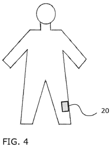
[0012] FIG. 4 shows a human figure that may be displayed by the tool with the
area of pain
indicated in the human figure.
[0013] FIG. 5 shows a flowchart illustrating an example of how spinal cord
neuromodulation
can be targeted based on the location of the pain on a patient's body.
[0014] FIG. 6 shows an example of spinal cord neuromodulation being targeted
to a specific
spinal cord level.
[0015] FIGS. 7A and 7B demonstrate an example of how the functional midline of
two
electrodes can be determined. FIG. 7C shows a slider bar that may be used by
the tool for
adjusting spinal cord neuromodulation.
[0016] FIGS. 8A and 8B show an example of how the functional midline of the
electrodes
can be aligned with the physiologic midline of the spinal cord.
[0017] FIG. 9 shows an example of how electrodes can be displayed with an
image of the
spinal cord.
[0018] FIG. 10 shows a flowchart illustrating an example of how the functional
midline of
two electrodes can be determined.
[0019] FIGS. 1 lA-l 1D show an example of how the tool can use the functional
midline for
targeting of spinal cord neuromodulation.
[0020] FIGS. 12A and 12B demonstrate an example of how the neuromodulation
settings
can be adjusted to accommodate for a change in electrode position. FIG. 12A
shows the
electrode prior to migration and FIG. 12B shows the electrode after migration.
3
CA 02802708 2012-12-13
WO 2011/159688 PCT/US2011/040329
[0021] FIG. 13 shows a flowchart illustrating an example of how the
neuromodulation
settings can be adjusted to accommodate for a change in electrode position.
DETAILED DESCRIPTION
[0022] The present invention provides a tool for assisting in the planning or
performing of
electrical neuromodulation of a patient's spinal cord (sometimes referred to
in the art as spinal
cord stimulation). In certain embodiments, the tool provides a simulation of
how much volume
of neural tissue is affected by the electrical neuromodulation. As used
herein, the term "volume
of activation" means a volume of neural tissue in which the neurons are
activated by the electric
field being applied to the neural tissue during electrical neuromodulation.
Neural activation may
have a stimulatory effect or an inhibitory effect on the neural tissue, or a
combination of both.
Although the volume refers to a three-dimensional space, the calculation,
analysis, and/or
displaying of the volume as described herein does not necessarily have to be
performed in three
dimensions. Such actions may be performed in two dimensions instead. For
example, the
volume of activation may be calculated in a two-dimensional plane and shown as
a two-
dimensional image.
[0023] The present invention may use any suitable method for calculating a
volume of
activation for neural tissue. For example, methods for calculating a volume of
activation suitable
for use in the present invention include those described in U.S. Patent No.
7,346,382 (McIntyre
et al.), U.S. Patent Application Publication No. 2007/0288064 (Butson et al.),
and U.S. Patent
Application Publication No. 2009/0287271 (Blum et al.), which are incorporated
in their entirety
by reference herein. In certain embodiments, to calculate a volume of
activation, the tool uses a
mathematical model of the electric field generated by one or more electrodes
positioned adjacent
the spinal cord of a patient. The mathematical model may be any suitable type
of model that can
be used to model an electric field created by an electrode, such as finite
element models of the
electrode(s) and the tissue medium.
[0024] The electric field generated by an electrode is dependent upon various
conditions of
the electrode itself, including the electrode position, electrode orientation,
electrode
configuration, electrode contact polarity, electrode contact selection,
electrode contact
capacitance, electrode contact impedance, and waveform parameters (e.g.,
shape, pulse width,
frequency, voltage, etc.). As used herein, "electrode neuromodulation
conditions" refers to one
4
CA 02802708 2012-12-13
WO 2011/159688 PCT/US2011/040329
or more of these factors. A set of electrode neuromodulation conditions may
include one or
more of these factors. For a given set of electrode neuromodulation
conditions, the tool can
calculate a volume of activation produced by the electrode. As used herein,
the term "electrode
neuromodulation settings" refers to a subset of electrode neuromodulation
conditions that relate
more specifically to the electrode contacts and can be adjusted during the
operation of the
electrode to vary the electric field. Examples of electrode neuromodulation
settings include
electrode contact selection and waveform parameters (e.g., shape, pulse width,
frequency,
voltage, etc.).
[0025] As used herein, the term "electrode" refers to the lead body along with
the electrode
contacts on the lead body. When referring to position, it is convenient to
refer to the electrode as
a whole, rather than referring to the position of the electrode contacts or
lead body individually
because the electrodes contacts are fixed on the lead body. Therefore, if the
position of the
electrode contacts relative to the lead body is known, then the position of
the electrode contacts
can be determined from the position of the lead body, and vice versa. Because
of this fixed
relationship, any reference to the position of the electrode is intended to
include the position of
the lead body and the electrode contacts as well. Also, when referring to the
"position" of the
electrode, this is intended to include the orientation of the electrode as
well.
[0026] The electric field model can be solved for the spatial and temporal
voltage
distribution that represents the electric field that is created in the tissue
medium by the electrode
according to a particular set of electrode neuromodulation conditions. In
certain embodiments,
the electric field model is coupled to a neuron model to determine whether the
electric potential
at a given point in space is sufficient to activate neurons in the tissue
medium. The boundaries
of neuronal activation predicted by the neuron model determines the volume of
activation.
Examples of such methods that can be used in the present invention include
those described in
U.S. Patent No. 7,346,382 (McIntyre et al.), U.S. Patent Application
Publication No.
2007/0288064 (Butson et al.), and U.S. Patent Application Publication No.
2009/0287271 (Blum
et al.), which are incorporated by reference herein. Where radiologic imaging
of the spinal
anatomy is available, the model axons of the neuron model can be aligned to
the orientation of
the spinal cord or spinal column.
[0027] Another way in which the volume of activation can be determined is by
calculating
the second order spatial derivative of the electric potential that is
distributed around the
CA 02802708 2012-12-13
WO 2011/159688 PCT/US2011/040329
electrode. The second spatial derivative is then compared against an
activation threshold. The
activation threshold is the threshold value at which a neuron is activated at
that particular point in
space for the tissue medium. If the second spatial derivative of the electric
potential exceeds the
activation threshold, then the neuron at that point in space is considered to
be activated. The
second order spatial derivative can be calculated by numerical or
approximation techniques. For
example, the second difference of the electrical potential can be used to
approximate the second
order derivative, as described in U.S. Patent No. 7,346,382 (McIntyre et al.),
U.S. Patent
Application Publication No. 2007/0288064 (Butson et al.), and U.S. Patent
Application
Publication No. 2009/0287271 (Blum et al.), which are incorporated by
reference herein.
[0028] These activation thresholds are determined from the application of the
calculated
electric field to the neuron model, as described above. However, the manner in
which the
activation thresholds are provided can vary according to different embodiments
of the present
invention. In some embodiments, these activation thresholds can be calculated
during the
operation of the tool. However, it is also possible to have these activation
thresholds calculated
prior to the operation of the tool. In this case, the activation thresholds
are predefined for use
during the operation of the tool. For example, based on the pre-calculations,
equations may be
formulated that give the activation thresholds as a function of distance from
the electrode and
one or more electrode neuromodulation conditions (such as pulse width and
voltage). Thus,
during operation of the tool, the tool may use one or more of these equations
to calculate the
activation thresholds by inputting the relevant values into the equation and
solving the equations
to obtain a spatial map of the activation thresholds. Thus, based on a given
set of
neuromodulation conditions, the spatial contour of the activation thresholds
can be established
and used to determine the volume of activation as the isosurface where the
second spatial
derivative is suprathreshold. In addition to these methods, other methods for
determining a
volume of activation by an electrode can be used in the present invention,
such as those methods
described in U.S. Patent Application Publication No. 2007/0288064 (Butson et
al.) and U.S.
Patent Application Publication No. 2009/0287271 (Blum et al.), which are
incorporated by
reference herein.
6
CA 02802708 2012-12-13
WO 2011/159688 PCT/US2011/040329
Electrode Registration
[0029] In certain embodiments, the tool may use a radiologic image in
performing the
functions that are described herein. The radiologic image may show the
electrodes and/or
various portions of the patient's spinal anatomy. As used herein, "spinal
anatomy" means the
anatomy relating to the spinal column, which includes the spinal cord, the
vertebral bodies,
nerves, and/or other soft or bony tissue of the spinal column. The radiologic
image may be any
type of body imaging used in medicine, such as x-rays (including conventional
film and
fluoroscopic x-rays), magnetic resonance imaging (MRI), computed tomography
(CT), positron
emission tomography (PET), etc. For example, the radiologic image may be an
anterior-
posterior view or a lateral view x-ray of the patient's spine. The radiologic
image may not
necessarily show all portions of the spinal anatomy. The portion of the
patient's spinal anatomy
that is visible on the radiologic image will depend upon the type of imaging
modality used. For
example, in x-ray images, only the bony structures may be visible in the image
(but not the
spinal cord itself). In MR images, the spinal cord itself may be visible, in
addition to the bony
and other soft tissue elements.
[0030] In the tool, the radiologic images are embodied as data structures
(e.g., digital
images). In some cases, the radiologic image may be used to register the
location of the
electrode. For example, the tool may register the electrode relative to a
landmark of the spinal
anatomy that is visible on the radiologic image. For example, in the case of x-
ray images, the
location of the electrode can be registered relative to the vertebral bodies
that are visible on the
image. As will be explained below, the location of the electrode relative to
the spinal cord itself
can be estimated based on the association between the vertebral level and the
spinal cord level.
[0031] As explained above, when referring to position, it is convenient to
refer to the
electrode as a whole, rather than referring to the position of the electrode
contacts or lead body
individually because the electrode contacts are fixed on the lead body. As a
result, if the position
of the lead body is registered by the tool, then the electrode contacts on the
lead body can also be
considered to be registered as well, and vice versa. Whether the tool will
locate the lead body or
the electrode contacts directly will depend on a variety of factors, such as
its visibility in the
radiologic image. Since the lead body is larger, in some cases, it may be more
practical to locate
the lead body and then locate the position of the electrode contacts based on
the lead body
position. In other cases, since the electrode contacts may be more radiopaque
and more readily
7
CA 02802708 2012-12-13
WO 2011/159688 PCT/US2011/040329
identifiable on CT or x-ray, it may be more practical to locate the electrode
contacts in the
image.
[0032] The electrode can be located automatically or manually in the
radiologic image.
Example methods of locating and registering an electrode that can be used in
the present
invention are described in U.S. Patent Application Publication No.
2009/0287271 (Blum et al.),
which is incorporated by reference herein.
[0033] Where there are multiple electrodes (two or more) present in the
radiologic image, the
tool may determine the position of the electrodes in relation to each other
and/or the spinal
anatomy. In some cases, three-dimensional positional information can be
reconstructed from
multiple (two or more) different two-dimensional views of the electrode and
the angle between
the different views. This three-dimensional reconstruction can be performed
using any suitable
technique known in the art.
[0034] For example, FIGS. IA and lB show x-ray images that can be used to
locate and
reconstruct the three-dimensional position of two electrodes 12 and 14 that
have been implanted
in a patient's spine. FIG. IA shows an anterior-posterior view of the spine
with electrodes 12
and 14 visible in the x-ray image. The tool registers the position of
electrodes 12 and 14 relative
to each other and/or the spinal anatomy.
[0035] FIG. lB shows a lateral view of the spine with electrodes 12 and 14
visible in the x-
ray image. The tool registers the position of electrodes 12 and 14 relative to
each other, and
optionally, with the spinal anatomy. Having these two different perspective
views (at a 90
angle) of electrodes 12 and 14, the tool can now reconstruct the three-
dimensional position of
electrodes 12 and 14 relative to each other, and optionally, the spinal
anatomy. Thus, the tool
can display a reconstructed three-dimensional view of electrodes 12 and 14
with respect to each
other and/or the spinal anatomy.
[0036] Thus, in certain embodiments, the tool may receive a first radiologic
image (e.g., an
anterior-posterior view x-ray) showing an electrode and the spinal anatomy of
the patient, and
receive a second radiologic image (e.g., a lateral view x-ray) showing the
electrode and the
spinal anatomy of the patient. The second radiologic image provides a
different view than the
first radiologic image so that they can be used to determine the three-
dimensional position of the
electrode in relation to the spinal anatomy. In some cases, the first and
second radiologic images
are used to determine the three-dimensional position of the multiple
electrodes in relation to each
8
CA 02802708 2012-12-13
WO 2011/159688 PCT/US2011/040329
other. Once the position of the electrodes is determined, a three-dimensional
image of the
electrodes and the spinal anatomy may be displayed to the user. The three-
dimensional image
may be rotated, panned, and zoomed to allow the user to precisely explore the
actual device
positioning in space.
Functional Images
[0037] In certain embodiments, in addition to anatomical structures, the
radiologic image of
the spinal anatomy may include information associating parts of the image to
one or more
neurologic functions (i.e., a functional image). The functional image may also
include other
symbolic information, such as structure names, object features, target volumes
generated from
previous patient data, anatomic landmarks, or boundaries. The neurologic
functions in the
functional image may be either motor or sensory functions. In some cases, the
functional image
may define different levels of the spinal cord in the image. For example, the
functional image
may include information that associates different parts of the image with the
dermatomes that are
innervated by the different spinal cord levels, as will be further explained
below.
[0038] Functional information can be incorporated into the image data using
any suitable
technique known in the art. In some cases, the functional information is
incorporated by
registering a patient-specific radiologic image to a standard atlas of the
same anatomy. A
standard atlas is an atlas of the spinal anatomy that is intended to represent
the typical or normal
anatomy that is present in human beings. As such, the standard atlas can be
derived from a
composite of the anatomy of multiple individuals to be representative of
"normal" or "typical"
human anatomy. The tool may have multiple standard atlases (e.g., variants of
normal anatomy)
and allow the user to select one that is a closest match to the patient being
treated.
[0039] Registration of the patient-specific image to the standard atlas may be
performed
using any suitable technique known in the art, including the methods described
in U.S. Patent
Application Publication No. 2009/0287271 (Blum et al.). For example, the image
registration
process may involve a transformation of the patient-specific image to match or
fit the standard
atlas, a transformation of the standard atlas to match or fit the patient-
specific image, or some
combination of both. In some cases, the image registration process may use
anatomic landmarks
that have been established in the image. These anatomic landmarks can be
identified manually
by a user or automatically by the tool. For example, in an x-ray of the spine,
the vertebral bodies
9
CA 02802708 2012-12-13
WO 2011/159688 PCT/US2011/040329
may be identified and registered into the image. Once the anatomic landmarks
are identified, the
patient-specific radiologic image can be scaled or morphed to fit the standard
atlas using the
transformation process described above.
[0040] For example, FIG. 2A shows an x-ray image of a patient's spine, which
is imported
into the tool. The spinal cord is not visible on the x-ray, but is located
within the vertebral spine
(i.e., spinal column), which is made up of a column of vertebral bodies
(vertebrae). As seen in
FIG. 2B, the user identifies the different vertebrae that are visible on the x-
ray image by drawing
a box around each of the vertebrae. The spinal cord itself is functionally
divided into segmental
levels defined by the spinal roots that enter and exit the spinal column
between each of the
vertebral body levels.
[0041] A dermatome is an area of the skin that is predominantly innervated by
nerves
originating from a single spinal level. FIG. 3 shows a dermatome map of the
human body.
Thus, the spinal cord can be divided functionally into segments that
correspond to different
dermatomes. The spinal cord segmental levels do not necessarily correspond to
the same level of
the vertebral body. Accordingly, the dermatomes innervated by the different
spinal cord levels
do not necessarily correspond to the vertebral levels. For example, the L5
dermatome level for
low back pain may correspond to the T10 vertebral level. However, based on
known anatomic
and physiologic relationships, the tool of the present invention can make the
appropriate
correlation between the dermatome levels, the spinal cord levels, and/or the
vertebral levels.
This association may be useful where the vertebral bodies are being used as a
reference for the
position of the electrode.
[0042] As seen in FIG. 2C, the association of these different vertebral levels
with their spinal
cord levels are registered into the image to create a functional image in
which spinal cord levels
T12, L1, and L2 are registered as functional regions in the image in
association with the vertebral
levels that are visible in the image. If electrodes are also present in the
image, the electrodes can
also be identified (either manually or automatically) and their position
registered in relation to
the functional regions.
[0043] As an alternative to having the user identify each vertebra, the
positions of the
vertebrae may be identified based on a user identification of a single
vertebra in an image. For
example, the user may input a vertebral outline, or part of a vertebral
outline, along with an
identification of the vertebra to which the outline corresponds (e.g., Ti).
The image is then
CA 02802708 2012-12-13
WO 2011/159688 PCT/US2011/040329
analyzed to extrapolate the positions of the remaining vertebrae based on
their relative positions
to the outlined vertebra.
Targeting of Neuromodulation
[0044] In certain embodiments, the tool can be used to select a region of the
spinal cord as a
target for electrical neuromodulation. The selection of the target region can
be provided in any
suitable manner. For example, the targeted region can be input by the user as
a specific anatomic
structure (such as a vertebral level), a segment of the spinal cord, a
dermatome level, or an area
of the body where the patient is experiencing pain or discomfort. In the
example where the user
indicates one or more dermatome levels as a targeted region, the tool may
determine the spinal
cord level(s) and/or vertebral level(s) that correspond to those dermatomes.
In the example
where the user indicates where the patient is experiencing pain or discomfort,
the tool may
determine the one or more dermatomes associated with that part of the body,
and then select one
or more spinal cord levels and/or vertebral levels that correspond to that
dermatome.
[0045] Having selected the targeted region, the tool can then find a set of
electrode
neuromodulation conditions that would direct the electrical neuromodulation to
that targeted
region by comparing the predicted volumes of activation against the targeted
region. For
example, the tool may use a scoring technique that measures the effectiveness
of the
neuromodulation based on how much of the predicted volume of activation
encompasses the
targeted region, how much of the targeted region is within the predicted
volume of activation,
how much of the predicted volume of activation is outside the targeted region,
how much of the
targeted region is outside the predicted volume of activation, how much of the
predicted volume
of activation encompasses neural tissue that would cause side effects, or a
combination thereof.
The tool may calculate multiple predicted volumes of activation under
different neuromodulation
conditions in order to find a suitable set of electrode neuromodulation
conditions. When a
combination of scoring factors is used, the different factors may be weighted
differently
according to their relative importance in determining the therapeutic
effectiveness of the
neuromodulation. In some cases, an improved or optimal set of neuromodulation
conditions can
be determined by using an optimization algorithm to find a set of electrode
neuromodulation
conditions that produces a volume of activation having the best score (e.g.,
highest or lowest
score).
11
CA 02802708 2012-12-13
WO 2011/159688 PCT/US2011/040329
[0046] For example, FIG. 4 shows a patient that is experiencing pain in area
20 of their body.
The user (e.g., the patient or a caretaker) enters the location of area 20
into the tool and the tool
correlates this area 20 with the L2 dermatome level on the left side, and then
correlates the left-
side L2 dermatome level with the corresponding region the spinal cord or the
vertebral level that
corresponds to the L2 level of the spinal cord. FIG. 6 shows an image of the
spinal cord 40 with
the spinal cord levels being represented as different functional regions in
the spinal cord (levels
T11-L4 being shown here). Adjacent the spinal cord 40 is an electrode 38
having three electrode
contacts 30, 32, and 34 fixed on a lead body 36. Based on the user's input,
the functional region
L2 of spinal cord 40 is selected as the target region for electrical
neuromodulation. Accordingly,
the tool determines a set of electrode neuromodulation settings that would
create a volume of
activation that is directed to functional region L2. In this instance, the set
of electrode
neuromodulation settings includes the selection of electrode contacts 32 and
34 for activation,
and electrode contact 30 for non-activation. Additionally, with this selected
set of electrode
neuromodulation settings, electrode 32 is predicted to create a volume of
activation 46 and
electrode 34 is predicted to create a volume of activation 48. Thus, with the
combination of
volume of activations 46 and 48, the selected set of electrode neuromodulation
settings create a
volume of activation that is directed to dermatome level L2 of the spinal
cord. FIG. 5 shows a
flowchart illustration of the above process.
[0047] Dermatome targeting using patient feedback about where the electrically-
induced
parasthesia is located in their body may not always be reliable because the
patient's sensory
perception may not be accurate or the patient may not sense sufficient
parasthesia from the
electrical neuromodulation. In certain embodiments, the dermatome location of
the electrical
neuromodulation can be localized more precisely using electromyography (EMG).
For EMG
localization of electrical neuromodulation, a number of EMG electrodes are
placed on the
patient's body. Electrical neuromodulation of the sensory fibers in the spinal
cord can elicit a
reflexive motor response and these motor responses can be detected as EMG
signals in the
specific dermatomes. Thus, by analyzing the EMG signals during electrical
neuromodulation,
the dermatome location of the electrical neuromodulation can be identified
more precisely, thus
allowing more accurate targeting of electrical neuromodulation.
[0048] In certain embodiments, the electrode used in the neuromodulation may
also have
recording electrodes which can sense neural signals passing through sensory
nerve fibers. This
12
CA 02802708 2012-12-13
WO 2011/159688 PCT/US2011/040329
can be useful for improved accuracy in identifying where the patient is
experiencing pain or
discomfort. The sensory signals passing through these sensory fibers may be
produced by
applying a sensory stimulation to the area where the patient is feeling the
pain or discomfort. A
variety of different kinds of sensory stimulations can be used, such as
applying a dull touch, a
sharp prick, or a slight electrical pulse to the skin. The recording electrode
could sense this
signal being transmitted along nearby sensory fibers as an increase in local
field potential. Based
on which recording contact records the strongest signal, or based on the
distribution of the signal
across multiple contacts, the fiber(s) carrying the sensory stimulation signal
from the afflicted
dermatome is identified. Moreover, the strength of the signal can be used to
determine the
magnitude of the patient's pain or discomfort in that area.
Cerebrospinal Fluid
[0049] One of the factors influencing the electric field generated by an
electrode is the
electrical conductivity of the surrounding tissue medium (e.g., the electrical
conductivity of the
spinal cord neural tissue or other body tissue in the vicinity of the
electrode, such as
cerebrospinal fluid, tissue membranes, encapsulation tissue around the
electrode, etc.). Thus, the
electric field model used by the tool may include a characterization of the
tissue electrical
conductivity. In some cases, different anatomical structures may be
represented as having
different electrical conductivities in the electric field model. One of the
tissue mediums that may
be relevant in spinal cord neuromodulation is the cerebrospinal fluid (CSF)
that surrounds the
spinal cord. The CSF is considered to be relatively more electrically
conductive compared to the
other surrounding tissue.
[0050] In certain embodiments, the electric field model may account for the
amount of CSF
that is present between the electrode and the spinal cord. For example, the
electric field model
may account for the thickness (in dimensional terms, not viscosity) of the CSF
between the
electrode and the spinal cord. The dimensional thickness of the CSF can be
determined using
various approaches. In some cases, the thickness of the CSF can be determined
by using a
radiologic image, such as an axial view MR image. In some cases, the thickness
of the CSF can
be approximated based on the electrode position relative to the spinal
anatomy. For example, the
thickness of the CSF can be approximated based on the vertebral level where
the electrode is
positioned or the size of the vertebrae where the electrode is positioned (in
general, the size of
13
CA 02802708 2012-12-13
WO 2011/159688 PCT/US2011/040329
the vertebral bodies progressively increase moving from the cervical to the
lumbar spine).
Accounting for the electrical conductivity of CSF may allow the tool to
calculate a more accurate
the volume of activation.
Total Potential Volume of Activation
[0051] In certain embodiments, the tool can show the total potential volume of
activation
capable of being produced by an electrode at a given position. The total
potential volume of
activation can be displayed as the overlap of the volume of activations
produced by the highest
tolerable amplitude anode/cathode pulse for each electrode. Knowing the total
potential volume
of activation may be useful during initial surgical implantation of the
electrode to help position
the electrode at a location that will meet both current and possible future
coverage needs (e.g.,
accounting for the possibility of electrode migration, worsening pain, or
wider extent of pain).
The feature can also be useful for quickly seeing how much area has been
tested by overlaying a
history of stimulated regions and the total potential volume of activation.
This feature can also
allow the user to view spaces that are outside the potential volume of
activation for a given
electrode placement. For example, if two electrodes are staggered or canted,
they may leave
regions of the spinal cord unable to be reached by electrical neuromodulation.
Displaying the
total potential volume of activation would allow this to be realized during
intraoperative or
postoperative programming.
[0052] This display of the total potential volume of activation can be turned
on and off, and
may appear in a variety of colors, gradients, and patterns to best suit
visualization. In addition, it
may be layered with current neuromodulation settings or previously trialed
settings to compare
the total potential volume of activation with volumes already tested. As with
other display
features, the total potential volume of activation can be displayed as a two-
dimensional area on a
spinal cord or as a three-dimensional volume. The total potential volume of
activation may also
be used to predict dermatome regions capable of neuromodulation, which would
then be
displayed on a two-dimensional or three-dimensional representation of the
spinal cord. The total
potential volume of activation could also be shown as all the dermatome
regions capable of
being affected by the neuromodulation, which could be displayed on an image of
a human figure.
14
CA 02802708 2012-12-13
WO 2011/159688 PCT/US2011/040329
Functional Midline
[0053] When multiple electrodes (two or more) are implanted into a patient,
the electrodes
are often not parallel to each other or not in level alignment with each other
(e.g., one is higher
than the other), and moreover, the position of the electrodes relative to the
spinal cord is often
not known since the spinal cord may not be visible on x-ray images. Where
multiple electrodes
are being modeled by the tool, the tool may determine a functional midline in
the
neuromodulation space around the electrodes. The functional midline is an
imaginary line
running in the neuromodulation space of the electrodes, which corresponds to
the sensory
midline of the patient's body, and which could be aligned to the physiologic
midline of the
patient's spinal cord. The functional midline is established by finding a set
of neuromodulation
settings that induces parasthesia in the center of the patient's body. The
functional midline can
then be derived from the relative pulse intensities between the multiple
electrodes. The tool may
also determine the functional midline for a paddle-type electrode having an
array of electrode
contacts on a single electrode lead or a single electrode that is implanted in
a lateral orientation.
[0054] An example of how this may be performed is illustrated in FIGS. 7A and
7B. FIG.
7A shows two electrodes, 50 on the left side and 51 on the right side, each
comprising a lead
body 58 connected to lead wires 60 and having three electrode contacts,
including top-most
contacts 52 and bottom-most contacts 56. The functional midline is determined
by finding the
functional midpoint between the left and right top-most electrode contacts 52,
and the left and
right bottom-most electrode contacts 56. The functional midpoint between the
left and right top-
most electrode contacts 52 is determined by varying the relative pulse
intensities (monopolar)
between the left and right top-most electrode contacts 52, and receiving
patient feedback of
where the parasthesia is being sensed. FIG. 7C shows how the stimulation field
can be shifted to
the left or right using a slider 70 displayed by the tool. Slider 70 is inside
a bar that represents
the left versus right relative pulse intensity. Area 72 in the bar corresponds
to the relative pulse
intensity for the electrode contact on the left electrode and area 74 in the
bar corresponds to
relative pulse intensity for the counterpart electrode contact on the right
electrode. Slider 70 can
be moved left or right to adjust the pulse intensity that is apportioned
between the left and right
electrode contacts. As an initial setting, the slider may be positioned in the
middle such that half
of a tolerable pulse intensity is sent to each of the counterpart electrode
contacts on the left and
right electrodes.
CA 02802708 2012-12-13
WO 2011/159688 PCT/US2011/040329
[0055] When the patient indicates that the parasthesia is being sensed in the
center of their
body, the relative pulse intensities of the left and right top-most electrode
contacts 52 gives the
proportionate distance of the functional midpoint from the respective left and
right electrode
contacts 52. As shown in FIG. 7B, the patient's parasthesia has been centered
for the top-most
electrodes 52 when the left top-most electrode contact has a pulse intensity
64 and the right top-
most electrode has a pulse intensity 65, with the functional midpoint being at
point 68. Pulse
intensities 64 and 65 do not represent actual activation fields, but is being
used only to help
illustrate how the left versus right relative pulse intensities can differ and
be used to find the
midpoint. The same process of varying the left/right relative pulse
intensities and receiving
patient feedback about the location of the parasthesia is repeated to find the
functional midpoint
for the bottom-most electrode contacts 56. In this instance, the patient's
parasthesia has been
centered for the bottom-most electrodes 56 when the left bottom-most electrode
contact has a
relative pulse intensity 66 and the right bottom-most electrode contact has a
relative pulse
intensity 67, with the functional midpoint being at point 69. An imaginary
line is drawn between
functional midpoints 68 and 69, and this imaginary line is the functional
midline 62 between
electrodes 50 and 51. FIG. 10 shows a flowchart illustration of the above
process.
[0056] Once the functional midline is determined, this information can be used
in various
ways to assist in electrical neuromodulation of a patient's spinal cord. One
use for the functional
midline is for aligning the electrodes with respect to the physiologic midline
of the spinal cord.
For example, FIG. 8A shows the two electrodes 50 and 51 again with their
functional midline 62.
Based on this functional midline 62, the position (including orientation) of
electrodes 50 and 51
can be aligned with a spinal cord. FIG. 8B shows a graphically rendered,
generic image of a
spinal cord 76 (not specific to any particular patient), with its physiologic
midline represented by
dotted line 78. By rotating the pair of electrodes 50 and 51, their functional
midline 62 is made
to be oriented parallel to physiologic midline 78 of spinal cord 76. The two
electrodes 50 and 51
are displayed over spinal cord 76 to give a more accurate representation of
how the electrodes 50
and 51 are oriented relative to the actual patient's spinal cord. FIG. 9 shows
another example of
how electrodes and a graphically rendered, generic image of a spinal cord
could be displayed by
the tool.
[0057] Thus, in certain embodiments, the tool receives a radiologic image of
the patient
showing one or more electrodes inside the patient and locates the one or more
electrodes in the
16
CA 02802708 2012-12-13
WO 2011/159688 PCT/US2011/040329
radiologic image. The one or more electrodes collectively have multiple
electrode contacts. The
tool determines the functional midline for the one or more electrodes and may
display on a
display screen, an image of a spinal cord and the one or more electrodes such
that the functional
midline of the one or more electrodes is aligned to the physiologic midline of
the spinal cord.
[0058] In some cases, the tool may receive information about the relative
electrical
neuromodulation intensity between a first electrode contact among the multiple
electrode
contacts and a first counterpart electrode contact among the multiple
electrode contacts. Based
on the relative electrical neuromodulation intensities, the tool can determine
a first midpoint
between the first electrode contact and the first counterpart electrode
contact. The tool may
further receive information about the relative electrical neuromodulation
intensity between a
second electrode contact among the multiple electrode contacts and a second
counterpart
electrode contact among the multiple electrode contacts. Based on the relative
electrical
neuromodulation intensities, the tool can determine a second midpoint between
the second
electrode contact and the second counterpart electrode contact. The functional
midline can be
established as the line between the first midpoint and the second midpoint.
This method may be
applied to a single electrode (e.g., a paddle-type electrode having multiple
electrode contacts
arranged in an array) or multiple separate electrodes.
[0059] In cases where there are multiple separate electrodes (which
collectively have
multiple electrode contacts), a functional midline may be found using a first
electrode contact
which is on a first one of the multiple electrodes and a first counterpart
electrode contact on a
second one of the multiple electrodes. Based on the relative electrical
neuromodulation
intensities, the tool can determine a first midpoint between the first
electrode contact and the first
counterpart electrode contact. Furthermore, the tool may receive information
about the relative
electrical neuromodulation intensity between a second electrode contact on the
first one of the
multiple electrodes and a second counterpart electrode contact on the second
one of the multiple
electrodes. Based on the relative electrical neuromodulation intensities, the
tool can determine a
second midpoint between the second electrode contact and the second
counterpart electrode
contact; and establish the functional midline as a line between the first
midpoint and the second
midpoint.
17
CA 02802708 2012-12-13
WO 2011/159688 PCT/US2011/040329
Adaptive Searching
[0060] The functional midline can also be used to assist in targeting of the
spinal cord
neuromodulation to the appropriate side of the body (right vs. left side).
Based on whether the
patient's symptoms are on the left or right side of their body, the electrical
neuromodulation to
the spinal cord can be directed to the same side (left or right) of the
functional midline. This
targeting may be implemented through a binary searching algorithm.
[0061] For example, FIGS. 1 lA-l 1D show one example of how this binary
searching
algorithm can be applied. In this particular example, two electrodes have been
implanted in the
patient's spine, and the tool has determined the functional midline between
the two electrodes in
the manner described above. The tool receives the location of where the
patient is experiencing
pain; in this particular case, the left thigh. As shown in FIG. 1 IA, an area
82 of the left thigh is
shown on the display screen as the area where the patient is experiencing
pain. With the pain
being located on the left side, one or more of the electrode neuromodulation
settings are
configured to apply neuromodulation to the left side of the spinal cord based
on the functional
midline of the two electrodes. The patient then indicates where the
neuromodulation-induced
parasthesia is being felt. In this instance, the patient indicates that the
parasthesia is felt on the
left abdomen, which is shown as parasthesia area 84 in FIG. 1 lB. Because the
parasthesia area
84 is too high above the targeted pain area 82, the electrode neuromodulation
settings are
adjusted to direct neuromodulation to an area lower on the spinal cord. After
this adjustment, the
patient again indicates where the neuromodulation-induced parasthesia is being
felt. In this
instance, as shown in FIG. 11 C, the patient indicates that the parasthesia
area 84 is being felt on
the left calf below the pain area 82. As shown in FIG. 11D, with further
adjustments to the
neuromodulation settings, the area of parasthesia 84 is now within the area of
pain 82. Since this
area of parasthesia 84 is not sufficient to cover the entire area of pain 82,
the pulse intensity may
need to be increased to achieve sufficient reduction in pain.
Electrode Migration
[0062] One of the problems associated with spinal cord neuromodulation is
changes in the
position of the electrode after its implantation. For example, the electrode
may migrate to a
different location (e.g., move downwards or move to the side in a "windshield-
wiper" fashion) or
change its orientation (e.g., the long axis of the electrode may tilt to a
different direction, or in
18
CA 02802708 2012-12-13
WO 2011/159688 PCT/US2011/040329
the case of a directional electrode contact, rotate towards a different
direction). This change in
the position of the electrode can result in a loss of therapeutic efficacy. In
certain embodiments,
the tool of the present invention can adjust the neuromodulation settings to
accommodate for the
change in electrode position. A change in the position of the electrode can be
detected on a
radiologic image, such as x-ray images, in the manner described above.
[0063] In some cases, the tool may compare the position of the electrode in a
radiologic
image taken prior to migration of the electrode (e.g., a post-operative x-ray)
to the position of the
electrode after migration. Based on the relative positioning of the electrode
before and after
migration, the tool can adjust one or more of the electrode neuromodulation
settings to redirect
the neuromodulation to the original target. In the example shown in FIG. 12A,
an electrode
comprising a lead body 96 and three electrodes 93, 94, and 95 are shown prior
to migration. At
this position, electrode contact 95 is activated to produce a volume of
activation 97 that is
directed to target site 92 on spinal cord 90.
[0064] FIG. 12B shows the same electrode after downward migration along spinal
cord 90
(see arrow 99 in FIG. 12A). Because of this migration, the prior
neuromodulation settings are
ineffective because the electrode has shifted relative to target site 92. But
by comparing the
relative position of the electrode before and after migration, the electrical
neuromodulation
settings may be adjusted to redirect the electrical neuromodulation to the
original target site 92.
Using the targeting methods described above, the tool finds a set of
neuromodulation settings
with the selection of electrode contact 93 that creates a volume of activation
98 that overlaps
with target site 92 or volume of activation 97. As a result, the tool has
accommodated the
electrical neuromodulation for electrode migration. FIG. 13 shows a flowchart
illustration of the
above process. Positional changes in the electrodes can also be determined
from means other
than by radiologic imaging. For example, the electrode may have an
accelerometer that detects
the position of the electrode. The tool may determine positional changes in
the electrode based
on the information from the accelerometer.
[0065] Thus, in certain embodiments, the tool receives a first radiologic
image of an
electrode inside a patient, wherein the electrode is in a first position. The
tool further receives a
second radiologic image of the electrode after a change in the electrode's
position, wherein the
electrode is in a second position. The tool determines the position of the
electrode in the second
position relative to the electrode in the first position and calculates a
first volume of activation
19
CA 02802708 2012-12-13
WO 2011/159688 PCT/US2011/040329
generated by the electrode in the first position. The tool can then determine
an electrode
neuromodulation setting for the electrode in the second position that produces
a second volume
of activation that at least partially encompasses the first volume of
activation. The tool may
display the second volume of activation on a display screen.
[0066] In some cases, the tool calculates multiple test volumes of activation
using different
electrode neuromodulation settings and compares the multiple test volumes of
activation to the
first volume of activation. Based on the comparison of the multiple test
volumes of activation,
the tool selects an electrode neuromodulation setting for the electrode in the
second position that
produces the second volume of activation.
Automated serial review of electrode contacts
[0067] In certain embodiments, the tool may also have a programming mode that
automates
the standard monopolar review process. In this mode, the user is asked to
identify the pain
location and severity. Then, each consecutive electrode contact is activated
at a tolerable
amplitude. The patient is asked to identify the location of the parasthesia
and what level of pain
they are currently feeling. This is repeated for each available electrode
contact. Once each
contact has been tested, the user may be given the option of having the tool
interpolate the
mapped data to predict the best neuromodulation settings. SFMs may be computed
and
displayed for each successive activation and displayed in real-time to the
user, together with real-
time display of the parasthesia locations on a three-dimensional model. Real-
time display of
SFMs and parasthesia locations may also be performed in other programming
modes (e.g., the
manual programming mode described below in connection with the interface
features).
Software and Machine Embodiments
[0068] The tool of the present invention may also be embodied as a computer-
readable
storage medium having executable instructions for performing the various
processes as described
herein. The storage medium may be any type of computer-readable medium (i.e.,
one capable of
being read by a computer), including non-transitory storage mediums such as
magnetic or optical
tape or disks (e.g., hard disk or CD-ROM), solid state volatile or non-
volatile memory, including
random access memory (RAM), read-only memory (ROM), electronically
programmable
memory (EPROM or EEPROM), or flash memory. The term "non-transitory computer-
readable
CA 02802708 2012-12-13
WO 2011/159688 PCT/US2011/040329
storage medium" encompasses all computer-readable storage media, with the sole
exception
being a transitory, propagating signal.
[0069] The tool of the present invention may also be embodied as a computer
system that is
programmed to perform the various processes described herein. The computer
system may
include various components for performing these processes, including
processors, memory, input
devices, and/or displays. The computer system may be any suitable computing
device, including
general purpose computers, embedded computer systems, network devices, or
mobile devices,
such as handheld computers, laptop computers, notebook computers, tablet
computers, and the
like. The computer system may be a standalone computer or may operate in a
networked
environment.
Interface Features
[0070] The tool may use any of a variety of interface features for interacting
with a user.
These interactions may include receiving inputs, producing outputs, displaying
information,
storing program settings, making selections (e.g., target sites,
neuromodulation settings, etc.),
and the like. The interface features may be adapted for any of the various
potential users of the
tool, including clinicians, care providers, technicians, salespeople, or the
patients themselves.
The interface may be provided through any suitable hardware devices, including
touch screens,
touch pads, mouse, trackball, buttons, wheels, dials, etc. For example, the
tool may display a
three-dimensional human figure the user may be able point to and select a part
of the human
figure by a touch screen or a mouse. Various types of interface features which
may be used by
the tool include those described in U.S. Patent Application Publication No.
2009/0287271 (Blum
et al.), which is incorporated by reference herein. The tool may display on a
display screen any
of the elements described above, including the volumes of activation, spinal
anatomy (e.g., of the
vertebrae, spinal cord, or both), radiologic images, electrodes, human
figures, and such, either
individually or in combination.
[0071] The tool may also have a manual programming mode in which previously
trialed
neuromodulation settings are displayed. Another feature may allow the user to
customize a
neuromodulation region, and then drag the region to the area of the spinal
cord for trial
simulations of neuromodulation; or allow the user to attempt neuromodulation
settings believed
to be advantageous by offering a specific visual history of previously
attempted settings. The
21
CA 02802708 2012-12-13
WO 2011/159688 PCT/US2011/040329
recorded results of the previously attempted settings may be displayed in two
or three-
dimensional space. For example, the patient's pain zone can be displayed on a
three-dimensional
model together with the parasthesia zones that resulted from a set of
attempted settings. The
three-dimensional model may be displayed in conjunction with the display of
SFMs calculated
for the set of attempted settings (e.g., in a separate display area that shows
a three-dimensional
model of the spinal cord). The patient's pain zone can be mapped on the human
figure and
distinguished in some way (by color, for example). The previous parasthesia
zones from trial
simulations can appear on the human figure. These zones may directly show a
result, such as
efficacy or indication of pain, by a different color or shade, or they may
have text that appears
inside them or in a pop-up when the user hovers or clicks the computer's
pointing mechanism
over the region. Example text may include Visual Analogue Scale (VAS) scores
and stimulation
settings. The corresponding volume of activation shown on the spinal cord
could also be
highlighted or identified when the user selects the affected dermatome. This
feature would allow
the user to easily see which dermatomes are impacted by the neuromodulation
zones, and vice
versa.
[0072] After viewing the results the user may wish to trial a volume of
activation that has not
been previously trialed. The manual programming mode in the tool can feature a
simple method
to trial an area of the spinal cord by entering a mode that displays a desired
volume of activation
that can be manipulated by the user. Alternatively, the user could start with
a previously trialed
volume of activation. The desired volume of activation may be resized and
dragged to the
desired location on the spinal cord image. An algorithm would then calculate
the closest actual
neuromodulation settings that would best fit the zone desired for
neuromodulation (i.e., adjusting
the settings associated with the previously trialed volume of activation to
levels that are
appropriate for the resized/re-located volume) and show the user the new
settings, who would
confirm and trial the neuromodulation. The calculation of the new settings may
be performed in
a similar fashion to the method previously described for adjusting settings in
response to
unintended electrode migration, i.e., creating a volume of activation that
overlaps with the new
volume. The algorithm may take into consideration factors pertaining to the
new location, such
as CSF thickness, when calculating the new settings. Since it may be
advantageous to view the
depth of tissue affected by the neuromodulation, a slidable bar can be
featured along the side of
the posterior spinal cord view. The bar may be positioned to the precise
location that a cross-
22
CA 02802708 2012-12-13
WO 2011/159688 PCT/US2011/040329
sectional view is desired. In the cross-sectional view, the slidable bar could
be used to
sequentially browse through different cross-sectional views. Once positioned,
the bar is selected
or clicked to bring a cross-sectional view that displays the desired volumes
of activation as well
as offers the same feature of using a desired volume of activation that can be
manipulated by the
user.
[0073] Once results of the manual programming mode are optimized, the final
settings may
be saved to memory, named, and the user is returned to the main programming
page. Saved
settings may be selected and displayed via an interface menu. Settings may be
merged to
combine a plurality of saved settings into a single set of saved settings. For
example, settings
targeting different pain zones may be combined in order to provide a custom
course of treatment
for a patient experiencing pain in more than one zone. Similarly, settings
that by themselves fail
to provide adequate pain zone coverage may be combined to provide sufficient
coverage.
[0074] The foregoing description and examples have been set forth merely to
illustrate the
invention and are not intended as being limiting. Each of the disclosed
aspects and embodiments
of the present invention may be considered individually or in combination with
other aspects,
embodiments, and variations of the invention. Further, while certain features
of embodiments of
the present invention may be shown in only certain figures, such features can
be incorporated
into other embodiments shown in other figures while remaining within the scope
of the present
invention. In addition, unless otherwise specified, none of the steps of the
methods of the
present invention are confined to any particular order of performance.
Modifications of the
disclosed embodiments incorporating the spirit and substance of the invention
may occur to
persons skilled in the art and such modifications are within the scope of the
present invention.
Furthermore, all references cited herein are incorporated by reference in
their entirety.
23