Note: Descriptions are shown in the official language in which they were submitted.
CA 02812254 2016-11-04
IMPLANT DEVICE AND TOOL RELATING TO TREATMENT OF PARANASAL
SINUSES
FIELD OF THE INVENTION
The invention relates to treatment of conditions of the paranasal sinuses,
including
with respect to implant devices, surgical tools and methods.
BACKGROUND OF THE INVENTION
In the United States alone, 35 million people a year are treated for sinus
infections, or
sinusitis, and 7 million of those will suffer from chronic sinusitis and will
have minimal
response to prescription drug therapies. Current surgical interventions may be
expected to, at
best, offer only moderate symptomatic improvement but no cure.
Current drug therapies include oral administration as pills and nasal topical
administration, neither of which is conducive to delivering adequate
concentration of
medication to the involved paranasal sinus. In addition to medication,
frequent sinus
irrigation can be helpful in flushing out debris, irritants and obstructing
viscous fluids, but
patients are generally not able to adequately perform this procedure at home.
For patients with particularly severe symptoms, surgical drainage may be the
only
additional option. An early surgical procedure was the Caldwell-Luc procedure,
which
involves creating a permanent fistula from the base of the paranasal sinus
into the oral cavity
above the front upper incisors. More recently, other surgical access points to
the paranasal
sinuses have been attempted. A variety of endoscopic techniques have been
developed that
access the paranasal sinuses through the nose, including functional endoscopic
sinus surgery
(FESS) and balloon sinuplasty. All attempt to increase drainage, but utilize
different routes
1
CA 02812254 2013-03-21
WO 2012/048278
PCT/US2011/055456
or tools. None of these surgical approaches has achieved wide-spread success,
and millions
of chronic sinusitis patients continue to suffer long-term disability and
discomfort.
SUMMARY OF INVENTION
A variety of medical treatments and medical procedures directed to the
paranasal
sinuses may be performed through a fistula that may be formed between the
lacrimal
apparatus and a paranasal sinus. Such a fistula provides direct access from
the lacrimal
apparatus to the paranasal sinus in a minimally invasive manner. Such direct
access permits
drugs to be conveniently administered for local treatment in the paranasal
sinus, rather than
having to rely on systemic drug treatments. Such direct access permits
irrigation fluids to be
conveniently introduced into the paranasal sinus. Such access permits fluids
to be
conveniently removed from the paranasal sinus. Such access permits a variety
of medical
procedures to be conveniently performed in the paranasal sinus.
A first aspect of the invention involves an implant device for implantation in
a human
to fluidly connect the lacrimal apparatus to a paranasal sinus through such a
fistula. The
implant device has a proximal end and a distal end located at opposite
longitudinal ends of
the device. A conduit extends from adjacent the proximal end to adjacent the
distal end. An
internal passage extends between the proximal end and the distal end, and
including through
the conduit. The internal passage has a first end open at the proximal end of
the implant
device and a second end open at the distal end of the implant device. The
implant device
includes a length longitudinally along the device between the proximal end and
the distal end
that is in a range of from 2 millimeters to 50 millimeters. A width of the
internal passage
transverse to the length is in a range of from 0.25 millimeter to 5
millimeters. The implant
device is configured to be implanted to fluidly connect the lacrimal apparatus
to the paranasal
sinus through the fistula so that when the implant device is implanted: the
proximal end is
disposed with the first end of the internal passage opening in the lacrimal
apparatus; the distal
end is disposed in the paranasal sinus with the second end of the internal
passage opening in
the paranasal sinus; and the conduit is disposed through the fistula.
A number of feature refinements and additional features are applicable to the
first
aspect of the invention. These feature refinements and additional features may
be used
individually or in any combination. As such, each of the following features
may be, but are
not required to be, used within any other feature or combination of features
of the first aspect
or any other aspects of the invention.
2
CA 02812254 2013-03-21
WO 2012/048278
PCT/US2011/055456
The conduit may be configured so that an exterior of the conduit comprises an
anchoring surface feature which assists to anchor the implant device when the
device is
implanted. The anchoring surface feature includes protrusion areas and recess
areas. The
implant device may be configured so that when implanted the conduit is
disposed through the
fistula with at least a portion of the recess areas disposed within the
fistula and with at least a
portion of the protrusion areas disposed in the fistula and engaging tissue
exposed within the
fistula to anchor the implant device. The structural and mechanical
characteristics of
protrusion occurrences in the protrusion areas may affect anchoring
performance of the
protrusion areas. The height of the protrusion areas relative to the recess
areas may affect
anchoring effectiveness when the implant device is implanted. A larger height
may provide
greater anchor effectiveness, but also may involve a larger overall width of
the implant
device that must be inserted into the fistula. The protrusion areas may have a
height relative
to the recess areas of at least 0.1 millimeter, at least 0.2 millimeter, at
least 0.25 millimeter or
at least 0.3 millimeter. The protrusions areas may have a height relative to
the recess areas of
no greater than 2 millimeters, no greater than 1.5 millimeter, no greater than
1 millimeter, no
greater than 0.75 millimeter, no greater than 0.5 millimeter or no greater
than 0.4 millimeter.
The height may be of particular protrusion occurrences relative to adjacent
areas of recesses.
Protrusion occurrences are also referred to herein as anchor protrusions. Such
anchor
protrusions may be configured to flexibly deform when the conduit is inserted
through the
fistula for implantation, for example to flexibly deform in a direction
opposite the direction of
insertion when the anchor protrusions contact tissue disposed in the fistula
during insertion.
After insertion, the anchor protrusions may over time return to their original
shape and extend
deeper into adjacent tissue to better anchor the implant device. The
mechanical properties of
the anchor protrusions may be influenced by materials of construction.
Preferred materials of
construction for the protrusion areas, and also for the portions of the
implant device, are
polymeric materials. The polymeric materials may preferably be medical grade
materials.
Some preferred polymeric materials are silicones and polyurethanes. For
enhanced
performance, the material of construction should have a rigidity that
interacts positively with
tissue in the vicinity of the fistula, for example to promote load sharing and
good anchoring.
One preferred material of construction is a polymeric material (e.g. silicone
or polyurethane)
having a durometer (Shore A) in a range having a lower limit of 50, 60, 70 or
80 and an upper
limit of 100, 80, 70 or 60, provided that the upper limit must be larger than
the lower limit.
One preferred range is for a durometer (Shore A) of 60-100, with a range of 80-
100 being
3
CA 02812254 2013-03-21
WO 2012/048278
PCT/US2011/055456
even more preferred. For some implementations the polymeric material has a
durometer
(Shore A) of about 60, of about 80 or of about 100. Mechanical properties of
the protrusion
occurrences of the protrusion areas will also be affected by the geometry of
the protrusion
occurrences. The protrusion occurrences may have a width that tapers, or
narrows, in a
direction from a base toward a top of the protrusion occurrences, with the
base being a
portion of a protrusion occurrence disposed toward the internal passage of the
conduit and a
top of the protrusion occurrence being the extremity of the protrusion
occurrence away from
the internal passage of the conduit. The width may be transverse to the length
of the conduit.
The protrusion occurrences may have a width at the base that is no larger than
2 millimeters,
no larger than 1.5 millimeters, no larger than 1.25 millimeters or no larger
than 1 millimeter.
One or more of the protrusion occurrences may have a width at the base that is
at least 0.2
millimeter, at least 0.3 millimeter, at least 0.5 millimeter, at least 0.75
millimeter or at least 1
millimeter. The protrusion occurrences may have a width adjacent the top that
is no larger
than 0.75 times width at the base, no larger than 0.5 times the width at the
base, or no larger
than 0.25 times the width at the base. The protrusion occurrences may have a
width midway
between the base and the top that is no larger than 0.8 times the width of the
base, no larger
than 0.7 times the width of the base, no larger than 0.6 times the width of
the base or no
larger than 0.5 times the width at the base.
The protrusion areas may be provided by a single protrusion occurrence feature
located to correspond with the interior of the fistula when the implant device
is implanted. In
more preferred implementations, the protrusion areas include multiple
protrusion occurrences
spaced on the exterior of the conduit. The protrusion occurrences may have a
center-to-
center spacing, in one or more directions, of at least 0.5 millimeter, at
least 0.75 millimeter, at
least 1 millimeter or at least 1.75 millimeters. The protrusion occurrences
may have a center-
to-center spacing of no greater than 2.5 millimeters, no greater than 2
millimeters or no
greater than 1.75 millimeters. The protrusion occurrences may have a center-to-
center
spacing longitudinally along the conduit. The protrusion occurrences may have
a center-to-
center spacing that is at least 0.5 times the base width of the protrusion
occurrences, or at
least 1 times the base width of the protrusion occurrences or at least 2 times
the base width of
the protrusion occurrences. The protrusion occurrences may have a center-to-
center spacing
that is no more than 5 times a base width of the protrusion occurrences, no
more than 3 times
a base width of the protrusion occurrences or no more than 2 times a base
width of the
protrusion occurrences.
4
CA 02812254 2013-03-21
WO 2012/048278
PCT/US2011/055456
The protrusion areas may be located on a longitudinal portion of the conduit
that
includes at least a portion of the conduit that will be disposed within a
fistula when the
implant device is implanted. The protrusion areas may be on a longitudinal
portion of the
conduit that extends for at least 2 millimeters along the length of the
implant device, that
extends for at least 3 millimeters along the length of the implant device,
that extends for at
least 4 millimeters along the length of the implant device, that extends for
at least 5
millimeters along the length of the implant device or that extends for at
least 8 millimeters
along the length of the implant device. A longitudinal portion of the conduit
including the
protrusion areas may be no longer than 20 millimeters, no longer than 15
millimeters or no
longer than 10 millimeters. A longitudinal portion of the conduit including
the protrusion
areas may be disposed at least 2 millimeters from the proximal end of the
device, at least 3
millimeters from the proximal end of the device, or at least 4 millimeters
from the proximal
end of the device. When the implant device has a head, a longitudinal portion
of the conduit
including the protrusions may be disposed at least 1 millimeter, at least 2
millimeters or at
least 3 millimeters from the head. Providing significant distance between the
head and
commencement of the protrusion areas permits the head to better "float" on the
surface of
tissue, which may enhance patient comfort and device performance. The
protrusion areas
may be disposed along a longitudinal portion of the conduit with the
protrusion areas
covering no more than 35% of the area along that longitudinal portion of the
conduit, no
more than 25% of the area along that longitudinal portion of the conduit or
not more than
20% of the area along that longitudinal portion of the conduit. Providing
significant spacing
between protrusion occurrences may permit better engagement of tissue by the
anchoring
surface feature.
The protrusion areas may comprise at least one circumferential ridge. By
circumferential ridge is meant a ridge that extends around an entire
circumference of the
conduit. The protrusion area may comprise at least two, at least three or at
least five
circumferential ridges. The protrusion areas may comprise a spiral ridge. Such
a spiral ridge
may extend along a longitudinal portion of the conduit. The protrusion areas
may comprise a
knob or may comprise multiple knobs. The anchoring surface feature may
comprise a
textured surface, with the protrusion areas comprising protruding portions of
the textured
surface and the recess areas comprising recess portions of the textured
surface.
The implant device may comprise a distal anchoring or retention feature that
will be
disposed in the paranasal sinus when implanted. Such a distal feature may
include, for
5
CA 02812254 2013-03-21
WO 2012/048278
PCT/US2011/055456
example, barbs or other features configured to be disposed distal of the
fistula and in the
paranasal sinus when the implant device is implanted and to provide a barrier
to removal of
the implant device from the fistula by withdrawal from the proximal end of the
fistula. Such
a feature may automatically deploy on insertion through the fistula. Such a
distal feature may
be used with or without use also of anchor protrusions to engage tissue within
the fistula, and
such a distal feature may extend peripherally beyond a peripheral extend of
such anchor
protrusions when the implant device also includes such anchor protrusions for
engaging
tissue within the fistula.
The length of the implant device may be selected within the general range
stated
above to provide sufficient conduit length for extending through the entire
length of the
fistula plus any extension distance desired in the lacrimal apparatus proximal
to the fistula
and in the paranasal sinus distal to the fistula. The length of the conduit
may be in a range
having a lower limit of 2 millimeters, 3 millimeters, 4 millimeters, 5
millimeters or 8
millimeters and an upper limit of 50 millimeters, 40 millimeters, 30
millimeters, 20
millimeters, 15 millimeters or 10 millimeters. One preferred range for some
implementations
when the fistula is between the orbit and the ethmoid sinus or the maxillary
sinus is for the
length of the implant device to be in a range of from 5 millimeters to 20
millimeters, with a
range of from 8 millimeters to 15 millimeters being more preferred. By length
of the implant
device it is meant the dimension longitudinally along the device from the
proximal end to the
distal end, and may be along a longitudinal axis through the internal passage.
The length may
be a straight line, for example when the internal passage is straight, or the
length may be
curvilinear or some other shape, for example when the internal passage is not
linear. When a
reference is made herein to transverse to the length, the reference is to a
right angle to the
longitudinal direction of the length at that point (e.g., right angle to a
line of the length or to a
line tangent to a curve of the length).
The implant device may advantageously be designed with a conduit of
appropriate
width dimensions to fit snuggly within a desired size of fistula. The implant
device may have
a first exterior width dimension defined by a maximum extent of the protrusion
areas
transverse to the length of the device, with the exterior width being within a
range having a
lower limit of 0.75 millimeter, 1 millimeter, 1.25 millimeters, 1.5
millimeters, 1.75
millimeters or 2 millimeters and an upper limit of 8 millimeters, 7
millimeters, 6 millimeters,
5 millimeters, 4 millimeters, 3 millimeters, 2 millimeters or 1.75
millimeters, provided of
course that the upper limit must be larger than the lower limit. The conduit
may have a
6
CA 02812254 2013-03-21
WO 2012/048278
PCT/US2011/055456
second width dimension defined by the minimum extent of the recess areas
transverse to the
length of the device, and which second exterior width dimension will be
smaller than the first
exterior width dimension defined by the protrusion areas. The second exterior
width
dimension defined by the recess areas may be smaller than the exterior width
dimension
defined by the protrusion areas by an amount within a range having a lower
limit of 0.2
millimeter, 0.25 millimeter, 0.35 millimeter or 0.5 millimeter and having an
upper limit of 1.5
millimeters, 1 millimeter or 0.75 millimeter. The height of the protrusion
areas may be one-
half the difference between the first exterior width and the second exterior
width. Either one
of or each one of the first exterior width and the second exterior width may
be the diameter of
a circle.
The implant device may comprise a plurality of apertures through a wall of the
conduit to provide fluid communication from outside of the conduit to the
internal passage in
the conduit. The apertures may be located on a portion of the conduit designed
to be distal to
the fistula and located in a paranasal sinus when the implant device is
implanted. Some or all
of the apertures may be located along the length of the device at least 5
millimeters from the
proximal end, at least 8 millimeters end from the proximal end or at least 10
millimeters from
the proximal end. The width of such an aperture may be equal to or may be
smaller than a
width of the portion of the internal passage into which the aperture opens.
The implant device may include a head adjacent to the conduit at the proximal
end of
the implant device. The implant device may be configured so that when the
implant device is
implanted, the head is disposed in the lacrimal apparatus, and preferably with
the head
located in the orbit. The head may beneficially keep the implant device from
migrating
through the fistula toward the paranasal sinus following implantation of the
implant device.
The head may comprise a flanged tissue engagement surface on a side of the
head disposed
toward the conduit and configured to engage tissue outside of and adjacent to
the fistula when
the implant device is implanted. The flanged tissue engagement surface may be
a flat
surface. The flanged tissue engagement surface may have non-flat surface
features
configured to improve seating of the surface against tissue, such as for
example to inhibit
rotation of the implant device within the fistula after implantation. The head
may have a face
surface opposite the flanged tissue engagement surface and also disposed away
from the
conduit and disposed away from tissue engaged by the flanged tissue engagement
surface
when the implant device is implanted. The face surface may be substantially
flat. The face
surface may be disposed at the proximal end of the implant device and the
internal passage
7
CA 02812254 2013-03-21
WO 2012/048278
PCT/US2011/055456
may open at the face surface. The separation distance between the face surface
and the
flanged tissue engagement surface may be in a range having a lower limit of
0.25 millimeter,
0.5 millimeter or 0.75 millimeter and having an upper limit of 2 millimeters,
1.5 millimeters
or 1 millimeter. Such separation distance need not be constant across the
flanged tissue
engagement surface and face surface. A maximum separation distance between the
face
surface and the flanged tissue engagement surface may be referred to as the
depth of the
head, and such depth may be in a range described above for the separation
distance between
the face surface and the flanged tissue engagement surface. The flanged tissue
engagement
surface need not be continuous and may be divided into multiple distinct
surface portions.
For example, the flanged tissue engagement surface may include a first flanged
portion
disposed to one side of the internal passage and a second flanged surface
portion disposed to
a second side of the internal passage that is opposite the first side. Each of
the face surface
and the flanged tissue engagement surface may have a length dimension that
represents a
maximum separation distance between points on an outer edge of the respective
surface, and
may each have a width dimension that is a maximum separation distance between
points on
the outer edge transverse to the length dimension. The length dimensions of
the face surface
and the flanged tissue engagement surface may be the same or may be different.
The width
dimensions of the face surface and the flanged tissue engagement surface may
be the same or
may be different. The face surface and the flanged tissue engagement surface
may have
corresponding outer edges. The length dimension of any or all of the face
surface, the
flanged tissue engagement surface and the head may be larger than a first
exterior width of
the conduit defined by an extent of the protrusion areas transverse to the
length of the implant
device, when the implant device includes an anchoring surface feature such as
summarized
above. The length dimension of any or all of the face surface, the tissue
engagement surface
and the head may be in a range having a lower limit of 1 millimeter, 2
millimeters, 3
millimeters, 4 millimeters or 5 millimeters and an upper limit of, 10
millimeters, 8
millimeters or 7 millimeters. The width dimension of any or all of the face
surface, tissue
engagement surface and the head may be in a range having a lower limit of 0.5
millimeter, 1
millimeter, 1.5 millimeters or 2 millimeters and an upper limit of 5
millimeters, 4 millimeters
or 3 millimeters. The length dimension of any or all of the face surface, the
flanged tissue
engagement surface and the head may be at least 1 millimeter, at least 2
millimeters, at least 3
millimeters or at least 4 millimeters larger than such first exterior width of
the conduit
defined by an extent of the protrusion areas, when the implant device includes
an anchoring
8
CA 02812254 2013-03-21
WO 2012/048278
PCT/US2011/055456
surface feature such as summarized above. A ratio of the length of any of or
all the face
surface, the flanged tissue engagement surface and the head to such a first
exterior width of
the conduit may be at least 2. Such a ratio may be smaller than 4. The width
of any or all of
the face surface, the flanged tissue engagement surface and the head may be
not larger than,
or may be smaller than (e.g., by at least 0.1 mm or by at least 0.2 mm), such
a first exterior
width of the conduit defined by an extent of the protrusion areas, when the
implant device
includes an anchoring surface feature such as summarized above. A ratio of the
length
dimension to the width dimension for any or all of the face surface, the
flanged tissue
engagement surface and the head may be in a range having a lower limit of 1,
1.5, 2 or 2.5
and an upper limit of 5, 4, 3 or 2.5, provided of course that the upper limit
must be larger than
the lower limit. Having a larger length dimension to width dimension on the
head is
particularly preferred when the head will be located in the orbit between the
lacrimal caruncle
and the plica semilunaris, because the length dimension may advantageously
align in a
vertical direction next to the eyeball and will help provide sufficient
flanged surface area to
effectively anchor the implant device on the proximal end and impede
conjunctival tissue
from covering the opening into the internal passage of the implant device,
compensating for
the narrower width. This is particularly advantageous when using polymeric
materials of
construction as described above.
The internal passage may have a substantially uniform shape along the entire
length
of the implant device, or may have a varying shape. The internal passage may
be
substantially straight from the proximal end of the device to the distal end
of the device. The
internal passage may have a cross-section available for flow (transverse to
the length of the
device) that is substantially uniform from the proximal end to the distal end
of the implant
device. The internal passage may have a substantially circular cross-section.
The internal
passage may have a substantially elliptical cross-section. The width of the
conduit
(maximum dimension across the cross-section of the internal passage available
for flow) may
be in a range having a lower limit of 0.25 millimeter, 0.5 millimeter or 0.75
millimeter and 1
millimeter and an upper limit of 5 millimeters, or 4 millimeters or 3
millimeters, 2
millimeters or 1.5 millimeters.
The implant device may be configured for implantation with the conduit passing
through a fistula between a location in a lacrimal apparatus within the orbit
and a paranasal
sinus selected from the group consisting of a frontal sinus, an ethmoid sinus,
a maxillary
sinus and a sphenoid sinus, with a frontal sinus, a maxillary sinus or an
ethmoid sinus being
9
CA 02812254 2013-03-21
WO 2012/048278
PCT/US2011/055456
preferred, with an ethmoid sinus or a maxillary sinus being more preferred,
and with an
ethmoid sinus being particularly preferred. The implant device may be
configured for
implantation with the conduit passing through a fistula between a location in
the lacrimal
apparatus within the nasolacrimal duct and a paranasal sinus selected from the
group
consisting of an ethmoid sinus and a maxillary sinus. The location within the
nasolacrimal
duct may be within the lacrimal sac.
The implant device may be disposed within a human body as implanted with the
conduit passing through a fistula between the lacrimal apparatus and the
paranasal sinus and
with the proximal end located within the lacrimal apparatus and the distal end
located within
the paranasal sinus, with a preferred implementation including the distal end
located in a
paranasal sinus selected from the group consisting of an ethmoid sinus, the
maxillary sinus
and a frontal sinus and the proximal end located in the orbit, and more
preferably with the
proximal end disposed between the plica semilunaris and the lacrimal caruncle.
The implant device is primarily configured for and described herein with
primary
reference to the implant device being implantable in a fistula that may be
formed between the
lacrimal apparatus and a paranasal sinus to provide a passage from the
lacrimal apparatus to
the paranasal sinus. The implant device is also implantable in a fistula that
may be formed
between the lacrimal apparatus (e.g., from the corner of medial portion of the
orbit between
the lacrimal caruncle and the plica semilunaris) and the nasal cavity, for
example for
enhanced drainage of lacrimal fluid, and such applications directed to the
nasal cavity are
within the scope of the different aspects of the invention.
A second aspect of the invention is provided by a surgical tool comprising an
implant
device and a carrier. The carrier includes a member with a distal tip, and the
member is
adapted to be disposed through a fistula between the lacrimal apparatus and a
paranasal
cavity with the distal tip located in the paranasal cavity. The carrier also
includes a hand-
manipulable handle connected to the member. The implant device is mounted on
the carrier
between the handle and the distal tip, with the member disposed through the
internal passage
and with a proximal end of the implant device disposed toward the handle and a
distal end of
the implant device disposed toward the distal tip. The carrier is
disengageable from the
implant device for implant placement of the implant device disposed through
the fistula.
A number of feature refinements and additional features are applicable to the
second
aspect of the invention. These feature refinements and additional features may
be used
individually or in any combination. As such, each of the following features
may be, but are
CA 02812254 2013-03-21
WO 2012/048278
PCT/US2011/055456
not required to be, used with any other feature or combination of features of
the second
aspect or any other aspects of the invention.
The implant device may be an implant device according to the first aspect of
the
invention. The implant device may be of a design other than according to the
first aspect of
the invention.
The distal tip of the member may be a piercing tip configured for piercing
tissue to
form a fistula (e.g., a sharp tip). The distal tip of the member may be a
blunt tip designed to
enter and advance through a fistula that has already been formed. The carrier
may have an
internal passage extending through the handle and the member and through which
passage a
guide wire may be passed to help guide the carrier to a location where an
implant device is to
be implanted in a fistula.
The member may be a solid member with the distal tip being a distal end of the
solid
member. The implant device may be mounted on the solid member with the solid
member
disposed through an internal passage of the implant device. The solid member
may comprise
a trocar or a stylet.
The member may be a hollow member. The implant device may be mounted on the
hollow member with the hollow member disposed through an internal passage of
the implant
device. The distal tip may comprise a distal end of the hollow member. The
hollow member
may be a hollow needle or a cannula. The carrier may comprise a syringe hub in
fluid
communication with the hollow interior of the member. The syringe hub may be
connected
with a proximal end of the hollow member. The syringe hub may be or comprise a
part of the
handle. The syringe hub may be adapted for connecting with a syringe to permit
performance
of at least one operation selected from the group consisting of injecting
fluid from the syringe
through the hollow member and aspiration of fluid through the hollow member
into a syringe.
Such a hub may be configured to make a luer connection with a syringe. The
carrier may
comprise another member, which may be a solid member disposed through such a
hollow
member. The distal tip of the carrier may comprise a distal end of the solid
member. The
solid member may be slidably removable from a proximal end of the hollow
member. The
solid member may be disengageable from the hollow member. The solid member may
be a
stylet or a trocar. The solid member may have a distal end that is in the form
of a blunt tip, or
that together with a distal end of the hollow member may form a blunt tip.
The distal end of the member, and the distal tip, may be located at least 0.3
centimeter, at least 0.5 centimeter, at least 0.75 centimeter or at least 1
centimeter from a
11
CA 02812254 2013-03-21
WO 2012/048278
PCT/US2011/055456
distal end of the implant device. The distal end of the member, and the distal
tip, may be
located not more than 5 centimeters, not more than 3 centimeters or not more
than 2
centimeters from the distal end of the implant device. The distal end of the
member, and the
distal tip, may be located at least 0.75 centimeter, at least 1 centimeter or
at least 2
centimeters from the proximal end of the implant device. The distal end of the
member, and
the distal tip, may be located not more than 7 centimeters, not more than 6
centimeters, not
more than 5 centimeters, not more than 4 centimeters or not more than 3
centimeters from the
proximal end of the implant device.
As with the first aspect, so also the surgical tool of the second aspect may
be used to
implant an implant device (e.g., of the first aspect) through a fistula
between the lacrimal
apparatus and the nasal cavity.
Other aspects of the invention are provided by various methods involving a
fistula
formed between the lacrimal apparatus of a human and a paranasal sinus. The
fistula
involved with any of these methods may be surgically formed by any appropriate
technique
between a location in the lacrimal apparatus of a human and a paranasal sinus.
The fistula
may be formed by a piercing or cutting instrument, such as for example a
needle, cutting
cannula, trocar or stylet. Other example techniques for forming the fistula
include drills,
lasers, radio frequency (RF) and ultrasound. The fistula may be formed using a
surgical tool
of the second aspect of the invention. The fistula may be formed by any
appropriate route
connecting a location in the lacrimal apparatus with the paranasal sinus of
interest. The route
of the fistula may be from the orbit to a frontal sinus, an ethmoid sinus or a
maxillary sinus.
The route may be subconjuctiyal from the orbit and through a wall of the
frontal, ethmoid or
maxilla bone, as the case may be. The fistula may be between the nasolacrimal
duct and
either a maxillary sinus or an ethmoid sinus. The location and the
nasolacrimal duct where
the fistula is formed may be in a top part of the nasolacrimal duct known as
the lacrimal sac
or may be in a location in the nasolacrimal duct below the lacrimal sac.
Although not
generally a preferred route, the fistula may be to the sphenoid sinus, such as
subconjunctiyally from the orbit and through a wall of the sphenoid bone to
the sphenoid
sinus. The fistula may be a durably patent fistula, for example when access to
the paranasal
sinus is desired over an extended period of time. The fistula may be more
temporary in
nature and formed to perform a single procedure after which it is desired that
the fistula will
quickly repair and close.
12
CA 02812254 2013-03-21
WO 2012/048278
PCT/US2011/055456
The fistula involved with methods of the invention may be formed by accessing
a
location in the lacrimal apparatus where the proximal end of the fistula is to
be located, and
the fistula is then formed through tissue into the target paranasal sinus. The
location in the
nasolacrimal duct may be accessed through the nasolacrimal duct, such as when
the location
where the fistula is to be formed is located in the nasolacrimal duct. The
location in the
lacrimal apparatus (e.g., lacrimal sac portion of nasolacrimal duct) may be
accessed through a
canaliculus. Access to the lacrimal duct or lacrimal sac may also be via a
percutaneous or
sub-conjunctival route, from which location a fistula may be formed from the
lacrimal duct or
lacrimal sac into the target paranasal sinus. Access to the nasolacrimal duct
may also be
through the buccal gingival reflection, passing through the maxillary sinus,
and the fistula
may then be formed from the nasolacrimal duct to the ethmoid sinus. When the
location
where the fistula will be formed is in the orbit, access may be directly to
the orbit. In some
situations the fistula may be formed surgically by first accessing the target
paranasal sinus
and then surgically forming the fistula from the paranasal sinus into a target
location in the
lacrimal system. For example, the maxillary sinus may be accessed
percutaneously, sub-
conjuntivally or through the buccal gingival reflection, and then from the
maxillary sinus a
fistula may be formed from the maxillary sinus to the nasolacrimal duct or
lacrimal sac. As
another example, the frontal sinus may be accessed percutaneously and then
from the frontal
sinus a fistula may be formed from the frontal sinus into the orbit or
lacrimal sac. For
situations when the fistula is between a location in the lacrimal apparatus
that is in the
nasolacrimal duct or the lacrimal sac and the paranasal sinus, all or a
portion of the lacrimal
apparatus from the puncta to the location in the nasolacrimal sac or the
nasolacrimal duct
may be intubated. Such intubation may, for example include a conduit that
extends from a
punctum through a canaliculus and to the location in the lacrimal sac or
nasolacrimal duct.
Such a conduit may be an integral part of an implant device that passes
through the fistula
into the paranasal sinus.
When a method involves a treatment formulation (also referred to as a
treatment
composition) the treatment formulation may be a drug formulation (also
referred to as a drug
composition), for example for treatment of sinusitis or some other condition.
Such a drug
formulation may include one or more than one drug. Some example drugs that may
be
included in such a drug formulation include anti-inflammatories,
antimicrobials, analgesics,
mucolytics, antivirals, decongestants, steroids, antihistamines, antibiotics
and anti-fungals.
Such a treatment formulation may be an irrigation fluid, for irrigating the
paranasal sinus.
13
CA 02812254 2013-03-21
WO 2012/048278
PCT/US2011/055456
Some specific methods of the invention involving a fistula between a lacrimal
apparatus of a human and paranasal sinus are summarized below.
A third aspect of the invention is provided by a method for providing access
to a
paranasal sinus of the human to permit performance of medical treatments or
procedures in
the paranasal sinus over an extended time. The method comprises creating a
surgically
formed, durably patent fistula between the lacrimal apparatus of the human and
the paranasal
sinus.
A number feature refinements and additional features are applicable to the
third aspect
of the invention. These feature refinements and additional features may be
used individually
or in any combination. As such, each of the following features may be, but are
not required
to be, used with any other feature or combination of the third aspect or any
other aspects of
the invention.
One or more techniques may be used to help maintain durable patency of the
fistula
for an extended period of time. One technique for imparting durable patency is
to, during the
creating, surgically form the fistula with a relatively large diameter, and
preferably with a
clean cut to form the fistula. Such large diameter openings of clean cut
tissue are highly
resistant to natural repair mechanisms and such a fistula may remain open for
a significant
amount of time, which may essentially be permanent. The fistula may be formed
with a
diameter of at least 2 millimeters, or at least 3 millimeters. The fistula may
be not greater
than 6 millimeters, not greater than 5 millimeters, not greater than 4
millimeters or not greater
than 3.5 millimeters. Another technique for imparting durable patency to the
fistula
comprises disposing through the fistula an implant device. The implant device
occupies
space within the fistula and prevents tissue from repairing and closing the
fistula. The
implant device may comprise an internal passage extending across the entire
length of the
fistula. A conduit of the implant device made to be disposed through the
fistula to maintain
patency. The implant device may be according to the first aspect of the
invention. The
implant device may be other than according to the first aspect of the
invention. The implant
device may be implanted using a surgical tool of a second aspect of the
invention. The fistula
may be formed using a surgical tool according to the second aspect of the
invention.
Forming the fistula may include formation of the fistula using one surgical
tool and
implanting the implant device with a different surgical tool. The fistula may
be dilated
between initial formation of the fistula and implantation of the implant
device. One or more
procedures may be aided by the use of a guide wire extending through the
fistula. For
14
CA 02812254 2013-03-21
WO 2012/048278
PCT/US2011/055456
example, implantation of the implant device may involve the use of such a
guide wire. As
another example, dilation of the fistula may involve the use of such a guide
wire. Another
technique that may be used to impart durable patency to the fistula comprises
mechanical
treatment of tissue adjacent to fistula to inhibit tissue repair and closing
of the fistula. One
mechanical treatment technique may be over-sewing tissue adjacent to the
fistula. Another
mechanical treatment technique may be stapling tissue adjacent to the fistula.
Another
technique for imparting durable patency to the fistula is treating tissue
adjacent the fistula
with a substance (e.g., a drug) effective to inhibit natural repair of the
fistula. The substance
may include an antigranulation agent or an anti-scarring agent. The substance
may comprise
a steroid. The substance may comprise Mitomycin C.
The method may include performing a procedure involving introduction of a
treatment formulation through the fistula into the paranasal sinus. Such a
treatment
formulation may include a drug formulation. Such a treatment formulation may
include an
irrigation fluid for irrigating the paranasal sinus. The method may comprise a
procedure
involving removal of fluid from a paranasal sinus. Such removal may be
effected by gravity
drainage when the fistula is to a location in the lacrimal apparatus at a
lower elevation than
the paranasal sinus (e.g., fistula from frontal sinus to orbit). Introducing a
treatment
formulation into the paranasal sinus or removing fluid from the paranasal
sinus, as the case
may be, may be performed through a hollow member disposed through the fistula.
Treatment
formulation may be injected into the paranasal sinus from such a hollow member
and fluid
may be removed by aspiration from the paranasal sinus through such a hollow
member. Such
a hollow member may be disposed through the fistula contemporaneously with
formation of
the fistula. The hollow member may be a hollow member of a surgical tool
according to the
second aspect of the invention. The invention may comprise performing a
procedure at a
later time not contemporaneous with forming the fistula. The method may
comprise
performing a treatment comprising administering a treatment formulation to the
vicinity of
the eye to flow from the lacrimal apparatus through the fistula into the
paranasal sinus. The
treatment formulation may be administered in the form of eye drops. The
treatment
composition may be an ophthalmic composition.
A fourth aspect of the invention is provided by a method for delivering a
treatment
formulation to a paranasal sinus of a human. The method comprises
administering the
treatment formulation for delivery to the paranasal sinus through a fistula
formed between the
lacrimal apparatus of a human and a paranasal sinus.
CA 02812254 2013-03-21
WO 2012/048278
PCT/US2011/055456
A number of feature refinements and additional features are applicable to the
fourth
aspect of the invention. These feature refinements and additional features may
be used
individually or in any combination. As such each of the following features may
be, but are
not required to be, used with any other features or combination of the fourth
aspect or any
other aspects of the invention.
The administering may comprise injecting the treatment formulation into the
paranasal sinus from the hollow member disposed through the fistula. Such a
hollow
member may be a hollow needle or cannula. The fistula may be a surgically
formed, durably
patent fistula. The fistula may be not durably patent. The hollow member may
be disposed
through the fistula for the purpose of delivering the treatment composition,
after which the
hollow member may be removed to permit the fistula to repair and close.
The administering may comprise administering the treatment formulation to the
vicinity of an eye to flow from the lacrimal apparatus through the fistula and
into the
paranasal sinus. The treatment composition may be administered in the form of
eye drops.
The eye drops may be an ophthalmic composition.
A fifth aspect of the invention is provided by a method for performing a
medical
procedure in a paranasal sinus. The method comprises aspirating fluid from or
injecting fluid
into the paranasal sinus through a conduit of a medical device while the
conduit is disposed
through the fistula between the lacrimal apparatus and the paranasal sinus.
A number feature refinements and additional features are applicable to the
fifth aspect
of the invention. These feature refinements and additional features may be
used individually
or in any combination. As such, each of the following features may be, but are
not required
to be used with any other feature or combination features of the fifth aspect
or any other
aspects of the invention.
The conduit may be a conduit of an implant device. The implant device may be
according to the first aspect of the invention. The implant device may be
other than as
according to the first aspect of the invention. The conduit may comprise a
hollow member
disposed through the fistula with the tip of the hollow member disposed within
the paranasal
sinus. Such a hollow member may be, for example, a hollow needle or a cannula.
The fluid
may comprise a treatment formulation.
A sixth aspect of the invention is provided by a method for treating a
paranasal sinus
of a human. The method comprises transmitting lacrimal fluid from the lacrimal
apparatus
16
CA 02812254 2013-03-21
WO 2012/048278
PCT/US2011/055456
through a surgically formed, durably patent fistula between the lacrimal
apparatus of the
human and a paranasal sinus.
A number of feature refinements and additional features are applicable to the
sixth
aspect of the invention. These feature refinements and additional features may
be used
individually or in any combination. As such each of the following features may
be, but are
not required to be, used with any other feature or combination of features of
the sixth aspect
or any other aspects of the invention.
Lacrimal fluid (tears) have significant therapeutic properties and providing a
supply
of lacrimal fluid to a paranasal sinus may have a beneficial effect concerning
sinus
conditions, such as sinusitis. The fistula may be maintained durably patent by
any
appropriate technique or techniques, such as those discussed previously. The
fistula may be
maintained as durably patent by an implant device with an internal passage
providing fluid
communication between the lacrimal apparatus and the paranasal sinus for
conducting
lacrimal fluid from the lacrimal apparatus to the paranasal sinus. The implant
device may be
according to the first aspect of the invention. The fistula may be between
locations in the
lacrimal apparatus and a paranasal sinus as previously described. One
preferred fistula route
is between the orbit and an ethmoid sinus. Another preferred fistula route is
between the
orbit and a maxillary sinus.
A seventh aspect of the invention is provided by a kit comprising multiple
surgical tools. The kit includes a first surgical tool designed for initially
forming a fistula and
a second surgical tool including an implant device and designed for
implantation of an
implant device in a fistula after the fistula has already been formed to a
desired size.
A number of feature refinements and additional features are applicable to the
seventh
aspect of the invention. These feature refinements and additional features may
be used
individually or in any combination. As such each of the following features may
be, but are
not required to be, used with any other feature or combination of features of
the seventh
aspect or any other aspects of the invention.
The kit may include a third surgical tool designed to dilate a fistula as
initially formed
using the first surgical tool. The kit may include a guide wire that may be
used to guide tools
to and through the fistula. The second surgical tool may be a surgical tool
according to the
second aspect of the invention. The implant device of the second surgical tool
may be
according to the first aspect of the invention. The implant device of the
second surgical tool
may be not according to the first aspect of the invention.
17
CA 02812254 2013-03-21
WO 2012/048278
PCT/US2011/055456
Still other aspects of the invention are summarized below:
The present methods and inventions described below propose a novel way to
treat
sinusitis that is much less invasive than even current advanced surgical
techniques. Natural
tears are rich in lysozymes and other agents that have potent antimicrobial
activity and anti-
inflammatory properties. The human eye produces an average of 300 micro liters
of tears per
day. These tears drain from the region of the medial canthus of the eye into a
collection
cistern, the naso-lacrimal sac (NLS) (also referred to herein as the lacrimal
sac) and are then
pumped through the nasal-lacrimal duct (NLD) into the nasal cavity, bypassing
the paranasal
sinuses. Tear outflow is governed in part by contraction of the orbicularis
oculi muscle plus
passive and/or active participation by a lacrimal pumping mechanism due to the
helical
arrangement of collagen and elastin fibers that make up the NLD. The NLS is
separated from
the ethmoid sinuses (a common site for isolated sinusitis) by a thin boney
wall. The mid
portion of the NLD is also separated from the maxillary sinus by a thin boney
wall.
For diagnostic and therapeutic purposes, the canaliculi of the lacrimal system
can be
cannulated with various probes. In addition, endoscopic or radiological
(fluoroscopic or
computed tomography (CT)) visualization can afford excellent navigational
guidance to
cannulate the NLS and NLD. The creation of a permanent or temporary
communication
between the NLS and the anterior ethmoid sinuses would allow direct constant
flow of
antibiotic tears into the sinuses. Similarly, the creation of a permanent or
temporary
communication more inferiorly between the NLD and the ipsilateral maxillary
sinus would
result in tear flow diversion into this sinus cavity. In addition to providing
a new method for
draining acute infections, this rerouting of lacrimal flow provides an
effective delivery
pathway for a host of ocular-safe active medications directly into the sinuses
bypassing a
major limitation to the medical treatment of sinusitis. Medications that are
highly likely to
prevent and/or treat acute/chronic sinusitis include antibiotics (such as
ofloxacin eye drops),
antihistamines, steroids, and even bacteriostatic saline eye drops (natural
tears).
Once an osteotomy between the NLS/NLD and the desired sinus cavity (the
general
term for this would be dacrocystosinotomy) is procured (either from the
lacrimal cannaliculi
or the nasal orifice), a temporary or permanent stent or portal could be
inserted to ensure
long-term patency. Such an otomy may be termed a dacrocystoethmoidotomy (NLS
to
ethmoid sinus) or a dacrocystomaxillotomy (NLD to maxillary sinus), analogous
to the
currently performed dacrocystorihnotomy (wherein the NLD is opened into the
nasal cavity
at a location superior to the normal drainage orifice). An alternate route of
access into the
18
CA 02812254 2013-03-21
WO 2012/048278
PCT/US2011/055456
NLD would be through the inferior meatus under the inferior turbinate. The
following
methods and instruments capitalize on functional anatomy and physiology to
optimize flow
diversion into desired areas and maintain patency of the osteotomy or
osteotomies without
compromising the normal pumping mechanism of the NLS/NLD. See figures 1-3 for
additional anatomic details
Some specific additional aspects of the invention, which may be combined in
any
combination with other aspects of the invention, or any features thereof,
contemplate:
1. A method to divert the tear duct pathway(s) from the superior and
inferior canaliculus
to the NLS & NLD and inferior meatus towards the targeted paranasal sinuses
and
creating a conduit for the purpose of treating a variety of sinus conditions
that
capitalizes on the natural pumping mechanism of the NLS/NLD system.
2. A method to suction and/or drain and/or irrigate the target sinus once
this conduit has
been established.
3. Tools or instruments to aid drainage of the target sinus, with or
without guidance
systems.
4. Methods and tool to maintain patency of the conduit otomy so the desired
drugs or
other materials can be delivered to the target sinus to treat a variety of
conditions,
including stents, drains, certain drugs, or energy sources, such as light,
acoustic, RF,
heat, or cryo devices.
5. A method whereby the natural antimicrobial and anti-inflammatory
properties of tears
can be diverted into the target sinus to reduce recurrence, relapse, or
chronicity of
infectious or inflammatory conditions.
6. If more than one sinus is targeted either on a single side or
bilaterally, a method and
tools to optimize flow dynamics preferentially into or away from a desired
sinus.
7. A method to perform this procedure as an office procedure without
general anesthesia
using fluoroscopic guidance, cross sectional imaging guidance, endoscopic
guidance,
unguided with tactile and directional feedback, or a combination of the above.
8. A method to perform this procedure in an operative environment as
an open or image
guided procedure as necessary.
9. A method to perform these procedures with 3D and or sterotactic
guidance.
10. Methods and tools to access the NLS and/or NLD via a percutaneous
or sub-
conjunctival route.
19
CA 02812254 2013-03-21
WO 2012/048278
PCT/US2011/055456
11. Methods and tools to access the maxillary sinus through the buccal
gingival reflection
and thereby create a portal into the NLD.
12. Specialized instruments to access the NLS through the superior or
inferior canaliculus
and thereby create a conduit or portal through which other instruments can be
delivered.
13. Special coatings of tools to improve navigation of both rigid and
flexible devises
throughout desire anatomic areas.
14. The channels from the superior or inferior canaliculus into the NLS are
mirror image
to one another; these specialized instruments are designed to be reversible
and
ergonomically efficient for accessing either portal of entry.
15. A method and tools to perform a dacrocystogram to aid fluoroscopic
guidance.
16. Specialized instruments to access the NLD through the inferior meatus
and thereby
create a conduit or portal through which other instruments can be delivered.
17. Method and instruments to pass a rigid, flexible, semi-flexible, or
steerable guide wire
or other navigational device through the NLS and NLD, past the valve of Hasner
to
provide access for instruments via the inferior meatus.
18. Method and instruments to pass a rigid, flexible, semi-flexible, or
steerable guide wire
or other navigational device through the NLS and NLD, from the inferior or
superior
canaliculus.
19. A method and instruments for creating an otomy between the NLS/NLD and
the
anterior ethmoid bulla.
a) Tools to create said otomy, include mechanical sources such as drills
and punches as well as energy sources (e.g. RF, LASER, and acoustic);
b) Specialized geometries of said instruments for optimize placement of
said otomy;
c) Specialized protective devises to maximize safety of otomy creation;
and
d) The use of balloons, hooks, or other friction devices to anchor the
device in the desired anatomy and thereby create a stable platform to
create the otomy.
20. A method and tools for navigating throughout the ethmoid sinuses
through said otomy
with fluoroscopic or endoscopic guidance, or other forms of imaging guidance
including 3D or sterotactic virtual guidance and navigation.
CA 02812254 2013-03-21
WO 2012/048278
PCT/US2011/055456
21. A method and tools for navigation throughout the ethmoid sinuses
to create and insure
patent communication between the ethmoid, sphenoid and frontal sinuses so that
each
of these sinuses can also be treated by the methods described herein.
22. A method and instruments for creating an otomy between the NLS/NLD
and the
maxillary sinus.
a) Tools to create said otomy, include mechanical sources such as drills
and
punches as well as energy sources (e.g. RF, LASER, and acoustic);
b) Specialized geometries of said instruments to optimize placement of said
otomy; and
c) Specialized protective devises to maximize safety of otomy creation.
23. Methods and instruments that capitalize on the natural functional
anatomy of the NLS
and NLD to optimize flow diversion to desired areas.
24. Methods, tools and instruments, including special materials and or
coatings or
capacity for drug elution to maintain patency of said otomies.
25. Techniques to provide drug elution capability of any temporary or
permanent implant
devices to aid healing patency, or optimize therapy.
26. Methods and tools to expand the otomy to a desired final diameter via
mechanical
methods such as a drill, punch, ronger, probe, or expandable balloon.
27. Methods and tools to expand the otomy to a desired final diameter via
energy sources
such as light, heat, RF, or acoustic devices.
28. In certain cases, a method for delivery of a balloon expandable or
selfexpandable stent
or conduit through the otomy to help maintain patency of said otomy.
29. Special design and techniques to manufacture said stent or conduit to
insure long term
patency.
30. Specialized geometries of said stent or conduit to optimize flow
diversion and help
maintain patency of sad pathways.
31. Methods and instruments to optimally occlude the NLD in a temporary or
permanent
manner to optimize flow diversion into the desired sinus cavities and away
from the
nasal cavity, including the use of energy sources to occlude the sinus or the
installation of temporary or permanent occlusive structures.
32. A method of therapy to divert the tear duct pathway(s) from the
nasallacrimal sac and
duct to targeted paranasal sinuses, the method having the steps:
21
CA 02812254 2013-03-21
WO 2012/048278
PCT/US2011/055456
a) inserting a surgical tool into the lacrimal sac or duct via the inferior
or superior
canaliculus;
b) guiding the tip of the surgical tool to a targeted spot adjacent to the
targeted
paranasal sinus;
c) using the surgical tool to open a fistula in the septum between the
lacrimal sac
or duct and the targeted paranasal sinus; and
d) removing said surgical tool, to create conduits for tear or
pharmaceutical flow
into the targeted paranasal sinuses for the purpose of treating a variety of
sinus
conditions.
33. The method of number 32 wherein the targeted paranasal sinus is an
ethmoid sinus.
34. A method and tools to cannulate the frontal sinus via its communication
with the
ethmoid sinus at the frontal-ethmoidal recess or by direct perforation into
this sinus
for the treatment of frontal sinusitis.
35. A method and tools to cannulate the sphenoid sinus via its
communication with the
ethmoid sinus at the spheno-ethmoidal recess or by direct perforation into
this sinus
for the treatment of sphenoid sinusitis.
36. The method of number 32 wherein the targeted paranasal sinus is a
maxillary sinus.
37. The method of number 32 wherein the diversion of the tear pathway
capitalizes on the
natural pumping mechanism of the nasal-lacrimal sac and duct system.
38. The method of number 32 wherein the procedure could be performed as an
office
procedure with or without general anesthesia, using fluoroscopic guidance,
cross
sectional imaging guidance, endoscopic guidance, unguided with tactile and
directional feedback, or a combination of the above.
39. The method of number 32 wherein surgical tools are used to cut or
excise tissue and
bone between the nasal-lacrimal sac and duct and the targeted paranasal
sinuses.
40. A method of therapy to divert the tear duct pathway(s) from the
nasallacrimal sac and
duct to targeted paranasal sinuses, the method having the steps:
a) inserting a surgical tool into the lacrimal duct via the valve of
Hasner;
b) guiding the tip of the surgical tool to a targeted spot adjacent to the
targeted
paranasal sinus;
c) using the surgical tool to open a fistula in the septum between the
lacrimal
duct and the targeted paranasal sinus;
22
CA 02812254 2013-03-21
WO 2012/048278
PCT/US2011/055456
d) removing said surgical tool, to create conduits for tear
flow into the targeted
paranasal sinuses for the purpose of treating a variety of sinus conditions.
41. The method of number 40 wherein the targeted paranasal sinus is an
ethmoid sinus &
by extension, methods and tools to treat both the frontal and sphenoid sinuses
through
the normal or created communication pathways of these sinus cavities as in
numbers
34 and 35.
42. The method of number 40 wherein the targeted paranasal sinus is a
maxillary sinus.
43. The method of number 40 wherein the diversion of the tear pathway
capitalizes on the
natural pumping mechanism of the nasal-lacrimal sac and duct system.
44. The method of number 40 wherein the procedure could be performed as an
office
procedure with or without general anesthesia using fluoroscopic guidance,
cross
sectional imaging guidance, endoscopic guidance, unguided with tactile and
directional feedback, or a combination of the above.
45. The method of number 40 wherein surgical tools are used to cut or
excise tissue and
bone between the nasal-lacrimal sac and duct and the targeted paranasal
sinuses.
46. A method of therapy to divert the tear duct pathway(s) from the
nasallacrimal sac and
duct to targeted paranasal sinuses, the method having the steps:
a) inserting a surgical tool directly into a maxillary sinus
via percutaneous
methods and then creating a communication into the lacrimal duct;
b) guiding the tip of the surgical tool to a targeted spot adjacent to the
targeted
paranasal sinus;
c) using the surgical tool to open a fistula in the septum between the
lacrimal
duct and the targeted paranasal sinus; and
d) removing said surgical tool, to create conduits for tear flow into the
targeted
paranasal sinuses for the purpose of treating a variety of sinus conditions.
47. The method of number 46 wherein the targeted paranasal sinus is an
ethmoid sinus &
by extension, methods and tools to treat both the frontal and sphenoid sinuses
through
the normal communication pathways of these sinus cavities.
48. The method of number 46 wherein the diversion of the tear pathway
capitalizes on the
natural pumping mechanism of the nasal-lacrimal sac and duct system.
49. The method of number 46 wherein the procedure could be performed
as an office
procedure with or without general anesthesia using fluoroscopic guidance,
cross
23
CA 02812254 2013-03-21
WO 2012/048278
PCT/US2011/055456
sectional imaging guidance, endoscopic guidance, unguided with tactile and
directional feedback, or a combination of the above.
50. The method of number 46 wherein surgical tools are used to cut or
excise tissue and
bone between the nasal-lacrimal sac and duct and the targeted paranasal
sinuses.
51. A method of therapy to divert the tear duct pathway(s) from the
nasallacrimal sac and
duct to targeted paranasal sinuses, the method having the steps:
a) inserting a surgical tool directly into the nasal lacrimal
sac or duct via
percutaneous or sub-conjunctival approaches and then creating a
communication from the lacrimal duct into the targeted sinus;
b) guiding the tip of the surgical tool to a targeted spot adjacent to the
targeted
paranasal sinus;
c) using the surgical tool to open a fistula in the septum between the
lacrimal
duct and the targeted paranasal sinus; and
d) removing said surgical tool, to create conduits for tear flow into the
targeted
paranasal sinuses for the purpose of treating a variety of sinus conditions.
52. The method of number 51 wherein the targeted paranasal sinus is an
ethmoid sinus &
by extension methods and tools to treat both the frontal and sphenoid sinuses
through
the normal communication pathways of these sinus cavities.
53. The method of number 51 wherein the targeted paranasal sinus is a
maxillary sinus.
54. The method of number 51 wherein the diversion of the tear pathway
capitalizes on the
natural pumping mechanism of the nasal-lacrimal sac and duct system.
55. The method of number 51 wherein the procedure could be performed as an
office
procedure with or without general anesthesia using fluoroscopic guidance,
cross
sectional imaging guidance, endoscopic guidance, unguided with tactile and
directional feedback, or a combination of the above.
56. The method of number 46 wherein surgical tools are used to cut or
excise tissue and
bone between the nasal-lacrimal sac and duct and the targeted paranasal
sinuses.
BRIEF DESCRIPTION OF THE DRAWINGS
Figure 1 is an illustration showing components of the lacrimal apparatus.
Figure 2 is an illustration showing general locations of paranasal sinuses.
Figure 3 is an illustration showing some example routes for fistulas between
the
lacrimal apparatus and the paranasal sinuses.
24
CA 02812254 2013-03-21
WO 2012/048278
PCT/US2011/055456
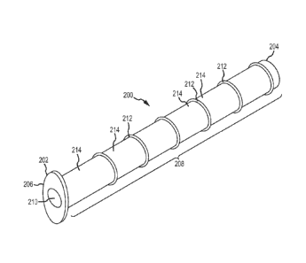
Figure 4 is perspective view of one embodiment of an implant device.
Figure 5 is a side view of the same embodiment of an implant device as shown
in
Figure 4.
Figure 6 is an end view of the same embodiment of an implant device as show in
Figure 4.
Figure 7 is a partial perspective view of the same embodiment of an implant
device as
shown in Figure 4.
Figure 8 is a partial side view of an embodiment of an implant device.
Figure 9 is a partial side view of an embodiment of an implant device.
Figure 10 is an illustration of cross-sections of various configurations for
anchor
protrusions for an implant device.
Figure 11 is an illustration of various head configurations for an implant
device.
Figure 12 is a perspective view of an embodiment of an implant device.
Figure 13 is an end view of the same embodiment of an implant device shown in
Figure 12.
Figure 14 is an illustration showing an embodiment for placement of an implant
device between the lacrimal caruncle and plica semilunaris.
Figure 15 is a side view of an embodiment of an implant device.
Figure 16 is a perspective view of an embodiment of a surgical tool.
Figure 17 is a perspective view of an embodiment of a surgical tool showing
some
components in exploded view.
Figure 18 is a perspective view of the same embodiment of a surgical tool
shown in
Figure 17, showing the surgical tool fully assembled.
Figure 19 is a perspective view showing a first carrier piece of the same
embodiment
of a tool shown in Figures 17 and 18, with the first carrier piece connected
with a syringe.
Figure 20 is an illustration showing use of a surgical tool to form a fistula
between the
orbit and an ethmoid sinus during a surgical procedure.
Figure 21 is an illustration showing insertion of a guide wire following
formation of
the fistula during a surgical procedure.
Figure 22 is an illustration showing a guide wire in place as a guide to a
fistula during
a surgical procedure.
Figure 23 is an illustration showing use of a surgical tool for implantation
of an
implant device during a surgical procedure.
CA 02812254 2013-03-21
WO 2012/048278
PCT/US2011/055456
Figure 24 is an illustration showing placement of an implant device following
implantation during a surgical procedure.
Figure 25 is an illustration showing use of a surgical tool to dilate a
fistula following
initial formation of the fistula during a surgical procedure.
DETAILED DESCRIPTION
The terms "lacrimal apparatus" and "lacrimal system" are used interchangeably
herein
to refer to the collection of physiological components that accomplish the
production and
secretion of lacrimal fluid to lubricate the eyeball, containment of lacrimal
fluid in a reservoir
of lacrimal fluid in the orbit and drainage of lacrimal fluid from the orbit
to the nasal cavity.
The lacrimal apparatus includes the lacrimal glands, the tear drainage system
and the
reservoir of lacrimal fluid located between the lacrimal glands and the tear
drainage system.
The reservoir of lacrimal fluid includes the eyelid margins and the
conjunctival sac (and
including the pool of tears in the lower conjunctival cul-de-sac that is
sometimes referred to
as the lacrimal lake). The tear drainage system includes the puncta,
canaliculi and
nasolacrimal duct (including the so-called lacrimal sac located at the top of
the nasolacrimal
duct) through which excess tears drain to Hasner's valve and into the nasal
cavity. Figure 1
shows generally the lacrimal apparatus. Lacrimal fluid is produced and
secreted from
lacrimal glands 102 to lubricate the surface of the eyeball 104 disposed
within the orbit.
Lacrimal fluid forms a coating over the eyeball 104 and is generally contained
within the
conjunctival sac (the space between the lower eyelid 106, upper eyelid 108 and
eyeball 104
that is lined by the conjunctiva). Excess lacrimal fluid is conducted to the
vicinity of the
medial canthus (medial corner of the eye) and drains through the lacrimal
puncta 110 into the
lacrimal canaliculi 112 and into the lacrimal sac 114 of the nasolacrimal duct
116. The
lacrimal fluid then drains from the nasolacrimal duct 116 through Hasner's
valve and into the
nasal cavity.
As used herein, a fistula between the lacrimal apparatus and a paranasal sinus
refers to
an artificially-created passage that fluidly connects the lacrimal apparatus
with the paranasal
sinus. The paranasal sinuses include the frontal sinuses, maxillary sinuses,
ethmoid sinuses
and sphenoid sinuses, which are cavities contained within frontal, maxilla,
ethmoid and
sphenoid bones, respectively. The paranasal sinuses drain into the nasal
cavity. Figure 2 is a
schematic of a human head showing generally the locations of the frontal
sinuses 122, the
maxillary sinuses 124 and the ethmoid sinuses 126. The sphenoid sinuses (not
shown) are
26
CA 02812254 2013-03-21
WO 2012/048278
PCT/US2011/055456
located generally behind the ethmoid sinuses 126. Figure 3 shows generally
some possible
routes for a fistula between the lacrimal system and a paranasal sinus.
Reference numerals
indicate the same features as shown in Figures 1 and 2, except as noted.
Figure 3 shows the
general proximity of the frontal sinus 122, maxillary sinus 124 and ethmoid
sinus 126 relative
to features of the lacrimal apparatus. Some example fistula routes are shown
in Figure 3 by
dashed lines. A first example fistula route 130 is from the orbit to the
frontal sinus. A
second example fistula route 132 is from the orbit to the ethmoid sinus 126. A
third example
fistula route 134 is from the orbit to the maxillary sinus 124. A fourth
example fistula route
136 is from the lacrimal sac 114 at the top of the nasolacrimal duct 116 to
the ethmoid sinus
126. A fifth example fistula route 138 is from the nasolacrimal duct 116 at a
location below
the lacrimal sac 114 to the ethmoid sinus 126. A sixth example fistula route
140 is from the
nasolacrimal duct 116 at a location below the lacrimal sac 114 to the
maxillary sinus 124.
The example fistula routes shown in Figure 3 are for purposes of general
illustration only and
not show precise locations where a fistula might be formed to connect a part
of the lacrimal
apparatus with the corresponding paranasal sinus. Although not shown in Figure
3, example
fistula routes to the sphenoid sinus include from the orbit to the sphenoid
sinus and from the
nasolacrimal duct 116 to the sphenoid sinus. Forming a fistula to connect to
the sphenoid
sinuses is generally not as preferred as forming a fistula to connect to the
ethmoid sinus, for
example because it is generally more convenient and direct to connect with the
ethmoid
sinus. Also, forming a fistula to either the ethmoid sinus 126 or the
maxillary sinus 124 is
generally preferred to forming a fistula to the frontal sinus 122, with one
reason being that a
fistula between the lacrimal system and either the ethmoid sinus 126 or the
maxillary sinus
124 may be formed in a way to obtain the benefit of gravity to assist drainage
of lacrimal
fluid from the lacrimal system into the corresponding paranasal sinus through
the fistula. The
frontal sinus is located generally above the orbit and will not benefit in the
same way from
gravity drainage of lacrimal fluid into the paranasal sinus. However, gravity
drainage may
beneficially assist drainage of fluid from the frontal sinus.
With continued reference to Figure 3, the first, second and third example
fistula routes
130, 132 and 134 are subconjuctival routes that penetrate the conjunctiva to
directly connect
the lacrimal fluid reservoir within the conjunctival sac to the corresponding
paranasal sinus.
A fistula along such a subconjunctival route may be surgically formed by a
surgical tool
piercing through the conjunctiva and the adjacent wall of the bone in which is
disposed the
corresponding paranasal sinus. For example, for the first example fistula
route 130, the
27
CA 02812254 2013-03-21
WO 2012/048278
PCT/US2011/055456
fistula would pass subconjunctivally from the orbit and through a wall of the
frontal bone into
the frontal sinus 122. For example, a fistula following second example fistula
route 132
would pass subconjuctivally from the orbit and through a wall of the ethmoid
bone into the
ethmoid sinus 126. For example, a fistula following the third example fistula
route 134
would pass subconjuctivally from the orbit through a wall of the maxilla bone
into the
maxillary sinus 124. Subconjuctival routes for a fistula such as those of the
first, second and
third example fistula routes 130, 132 and 134 are generally preferred as being
formed at
locations that are relatively easy to access. In a preferred implementation of
the first, second
and third example fistula routes 130, 132 and 134, the proximal end of the
fistula opening
into the orbit is located between the lacrimal caruncle 142 and the plica
semilunaris 144,
shown in Figure 3.
Continuing with reference to Figure 3, a fistula at the fourth, fifth or sixth
example
fistula routes 136, 138 and 140 will have a proximal end opening into a
location within the
nasolacrimal duct 116. Formation of a fistula in such a location requires
insertion of a
surgical tool into the lacrimal drainage system, such as through the puncta
110 and canaliculi
112 to access the nasolacrimal duct 116 or through the nose to access the
nasolacrimal duct
116. For example, a fistula at the fourth example fistula route 136 may be
formed by a
piercing instrument (e.g., a trocar or trocar/cannula assembly) inserted into
one of the puncta
110, through one of the canaliculi 112 and across the lacrimal sac 114 to
pierce a hole at the
location of the fourth example fistula route 136. As another example, a
fistula may be
formed at one of the fourth, fifth and sixth example fistula routes 136, 138
and 140 using a
guide wire inserted into one of the puncta 110, through one of the canaliculi
112, into the
lacrimal sac 114 and downward through the nasolacrimal duct 116. The guide
wire may be
used to engage a surgical tool and to guide the surgical tool from the nose
through Hasner's
valve (not shown) and to the appropriate location within the nasolacrimal duct
116 to permit
performance of a surgical operation at that location to form the desired
fistula.
Figures 4-7 show one embodiment of an implant device. As shown in Figures 4-7,
an
implant device 200 has a proximal end 202 and a distal end 204 located on
opposite
longitudinal ends of the implant device 200. The implant device 200 includes a
head 206 at
the proximal end 202 and a conduit 208 extending from the head 206 to the
distal end 204.
An internal passage 210 extends from the proximal end 202 to the distal end
204, passing
through the head 206 and the conduit 208. The internal passage 210 opens at
the proximal
end 202 and the distal end 204, thereby providing a passage through the entire
longitudinal
28
CA 02812254 2013-03-21
WO 2012/048278
PCT/US2011/055456
length of the implant device 200. The internal passage 210 of the embodiment
shown in
Figure 4 has a cylindrical shape with a uniform circular cross-section
(transverse to the length
of the implant device 200), and the width of the internal passage is equal to
the diameter of
the circle of the cross-section and is uniform along the length of the implant
device 200. The
length of the implant device 200 is the minimum distance longitudinally along
the implant
device 200 between the proximal end 202 and the distal end 204, and will
typically be equal
to the distance along an axis of the internal passage 210 from the proximal
end 202 to the
distal end 204. The implant device 200 includes multiple anchor protrusions
212 on an
exterior of the conduit 208. In the embodiment shown in Figures 4-7, the
anchor protrusions
212 are in the form of spaced circumferential ridges that each extends around
the entire
circumference of the conduit 208. Adjacent the circumferential ridges of the
anchor
protrusions 212 are areas of recess 214 on the exterior of the conduit 208.
With continued reference to Figures 4-7, when the implant device is implanted
to
fluidly connect the lacrimal apparatus to a paranasal sinus through a fistula,
the head 206 is
disposed in the lacrimal apparatus and the proximal end 202 is disposed in the
paranasal
sinus, and with at least a portion of the conduit 208 disposed through the
fistula with at least
one, and preferably more than one, of the anchor protrusions 212 engaging
tissue within the
fistula to anchor the implant device 200. When implanted in this manner, the
internal
passage 210 opens into the lacrimal apparatus at the proximal end 202 and into
the paranasal
sinus at the distal end 204. The head 206 has a flanged tissue engagement
surface 216 on a
side of the head 206 disposed toward the conduit 208, and which flanged tissue
engagement
surface 216 is advantageously configured to engage tissue adjacent the
proximal end of
fistula and to prevent the proximal end 202 of the implant device 200 from
migrating into the
fistula following implantation. On the side of the head 206 opposite the
flanged tissue
engagement surface 216 is a face surface 218 of the head 206, which face
surface 218 is
disposed away from tissue engaged by the flanged tissue engagement surface 216
when the
implant device is implanted. The head 206 has a first dimension 220 and a
second dimension
222 on both the flanged tissue engagement surface 216 and the face surface
218. The first
dimension 220 is the length of the respective surface and the second dimension
is the width
of the respective surface. Such length and width dimensions may also be
referred to as major
and minor dimensions. The first dimension 220 of a surface 216 or 218
corresponds to the
maximum separation distance between points on the outer edge of the surface,
and the second
dimension 222 of the surface 216 or 218 corresponds to the maximum separation
distance
29
CA 02812254 2013-03-21
WO 2012/048278
PCT/US2011/055456
between points on the outer edge of the surface that are on a line transverse
to the first
dimension. Conveniently, the face surface 218 and the flanged tissue
engagement surface
216 may be made with corresponding outer edges, so that the opposing surfaces
216 and 218
have substantially equal length and width dimensions, although such is not
required. The
first dimension 220 and the second dimension 222 may be referred to generally
as the length
and width, respectively, of the head 206 when the surfaces 216 and 218 have
corresponding
shapes, as is the case for the embodiment shown in Figures 4-7. When the
surfaces 216 and
218 do not have corresponding shapes, the length and width dimensions of the
head will be
different from one or more of the length and width dimensions of the surfaces
216 and 218.
The head 206 has a depth dimension 223 between surfaces 216 and 218. The depth
dimension 223 should preferably be kept to a small value so that the head 206
will have a low
profile adjacent the proximal end of the fistula when the implant device 200
is implanted with
the flanged tissue engagement surface engaging tissue adjacent the proximal
end of the
fistula.
With continued reference to Figures 4-7, the conduit 208 has a first exterior
width 224
that is a maximum exterior width of the conduit 208 as defined by the maximum
extents of
the anchor protrusions 212 transverse to the length of the conduit 208. The
conduit 208 has a
second exterior width 226 that is a minimum exterior width of the conduit 208
defined
between the most recessed portions of the areas of recess 214. In the
embodiment shown in
Figures 4-7, the height of the anchor protrusions 212 is equal to one-half the
difference
between the first exterior width 224 and the second exterior width 226 of the
conduit 208. In
the configuration of the head 206 shown in Figure 4-7, the first dimension 220
of the head is
larger than both the first exterior width 224 and the second exterior width
226 of the conduit
208, while the second dimension 222 of the head is approximately equal to the
second
exterior width 224 of the conduit 208.
With continued reference to Figures 4-7, the anchor protrusions 212 are in the
form of
circumferential ridges having a width that is at a maximum at the bottom of
the ridges located
adjacent the areas of recess 214, and which width tapers to a minimum at the
top of the ridges
212 located away from the recess areas 214. Other configurations for anchor
protrusions are
possible, and all anchor protrusions on an implant device need not be of the
same size,
geometry or height. Likewise, areas of recess may have varying configurations,
and not all
recesses on an implant device need to be the same size or configuration. The
implant device
CA 02812254 2013-03-21
WO 2012/048278
PCT/US2011/055456
200 has a length 228 including the depth 223 of the head 206 and the length of
the conduit
208. The anchor protrusions 212 are on a longitudinal portion 230 of the
conduit 208.
Referring now to Figure 8, an alternative embodiment is shown of a conduit 240
of an
implant device having anchor protrusions 242 in the form of knobs, or buttons,
and areas of
recess 244 adjacent the anchor protrusions 242. The conduit 240 has a first
exterior width
246 defined by the anchor protrusions 242 and a smaller, second exterior width
248 defined
by the areas of recess 244. An example of another configuration for anchor
protrusions is
shown in Figure 9. As shown in Figure 9, a conduit 250 of an implant device
has anchor
protrusions 252 and areas of recess 254 on the exterior surface of the conduit
250. The
anchor protrusions 252 are in the form of a continuous spiral ridge extending
along a portion
of the longitudinal length of the conduit 250. The conduit 250 has a first
exterior width 256
defined by the anchor protrusions 254 and a smaller, second exterior width 258
defined by
the areas of recess 254. As with the embodiments shown in Figures 4-7, the
conduit
embodiment shown in Figures 8 and 9 include a height of the anchor protrusions
that is equal
to one half the difference between the larger and smaller outer diameters of
the respective
conduits. As will be appreciated from the embodiments of Figures 8 and 9, the
first exterior
width is determined as the width of an envelope volume that contains the
anchor protrusions.
Figure 10 shows examples of some shapes for anchor protrusions that include a
tapering width in a direction from the base of the anchor protrusion toward a
top of the
anchor protrusion. Figure 10 shows cross-sections of anchor protrusion
configurations
(designated A-D), each having a greater width at the base than at the top. The
height (H) and
base width (W) of the anchor protrusions are indicated in Figure 10. The cross-
sections
shown in Figure 10 may, for example, be across a ridge (e.g., circumferential
ridge, spiral
ridge), a knob protrusion or other anchor protrusion form. All of the anchor
protrusion
configurations A-D in Figure 10 are shown with a leading side of the anchor
protrusion on
the right side and a trailing side on left side of the anchor protrusion. By
leading side it is
meant a side that enters the fistula first when a conduit containing the
anchor protrusion is
inserted into the fistula for implantation. By trailing side it is meant the
side opposite the
leading side and that enters the fistula after the leading side. As will be
appreciated, forces
applied to the anchor protrusions by tissue contacting the anchor protrusions
during insertion
into a fistula will impart stresses to the anchor protrusions and, to an
extent as permitted by
the material of construction of the anchor protrusion, such stresses will tend
to deform the
anchor protrusion in a direction toward the trailing side. Such deformation
aids insertion, and
31
CA 02812254 2013-03-21
WO 2012/048278
PCT/US2011/055456
is generally preferred to some degree. The different shapes of the
configurations A-D affect
the relative ease of insertion of a conduit into and removal of the conduit
from a fistula.
Configuration A is designed to be equally easy to insert and removable from a
fistula while
each of configurations B-D are designed to be more easy to insert into a
fistula and more
difficult to remove from the fistula. Configurations B and C are angled in a
way to promote
more easy insertion and more difficult removal from a fistula. Configuration D
includes a
hooked end to engage tissue on the trailing side to make removal from a
fistula more difficult
than insertion.
Figure 11 shows some different example configurations (designated E-H) for a
head
for an implant device. For each head configuration, the length dimension (L)
and width
dimension (W) of the head configurations are shown. The heads of
configurations E-H are
shown on end showing the face surface (surface facing away from the fistula
when
implanted) and the opening of the internal passage at the proximal end of the
implant device.
For each of the head configurations E-H, the length and width of the face
surface and the
opposing flanged tissue engagement surface are the same. As shown in Figure
11, head
configuration E has a circular outer edge, and thus has equal length and width
dimensions.
Head configuration F has an elongated length dimension relative to width
dimension, similar
to that shown in the implant device embodiment described with reference to
Figures 4-7.
Head configuration G has an elongated length dimension relative to the width
dimension,
similar to configuration F, but for configuration G the internal passage
opening at the
proximal end of the implant device has an elliptical cross-section, rather
than a circular cross-
section as is the case for configurations E and F. Head configuration H has a
crescent-shaped
head with a significantly larger length dimension than width dimension. The
internal passage
for configuration H is also shown with an elliptical cross-section.
Configurations F-H, with a
larger length than width, are advantageously configured for use with fistulas
opening into the
orbit between the plica semilunaris and the lacrimal caruncle, with the length
dimension of
the head extending generally in a direction from the bottom of the orbit
toward the top of the
orbit next to the eyeball, and for configuration H with the concave side of
the crescent
disposed toward the eyeball and the convex side of the crescent disposed
towards the lacrimal
caruncle.
Figures 12 and 13 show another embodiment for an implant device. As shown in
Figures 12 and 13, an implant device 300 has a proximal end 302 and a distal
end 304, with a
head 306 located at the distal end 304 and a conduit 308 extending from the
head 306 to the
32
CA 02812254 2013-03-21
WO 2012/048278
PCT/US2011/055456
distal end 304. The conduit 308 includes an internal passage 310 with a
cylindrical shape and
opening at the proximal end 302 and the distal end 304. The conduit 310 has an
exterior
surface including anchor protrusions 312, in the form of circumferential
ridges with tapering
width, and areas of recess 314 adjacent the anchor protrusions 312. The head
306 has an
elongated shape with a significantly larger length dimension 316 than width
dimension 318.
As seen in Figure 12, a flanged tissue engagement surface 320 has a beveled
configuration
(beveled halves extending from central line) to help seat against tissue in a
manner to prevent
rotation of the implant device 300 when implanted. The face surface 322 is a
flat surface to
provide a low profile to the head 306 when the implant device 300 is
implanted. The
configuration of the head 306 is well suited for placement between the plica
semilunaris and
lacrimal caruncle for use with a subconjunctival fistula route from the orbit
where the
opening of the fistula into the orbit is located between the plica semilunaris
and the lacrimal
caruncle. The length dimensions 316 and width dimension 318 represents the
length and
width of each of the face surface 322 and the flanged tissue engagement
surface 320.
Figure 14 shows an example of an implant device with a conduit passing through
a
fistula formed subconjunctivally between the lacrimal caruncle 350 and the
plica semilunaris
352, and showing an example location for the head 354 of the implant device.
The head 354
is shown with an elongated configuration, such as for example the head
configuration shown
in Figures 4-7, one of the head configurations F-H shown in Figure 11 or the
head
configuration shown in Figures 12 and 13.
Figure 15 shows another embodiment of an implant device. As shown in Figure
15,
an implant device 400 has a proximal end 402 and a distal end 404. The implant
device 400
includes a head 406 at the proximal end 402 and a conduit 408 extending from
the head 406
to the distal end 404. The conduit 408 has an exterior surface with anchor
protrusions 412
and areas of recess 414 adjacent the anchor protrusions 412. An internal
passage 410 (shown
by dashed lines) extends from the proximal end 402 to the distal end 404. A
distal
longitudinal portion of the conduit 408 includes apertures 415 through the
wall of the conduit
408 and providing fluid communication from the internal passage 410 to outside
of the
conduit 408. The apertures 415 provide a route for drug formulations,
irrigation solutions or
other treatment compositions to exit from the internal passage into different
locations within
a paranasal sinus when the implant device 400 is implanted. When the implant
device 400 is
implanted, at least one or more of the anchor protrusions 412 will be located
within the fistula
to engage tissue for anchoring and at least some, and preferably all, of the
apertures 415 will
33
CA 02812254 2013-03-21
WO 2012/048278
PCT/US2011/055456
be disposed beyond the distal end of the fistula inside of a paranasal cavity.
The
configuration shown in Figure 15 is particularly advantageous for situations
when the conduit
408 extends through multiple cavities of a paranasal sinus or when the conduit
408 extends
from one paranasal sinus into another paranasal sinus. The embodiment shown in
Figure 15
does not include the anchor protrusions 412 on the longitudinal portion of the
conduit 408
where the apertures 415 are disposed. As an alternative configuration, the
longitudinal
portion of the conduit 408 including the apertures 415 could include anchor
protrusions, of
the same configuration as those of the anchor protrusions 412 or of different
configurations.
Figure 16 shows one embodiment of a surgical tool. As shown in Figure 16, a
surgical tool 500 includes an implant device 502 having a head 504 and a
conduit 506, for
example as previously described with respect to any of the Figures 4-15. The
implant device
502 is mounted on a carrier 510. The carrier 510 comprises a handle 511
adjacent a proximal
end of the surgical tool 500. The carrier 510 includes a working member 512
connected to
the handle 511. The working member 512 extends from the handle 511 through the
internal
passage of the implant device 502 and to a distal end of the surgical tool
500. At the distal
end of the working member 512 is a distal tip 514. The handle 511 may be made
of any
convenient material of construction, for example plastic or metallic
compositions. The
working member 512 may be made for example of a medical-grade metallic
composition,
such as a medical-grade stainless steel. In general when a member is referred
to herein as a
"working member", the term indicates that the member is such that at least a
portion of the
member is designed for being disposed within or through a fistula when a tool
containing the
member is used, for example during formation of a fistula or during
performance of some
procedure in or through a fistula. Some examples of working members include
various
hollow members (e.g., hypodermic needles, cannulas) and various solid members
(e.g.,
trocars, stylets, dilating members, implant delivery members). Such a working
member may
be disposed in or through the fistula in a manner that the member contacts
tissue in the fistula
or in a manner not to contact tissue in a fistula (e.g., inside of a passage
of an implant device
passing through the fistula).
With continued to reference to Figure 16, the implant device 502 is mounted on
the
carrier 510 with the working member 512 disposed through the internal passage
of the
implant device 502. The width of the working member 512 disposed through the
internal
passage of the implant device 502 may advantageously be sized to be just
smaller than the
internal passage of the implant device for a close fit between them, provided
that the fit is not
34
CA 02812254 2013-03-21
WO 2012/048278
PCT/US2011/055456
so tight that the implant device 502 is difficult to slide down the working
member 512 toward
the distal tip 514.
Continuing to refer to Figure 16, the surgical tool 500 may be used to form a
fistula
between the lacrimal system and a paranasal sinus and to facilitate
implantation of the
implant device 502 in the fistula. A surgeon may manipulate the surgical tool
500 by hand-
grasping the handle 511. The surgeon may advance the distal tip 514 to a
location within the
lacrimal apparatus where the fistula is to be formed to a target paranasal
sinus. The surgeon
may then force the distal tip through tissue separating the lacrimal apparatus
and the target
paranasal sinus to form the fistula. With a leading portion of the working
member 512
disposed through the fistula, a surgeon may slide the implant device 502 along
the working
member 512 toward the distal tip 514 until the implant device 502 is
positioned for
implantation with the conduit 506 disposed through the fistula and a flanged
tissue
engagement surface of the head 506 disposed against tissue adjacent the
proximal end of the
fistula in the lacrimal apparatus, or the carrier may continue to be advanced
to push the
conduit 506 into the fistula. After the implant device 502 is positioned for
implantation, the
surgeon may then manipulate the handle 504 to retract the working member 512
to withdraw
the working member from the internal passage of the implant device 502 and to
fully
disengage the carrier 510 from the implant device 502, leaving the implant
device 502
implanted with the conduit 506 extending through the fistula and into the
paranasal sinus.
With continued reference to Figure 16, the working member 512 may be a solid
member (e.g., trocar, stylet) or may be a hollow member (e.g., a hollow
needle, cutting
cannula). If the working member 512 is a hollow member with an opening at the
distal tip
514, then tissue will tend to be cored and collected in the hollow interior of
the working
member 512 when the surgical tool 500 is used to form a fistula. If the
working member 512
is a solid member, then tissue coring should not occur. In many instances, it
may be
preferred to have the working member 512 be a solid member that does not core
tissue,
because the implant device may tend to be held more securely within a fistula
formed without
tissue coring. The surgical tool 500 shown in Figure 16 is particularly well
adapted for
forming a fistula from the orbit subconjunctiyally to a paranasal sinus, and
particularly when
the fistula is formed at a location in the orbit between the plica semilunaris
and the lacrimal
caruncle.
Figures 17 and 18 show another surgical tool. Figure 17 shows an expanded view
of
some features of a surgical tool 520 and Figure 18 shows the same surgical
tool 520 as the
CA 02812254 2013-03-21
WO 2012/048278
PCT/US2011/055456
surgical tool 520 appears fully assembled. As shown in Figure 17, the surgical
tool 520
includes an implant device 522 with a head 524 and a conduit 526, for example
as described
previously with respect to any of Figures 3-16. The surgical tool 520 includes
a carrier with
two pieces, a first carrier piece 530 and a second carrier piece 532. The
first carrier piece 530
has a syringe hub 534 (e.g., for making a luer connection) and a hollow
working member 536
(e.g., hollow needle, cannula) connected with the hub 534. The hollow working
member 536
has a distal tip 538. The second carrier piece 532 has a handle 540 and a
solid working
member 542 (e.g., stylet, trocar) connected with the handle 540. The solid
working member
542 has a distal tip 544. As assembled, the surgical tool 520 includes the
solid working
member 542 inserted through the interior of the hub 534 and through the hollow
interior of
the hollow working member 536. As assembled, the handle 540 of the second
carrier piece
532 is disposed distal of the hub 534 with an engagement member 544 inserted
into the
interior of the hub 534. As will be appreciated, features of the hub 534
and/or the
engagement member 544 and/or the handle 540 may contain keying and engagement
features
to align and/or permit detachable engagement of the first carrier piece and
the second carrier
piece when assembled. Figure 18 shows the same surgical tool 520 as it appears
fully
assembled. As shown in Figure 18, the first carrier piece 522 and the second
carrier piece
532 are engaged with the solid working member 542 disposed through the hollow
interior of
the hollow working member 536.
With continued reference to Figures 17 and 18, the surgical tool 520 may be
used to
form a fistula between the lacrimal apparatus and a paranasal sinus. The
distal tips 538 and
544 of the first and second carrier pieces 530 and 532 form a distal tip that
will not
significantly core tissue. A surgeon may grasp the handle 540 and advance the
distal tip to a
location in the lacrimal apparatus where the fistula is to be formed (e.g., in
the orbit, in the
nasolacrimal duct) and the distal tip may then be forced through tissue into a
paranasal sinus
to form the fistula to the target paranasal sinus. With a leading portion of
the hollow working
member 536 disposed through the fistula, the implant device 522 may be slid
down the
hollow working member 536 and into position for implantation with the conduit
526 disposed
through the fistula and the head 524 disposed adjacent the proximal end of the
fistula, or the
hollow working member 536 may be further advanced to push the conduit 526 into
the
fistula. The hollow working member 536 may then be retracted and disengaged
from the
implant device 522 to leave the implant device 522 in the implanted position.
36
CA 02812254 2013-03-21
WO 2012/048278
PCT/US2011/055456
Continuing with reference to Figures 17 and 18, the hollow working member 536
facilitates performance of an ancillary medical procedure involving aspirating
fluid from or
introducing fluid into the paranasal sinus. For example, before or after
positioning the
implant device 522 in the proper location for implantation, the second carrier
piece 532 may
be disengaged from the first carrier piece 534 to remove the solid working
member 542 from
the hollow interior of the hollow working member 536. The hollow working
member 536 is
then available for aspiration of fluid from or injection of fluid into the
paranasal sinus. The
hub 534 may be engaged with a corresponding connection structure of a syringe
and the
syringe may be operated to aspirate fluid from the paranasal sinus into the
syringe or to inject
fluid from the syringe into the paranasal sinus. Fluids that may be injected
into the paranasal
sinus include irrigation fluid or treatment compositions containing a drug,
for example to
inject a drug bolus for treatment of sinusitis. As used herein, "fluid"
includes flowable
compositions, including compositions that may have a solid material dispersed
or suspended
in a fluid medium. After the implant device has been properly positioned for
implantation
and after performing any desired ancillary medical procedure, the first
carrier pierce may be
retracted to disengage the hollow working member 536 from the internal passage
of the
implant device 522 and to leave the implant device 522 as an implant. Figure
19 shows the
first carrier piece 536 of the surgical tool 500 connected with a syringe 550.
Referring now to Figures 20-25, some additional examples of surgical
procedures
involving forming a fistula and implanting an implant device, and some example
surgical
tools for use therewith, will now be described.
In Figure 20 a surgical tool in the form of an entry tool 600 is shown in the
process of
making a fistula through tissue between the lacrimal caruncle 142 and the
plica semilunaris
144. Numbering of anatomical parts is the same as in Figures 1 and 3. The
fistula is formed
through tissue between the conjunctival sac in the orbit and the ethmoid sinus
126. The route
for the fistula would be consistent with general fistula route 132 as shown in
Figure 3. The
entry tool 600 includes a first piece 602 and a second piece 604. The first
piece 602 includes
a hollow working member 606 and a hub 608. The second piece 604 includes a
solid
working member (not shown) disposed through a hollow interior of the hollow
working
member 606. A distal tip portion of the hollow working member 606 of the first
piece 602
and a distal tip portion of the solid working member of the second piece 604
form a distal tip
610 with a shape suitable for insertion through the tissue to form a fistula
from the
conjunctival sac to the ethmoid sinus 126. The second piece 604 includes a
hand-
37
CA 02812254 2013-03-21
WO 2012/048278
PCT/US2011/055456
manipulable handle 612. The hub 608 may be configured for connecting with a
syringe or
other fluid manipulation device, such as through a luer connection. The handle
612 may be
retracted relative to the hub 608 to remove the solid working member from the
interior of the
hollow working member 606 and to disengage the second piece 604 from the first
piece 602.
As shown in Figure 20, the distal tip 610 has been advanced from a location in
the
conjunctival sac between the caruncle 142 and the plica semilunaris 144 to
form a fistula
between the conjunctival sac and the ethmoid sinus 126. As shown, the fistula
passes behind
the caruncle 142, canaliculi 112 and nasolacrimal duct 116 to access the
ethmoid sinus 126.
The first piece 602 of the entry tool 600 includes a collar stop 614 to
prevent the hollow
working member 606 from being advanced through tissue beyond a certain
distance. The
first piece 602 and the second piece 604 may, for example, be substantially
the same as the
first carrier piece 530 and the second piece 532 of the tool assembly 520 of
Figure 17, but
with the added collar stop 614 and not including an implant device mounted
thereon.
After the entry tool 600 has been used to initially form a fistula to the
ethmoid sinus
126, then the second piece 604 may be disengaged from the first piece 602 and
a guide wire
inserted through the internal passage through the hollow working member 606.
Figure 21
shows the first piece 602 after disengagement of the second piece 604 and
after insertion of a
guide wire 620 through the first piece 602 and exiting from a distal end of
the first piece 602
in the ethmoid sinus 126. After insertion of the guide wire 620, the first
piece 602 may be
retracted and removed from the fistula, leaving the guide wire 620 in place as
a guide to and
through the fistula. Figure 22 shows the guide wire 620 disposed through the
fistula after
removal of the first piece 602. The guide wire 620 is now available for
guiding additional
tools to and through the fistula into the ethmoid sinus 126.
With reference now to Figure 23, the guide wire 620 has been used to guide a
surgical
tool, in the form of an implant tool 624. The implant tool 624 includes a
hollow working
member 626 and a hand-manipulable handle 628. The implant tool 624 includes an
internal
passage passing through the handle 628 and the hollow working member 626. As
shown in
Figure 23, the guide wire 620 has been threaded through the internal passage
of the implant
tool 624 to guide the hollow working member 626 to and through the fistula and
into the
ethmoid sinus 126. The implant tool 624 also includes an implant device 630
mounted on the
hollow working member 626. Figure 23 shows the implant tool 624 advanced to a
point
where the distal end of the implant device 630 is in the vicinity of the
proximal end of the
fistula opening into the conjunctival sac. From this position, the implant
device 630 may be
38
CA 02812254 2013-03-21
WO 2012/048278
PCT/US2011/055456
advanced into the fistula with a head of the implant device 630 disposed
adjacent the
conjunctiva in the conjunctival sac and a distal end of the implant device 630
extending into
the ethmoid sinus 626. The implant tool 624 may, for example, be a tool of the
design such
as that shown for the surgical tool 500 in Figure 16, with a hollow needle for
the working
needle 518. The implant device 630 of the implant tool 624 may, for example,
have features
as described with respect to any of Figures 4-19. With the continued reference
to Figure 23,
the hollow working member 626 of the tool 624 preferably includes a blunt tip.
The handle
628 and the hollow working member 626 form a carrier for the implant device
630. The
handle 628 may be retracted and the hollow working member 626 disengaged from
the
implant device 630 after the implant device has been appropriately positioned
for
implantation through the fistula. As an alternative to the configuration of
the implant tool 624
as shown in Figure 23, the implant tool 624 could be configured to include a
hub for
connection (e.g., through a luer connection) with a syringe of other fluid
manipulation device.
For example, the implant tool 624 could be configured with a hub in a manner
similar to the
configuration of the first piece 602 shown in Figure 21 and with the implant
device
appropriately mounted for implantation. As another variation on the
configuration of the
implant tool 624, the working member 626 could be fitted with a collar stop
(e.g., as shown
in Figure 21) or other mounting aid against which the implant device 630 could
be disposed
to provide some additional distance between a proximal end of the implant
device 630 and
the handle 628. Figure 24 shows the implant device 630 as implanted and
following
disengagement of the hollow working member 626 of the implant tool 628. As
implanted, a
head 632 at the proximal end of the implant device 630 is located adjacent the
conjunctiva in
the conjunctival sac within the orbit between the caruncle 142 and the plica
semilunaris 144
and the distal end 634 of the implant device 630 is located in the paranasal
sinus 626. Some
of anchor protrusions 636 are disposed within the fistula to engage tissue and
help anchor the
implant device 630.
The procedure as described with reference to Figures 20-24 permits the working
member 606 of the entry tool 600 to have a larger diameter working member 626
to form a
fistula of appropriate size for accommodating the implant device 630 which is
then implanted
in a separate step using the implant tool 624 with the implant device 630
carried on to the
working member 626, which may advantageously have a smaller diameter then the
working
member 606 used to form the fistula. As an alternative, an intermediate step
to dilate the
fistula to a desired size for implantation may be performed between initially
forming the
39
CA 02812254 2013-03-21
WO 2012/048278
PCT/US2011/055456
fistula with the entry tool 600 and implanting the implant device 630 using
the implant tool
624. Figure 25 shows a surgical tool in the form of a dilator tool 640 having
a hollow
working member 642 and a hand-manipulable handle 644. The working member 642
is
disposed through the fistula, guided by the guide wire 620 passing through an
internal
passage through the dilator tool 640. As shown in Figure 25, the working
member 642 has
been advanced to the point where a stop collar 646 attached to the working
member 642 has
engaged conjunctival tissue in the conjunctival sac adjacent a proximal end of
the fistula. For
this alternative implementation, the hollow working member 642 of the dilator
tool 640
would have a larger diameter than the hollow working member 606 of the entry
tool 600
shown in Figures 20 and 21. The hollow working member 642 of the dilator tool
640,
therefore widens the fistula further to a desired size to accommodate easier
insertion of the
implant device 630. Although the intermediate step of dilation as shown is not
required, it
permits the use of a smaller-diameter working member 606 during initial
formation of the
fistula. The use of a smaller diameter for the working member 606 to initially
form the
fistula permits better visibility and procedural control for a surgeon
performing the
procedure. The working member 642 may preferably include a blunt tip.
In a method for providing access to a paranasal sinus to a human to permit
performance of medical treatments or procedures in the paranasal sinus over an
extended
time, a surgically formed, durably patent fistula may be created between the
lacrimal
apparatus of the human and the paranasal sinus. By surgically formed, it is
mean that the
fistula is an artificial passage through tissue that is intentionally formed
by a surgical
operation. For example, the fistula may be formed using a trocar, stylet,
needle or cannula.
The fistula may be formed by a surgical tool as described with reference to
any of Figures 16-
19. By "durably patent" it is meant that the fistula is resistant to closure
by natural tissue
repair mechanisms and remains open (patent) for an extended period of time to
provide
access into the paranasal sinus over the extended period of time. The extended
period of time
may be any period of time sufficient for performing through the fistula any
desired medical
treatments or procedures. The extended period of time may, for example, be at
least 7 days,
at least 14 days, at least 30 days, at least 180 days, or longer. The extended
period may be
permanent.
A fistula may be maintained as durably patent for an extended period of time
by a
variety of techniques. As one example for maintaining fistula patency, an
implant device
may be disposed through the fistula to prevent the fistula from closing, and
the implant
CA 02812254 2013-03-21
WO 2012/048278
PCT/US2011/055456
device may include an internal passage for providing access through the
fistula into the
paranasal sinus. When access to the paranasal sinus is no longer required, the
implant device
may be removed to permit tissue to repair and close the fistula. The implant
device may, for
example, have a configuration as described with respect to any of Figures 4-19
or may have a
different configuration. As another example for maintaining fistula patency,
the fistula may
be formed initially with a relatively large diameter, and preferably with a
clean cut. A large,
cleanly cut hole will naturally tend to remain patent and not repair for at
least a significant
time. The relatively large diameter of the fistula may, for example be at
least 2 millimeters or
larger, as described above. When the fistula is formed with such a large
diameter, the fistula
will preferably be formed at a location in the nasolacrimal duct. As another
example for
maintaining fistula patency, after the fistula is formed the tissue adjacent
the fistula may be
mechanically treated to form a mechanical impediment to tissue repair that
would close the
fistula. The mechanical treatment could involve, for example over-sewing
tissue adjacent the
fistula or stapling tissue adjacent the fistula to mechanically retain the
tissue in a manner to
inhibit tissue repair that would close the fistula. As another example for
maintaining fistula
patency, tissue adjacent the fistula may be treated with a substance (e.g., a
drug) effective to
inhibit natural tissue repair and closure of the fistula, such as for example
treatment with an
antigranulation or anti-scarring agent (e.g., steroids, Mitomycin C).
A variety of medical treatments and procedures may be performed through a
fistula
formed between the lacrimal apparatus and a paranasal sinus, whether or not
the fistula is
durably patent. One or more medical devices may be inserted into the paranasal
sinus
through the fistula. For example a hollow working member (e.g., hollow needle,
cannula)
may be inserted through the fistula into the paranasal sinus to permit
aspiration of fluid from
or injection of a treatment formulation (e.g., drug formulation, irrigation
fluid) into the
paranasal sinus. As another example, a treatment formulation (e.g., drug
formulation,
irrigation fluid) may be transmitted through the fistula into the paranasal
sinus by natural
flow from the lacrimal system. A treatment formulation may be administered to
the vicinity
of the eye (e.g., as eye drops) to naturally flow from the lacrimal apparatus
through the fistula
and into the paranasal sinus. The fistula may, but need not necessarily be, a
durably patent
fistula. For example, a conduit of a medical device be inserted from the
lacrimal apparatus
through tissue and into the paranasal sinus, fluid may be aspirated through or
injected from
the conduit, and the conduit may then be removed to allow the fistula formed
by insertion of
the conduit to quickly repair. Such a conduit may, for example, be a
hypodermic needle or
41
CA 02812254 2013-03-21
WO 2012/048278
PCT/US2011/055456
cannula (e.g., connected to a syringe, drip system or other fluid
injection/aspiration system).
The fistula may be formed by insertion of a member including the needle or
cannula and may
naturally repair and close quickly following removal of the conduit. For
example, the fistula
may be formed by insertion of a hypodermic needle, a fluid may be injected or
aspirated
through the hypodermic needle and the hypodermic needle may then be removed to
permit
the fistula to repair. As another example, the fistula may be formed by a
trocar/cannula
assembly, the trocar may then be removed, a medical procedure performed
through the
cannula (e.g., fluid injection or aspiration), and the cannula may then be
removed to permit
the fistula to repair. As another example, the fistula may be formed by a
cutting cannula, a
medical procedure performed through the cannula (e.g., fluid injection or
aspiration), and the
cannula may then be removed to permit the fistula to repair.
A surgically created, durably patent fistula may be advantageously located for
transmitting lacrimal fluid (tears) to a paranasal sinus. Lacrimal fluid from
the lacrimal
apparatus may be permitted to drain into the paranasal sinus. In one preferred
implantation,
the surgically-created, durably patent fistula is from either the orbit or the
nasolacrimal duct
to either the ethmoid sinus or the maxillary sinus, with a fistula route from
the orbit being
more preferred.
The foregoing discussion of the invention and different aspects thereof has
been
presented for purposes of illustration and description. The foregoing is not
intended to limit
the invention to only the form or forms specifically disclosed herein.
Consequently,
variations and modifications commensurate with the above teachings, and the
skill or
knowledge of the relevant art, are within the scope of the present invention.
The
embodiments described hereinabove are further intended to explain best modes
known for
practicing the invention and to enable others skilled in the art to utilize
the invention in such,
or other, embodiments and with various modifications required by the
particular applications
or uses of the present invention. It is intended that the appended claims be
construed to
include alternative embodiments to the extent permitted by the prior art.
Although the
description of the invention has included description of one or more possible
implementations
and certain variations and modifications, other variations and modifications
are within the
scope of the invention, e.g., as may be within the skill and knowledge of
those in the art after
understanding the present disclosure. It is intended to obtain rights which
include alternative
embodiments to the extent permitted, including alternate, interchangeable
and/or equivalent
structures, functions, ranges or steps to those claimed, whether or not such
alternate,
42
CA 02812254 2013-03-21
WO 2012/048278
PCT/US2011/055456
interchangeable and/or equivalent structures, functions, ranges or steps are
disclosed herein,
and without intending to publicly dedicate any patentable subject matter.
Furthermore, any
feature described or claimed with respect to any disclosed implementation may
be combined
in any combination with one or more of any other features of any other
implementation or
implementations, to the extent that the features are not necessarily
technically incompatible,
and all such combinations are within the scope of the present invention.
The terms "comprising", "containing", "including" and "having", and
grammatical
variations of those terms, are intended to be inclusive and nonlimiting in
that the use of such
terms indicates the presence of some condition or feature, but not to the
exclusion of the
presence also of any other condition or feature. The use of the terms
"comprising",
"containing", "including" and "having", and grammatical variations of those
terms in
referring to the presence of one or more components, subcomponents or
materials, also
include and is intended to disclose the more specific embodiments in which the
term
"comprising", "containing", "including" or "having" (or the variation of such
term) as the
case may be, is replaced by any of the narrower terms "consisting essentially
of' or
"consisting of' or "consisting of only" (or the appropriate grammatical
variation of such
narrower terms). For example, a statement that some thing "comprises" a stated
element or
elements is also intended to include and disclose the more specific narrower
embodiments of
the thing "consisting essentially of' the stated element or elements, and the
thing "consisting
of' the stated element or elements. Examples of various features have been
provided for
purposes of illustration, and the terms "example", "for example" and the like
indicate
illustrative examples that are not limiting and are not to be construed or
interpreted as
limiting a feature or features to any particular example. The term "at least"
followed by a
number (e.g., "at least one") means that number or more than that number. The
term at "at
least a portion" means all or a portion that is less than all. The term "at
least a part" means all
or a part that is less than all.
43