Note: Descriptions are shown in the official language in which they were submitted.
RETRIEVAL SYSTEMS AND METHODS FOR USE THEREOF
RELATED APPLICATIONS
[0001( This application is a non-provisional of U.S. Provisional
Application Nos. 61/489,183
filed May 23, 2012 entitled RETRIEVAL SYSTEMS AND METHODS FOR USE THEROF,
and 61/489,254 filed May 24, 2012 entitled STENT RETRIEVER WITH INTEGRATED
PROTECTION .
FIELD OF THE INVENTION
10002.1 The devices described herein are intended to retrieve obstructions
from the body.
Such devices have applicability throughout the body, including clearing of
blockages within
body lumens and providing passive protection of such, such as the vasculature,
by providing a
capturing portion that can translate and/or mobilize the obstruction within
the body lumen.
BACKGROUND OF THE INVENTIO.N
.100031 .. A large number of medical procedures require the use of medical
device(s) to remove
an obstruction from a body lumen, vessel, or other organ. An inherent risk in
such procedures
is that mobilizing or otherwise disturbing the obstruction can potentially
create further harm if
the obstruction or a fragment thereof dislodges from the retrieval device. If
a particle or the
obstruction breaks free from the device and flows downstream, it is highly
likely that the
particle or obstruction will become trapped in smaller and more tortuous
anatomy. In many
cases, the physician will no longer be able to use the same retrieval device
to again remove the
obstruction because the size of the device may prevent advancing the device to
the site Idle
= neW obstruction.
100041 Even in successful procedures, a physician must proceed with caution
to prevent the
walls of the vessel or body lumen from imparting undesired forces to shear or
dislodge the
obstruction as it is translated through the body during removal. These forces
have the
potential of breaking portions or fragments of the obstruction away. In some
cases, the
obstruction can simply break free from the retrieval device and can lodge in a
new area
causing more concern than the original blockage.
100051 Procedures for restoring flow within the cerebral vasculature as a
result of ischemic
stroke are one example of where these issues present a concern. The brain
relies on its arteries
and veins to supply oxygenated blood from the heart and lungs and to remove
carbon dioxide
1
CA 2874586 2018-08-28
CA 02874586 2014-11-24
WO 2012/162437 PCMJS2012/039216
and cellular waste from brain tissue. Blockages that 'interfere with this
supply eventually
cause the brain tissue to stop functioning. If the disruption in supply occurs
for a sufficient
amount of time, the continued lack of nutrients and oxygen causes irreversible
cell death
(infarction). Accordingly, immediate medical treatment of an ischemic stroke
is critical for
the recovery of a patient. To access the cerebral vasculature a physician
typically advances a
catheter from a remote part of the body (typically a leg) through the
vasculature and into the
cerebral region of the vasculature. Once within the cerebral region, the
physician deploys a
device for retrieval of the obstruction causing the blockage. Concerns about
dislodged
obstructions or the migration of dislodged fragments increases the duration of
the procedure at
time when restoration of blood flow is paramount. Furthermore, a physician
might be
unaware of one or more fragments that dislodge from the initial obstruction
and cause
blockage of smaller more distal vessels.
100061 Many physicians currently use stents to perform thrombectomy (i.e.
clot removal) to
resolve ischemic stroke. Typically, the physician deploys the stent into the
clot, in an attempt
to push the clot to the side of the vessel and re-establish blood to flow.
Tissue plasminogen
activator ("Tpa") is often injected into the bloodstream through an
intravenous line. The TPA
'must travel in the blood stream until it reaches the clot that is causing the
blockage. Once the
Tpa contacts the clot, it begins to break up the clot with the hope of
restoring blood flow to the
affected areas. Tpa is also often administered to supplement the effectiveness
of the stent.
Yet, if attempts at clot dissolution are ineffective or incomplete, the
physician can attempt to
remove the stein while it is expanded against or enmeshed within the clot. In
doing so, the
physician must effectively drag the clot from the vessel, in a proximal
direction, into a guide
catheter located within vessels in the patients neck (typically the carotid
artery). While this
procedure has been shown to be effective in the clinic and easy for the
physician to perform,
there remain some distinct disadvantages using this approach.
100071 The stein may not sufficiently hold onto the clot as it drags the
clot to the canner. In
such a case, the clot might not move from the vessel. Another risk is that use
of the stent
might mobilize the Clot might from the original blockage site, but the clot
might not adhere to
the stein during translation toward the catheter. This is a particular risk
when translating
through bifurcations and tortuous anatomy. .Furthermore, blood flow can
migrate the clot (or
fragments of the clot) into a branching vessel at a bifurcation. If the clot
is successfully
brought to the guide catheter in the carotid artery, yet another risk is that
the clot may be
"stripped" or "sheared" from the stent as the stent enters the guide catheter.
Regardless,
simply dragging an expanded stent (either fully or partially expanded) can
result in undesired
2
CA 02874586 2014-11-24
WO 2012/162437 PCMJS2012/039216
trauma to the vessel. In most cases, the stent is oversized compared to the
vessel. Dragging a
fixed metallic (or other) structure can pull the arteries and/or strip the
cellular lining from the
vessel, causing further trauma such as a hemorrhagic stroke (leakage of blood
from a cerebral
vessel). Also, the stent can become lodged on plaque on the vessel walls
resulting in further
vascular damage.
100081 In view of the above, there remains a need for improved devices and
methods that can
remove occlusions from body lumens and/or vessels. While the discussion
focuses on
applications in the cerebral vasculature, the improved devices and methods
described below
have applications outside of the area of ischemic stroke.
SUMMARY OF THE INVENTION
100091 The examples discussed herein show the inventive device in a form
that is suitable to
retrieve obstructions or clots within the vasculature. The term obstructions
may include blood
clot, plaque, cholesterol, thrombus, naturally occurring foreign bodies (i.e.,
a part of the body
that is lodged within the lumen), a non-naturally occurring foreign body
(i.e., a portion of a
medical device or other non-naturally occurring substance lodged within the
lumen.)
However, the devices are not limited to such applications and can apply to any
number of
medical applications where elimination or reduction of the number of
connection points is
desired.
100101 The devices discussed herein include interventional medical devices
for retrieving and
securing an obstruction within a vessel lumen. In one example the device
comprisesa shaft
having a flexibility to navigate through tortuous anatomy, the shaft a distal
portion and a
proximal portion; a capturing structure located at a distal portion of the
shaft comprising a
plurality of struts, the capturing stnicture having a reduced profile for
positioning in or
adjacent to the obstruction and an expanded profile, such that when expanded
into the
obstruction, the struts at least partially enmesh with the obstruction such
that subsequent
movement of the capturing structure permits dislocation of at least a portion
of the obstruction
from the lumen; an eversible cover having a fixed section affixed relative to
a proximal end of
the capturing structure, a free section extending in a proximal direction from
the distal section
and a cover wall extending from the fixed section to the free section, where
the eversible cover
is expandable such that at least a portion of the eversible cover has a
diameter equal to or
greater than the capturing structure, the eversible cover being axially
compliant such that
when the shaft is moved proximally within the lumen, in some cases friction
between the
cover wall and the lumen resists proximal movement of the cover wall to cause
the eversible
3
CA 02874586 2014-11-24
WO 2012/162437 PCMJS2012/039216
cover to evert over the capturing structure allowing for the free section of
the cover to be distal
to the fixed end of the capturing portion. .Evert or eversible generally
includes movement of
the device within the cover causing the cover to turn inside out as it
protects and covers the
-retrieval device. The covers disclosed herein can be expandable through self-
expanding
configurations, or via actuated expansion (e.g., a shape memory alloy, spring
expansion, or
other actuation).
100111 In one example, the device includes a configuration where the
eversible cover,
capturing structure, and shaft are a unitary structure. The device can also be
configured so
that each wire located at an end of the free section loops back to the cover
causing the wires at
the end of the -free section to be continuous.
100121 In another variation at least a portion of the cover wall adjacent
to the distal end has a
set shape that is everted upon expansion. The device can also optionally
include a catheter
body, Where in a delivery configuration, the shaft, capturing structure and
eversible cover are
located within the catheter body and where the capturing structure is
advanceable in and out of
a distal end of the catheter body.
100131 The device can also be configured so that the -Fixed section of the
eversible cover
comprises a pre-set shape to reduce a force required to evert the evertable
cover.
100141 The retrieval devices can comprise any number of capturing or
retrieval device such
as a .filter, an artherectomy device, a rotational cutter, an aspiration
device, stent based
retrievers and retrieval baskets.
1.00151 The methods described herein can include methods of securing an
obstruction within a
vessel. In one example, the method can comprise: positioning a catheter within
a vessel;
advancing a shaft having a retrieval device affixed thereto out of the
catheter; advancing an
eversible cover out of the catheter such that a fixed end of the eversible
cover is affixed
adjacent to a proximal end of the retrieval device and a free end of the
eversible cover is
moveable relative to the shall and retrieval device; expanding a at least a
portion of the
eversible cover against a portion of a wall of the vessel; manipulating the
retrieval device to
'become at least partially enmeshed with the obstruction; and proximally
translating the shaft
and retrieval device with at least a portion of the obstruction affixed
thereto such that
resistance of the eversible cover against the vessel resists movement of the
eversible cover
causing the free section of the eversible cover to even over the proximally
translated retrieval
device.
100161 In another variation, the methods can include further withdrawing
the shaft from the
vessel such that during withdrawal the eversible cover forms a protective
barrier over the
4
CA 02874586 2014-11-24
WO 2012/162437 PCT/US2012/039216
obstruction to lessen shearing forces caused by the vessel and reduce
dislodging portions of
the obstruction from the retrieval device.
100171 Another variation of a method includes a method of preparing a
retrieval device
comprising: providing a retrieval device having been previously removed from a
body of a
patient where the retrieval device includes a protective cover where a fixed
end of the
protective cover is affixed adjacent to a proximal end of the retrieval device
and where a free
end is located distally to the fixed end covering the retrieval device and is
moveable relative to
the second end; reversing the protective cover by moving the free end
proximally of the fixed
while the fixed end remains affixed adjacent to the proximal end of the
retrieval device;
inserting the retrieval device and cover into a catheter where the free end of
the cover is
proximal to the fixed end or the cover and retrieval device such that upon
deployment from
the catheter, the free end of the cover deploys proximally to the fixed end of
the cover.
100181 In another example, the devices described 'herein can include
medical device retrieval
systems for securing an obstruction within a vessel lumen and for use with a
catheter
configured to be navigated through the vasculature. In one variation, the
device comprises an
elongated stent comprising a plurality of struts, the stent being collapsible
for positioning in
the catheter during delivery and having an expanded profile such that when
expanded the
struts are configured to engage the obstruction; a shaft fixedly attached to
the elongated stent
and having a :flexibility to navigate through tortuous anatomy; a fluid
permeable cover having
a distal end coupled to a proximal end of the elongated stem a cover wall
defining a cavity and
extending along the shaft, and a proximal end being moveable relative to the
shaft, where the
fluid permeable cover :is collapsible for :positioning in the catheter during
delivery and is
expandable upon deployment from the catheter such that at least a portion of
the fluid
permeable cover is expandable; where the fluid permeable cover is axially
pliable such that
when the device is deployed in the vessel the frictional forces between the
vessel and the fluid
.permeable cover permit proximal movement of the shaft and elongated stem to
cause
inversion of the fluid permeable cover wall such that the :fluid permeable
cover wall everts
over the elongated stem.
100191 Another variation of a device includes an interventional medical
device for use with a
catheter configured for delivery through vasculature for securing an
obstruction within a
vessel lumen. For example, the device can comprise a shaft having a
flexibility to
navigate through tortuous anatomy, the shaft having a distal portion and a
proximal portion; a
capturing device comprising a sidewall, the capturing device fixedly located
at a distal portion
of the shaft and having a reduced profile for positioning in the catheter and
an expanded
CA 02874586 2014-11-24
WO 2012/162437 PCT/US2012/039216
profile, such that upon deployment from the catheter, the capturing device
expands to force a
portion of the sidewall into the obstruction to at least partially attach to
the obstruction; a
cover having a distal end coupled adjacent to a proximal end of the capturing
structure, a
proximal end and a cover wall extending therebetween, where the proximal end
of the cover is
slidable relative to the distal end, where the cover is expandable such that
when located in the
catheter the cover is in a reduced delivery state and upon advancement :from
the catheter the
cover expands with the proximal end located proximally of the distal end,
where the cover
wall is compliant such that when deployed from the catheter and the shaft is
pulled in a
proximal direction frictional forces between the vessel and the cover wall or
proximal end
cause the cover to invert as the cover wall inverts over the capturing device
to surround the
capturing device.
100201 Another variation of the device include an interventional medical
device for securing
a retrieval device having one or more obstructions located therein for removal
from a body. In
one such example the medical device includesa sheath having a flexibility to
navigate through
tortuous anatomy, the sheath a distal portion and a proximal portion and a
lumen extending
therethrough; an eversible cover having a fixed section affixed to the distal
:portion of the
sheath, a free section extending in a proximal direction from the fixed
section and a cover wall
extending from the fixed section to the free section, where the eversible
cover is expandable,
the eversible cover being axially compliant such that when the retrieval
device is positioned
through the sheath lumen moved in a proximal direction against the eversible
cover, the
eversible cover everts over the retrieval device allowing for the free section
of the cover to be
distal to the retrieval device.
100211 Another variation of the method includes advancing a shaft having a
retrieval device
affixed thereto to the obstruction; advancing a protective device over the
shaft, the .protective
device comprising a sheath having an eversible cover, where a fixed end of the
eversible cover
is affixed to a distal portion of the sheath and a free end of the eversible
cover is located
:proximal to the fixed end; positioning the fixed end of the eversible cover
adjacent to the
retrieval device and expanding at least a portion of the eversible cover
against a portion of a
wall of the vessel; proximally translating the shaft and retrieval device with
at least a portion
of the obstruction affixed thereto such that resistance of the eversible cover
against the vessel
resists movement of the eversible cover causing the free section of the
eversible cover to evert
over the proximally translated retrieval device.
100221 The capturing portions described herein can include a stent -
retrieval device for
expanding against one or more occlusive bodies in a vasculature, in one
example, the stent
6
CA 02874586 2014-11-24
WO 2012/162437 PCMJS2012/039216
retrieval device includes an elongate shaft having a flexibility to navigate
through tortuous
anatomy, the elongate shaft having a distal portion and a proximal portion; a
plurality of
filaments that diverge from the distal portion of the elongate shaft to form
an expandable
elongated stent body having a open distal end and a fluid permeable closed
proximal end and a
cavity therebetween, where divergence of the filaments at the distal portion
of the elongate
shaft forms the fluid permeable closed proximal end; where the plurality of
filaments
extending along the shaft are free of any connection joints in the distal
portion to permit
increased flexibility of the distal portion as it navigates though tortuous
anatomy; and one or
more connection joints proximal to the distal portion where the connection
joints secure the
plurality of filaments to the shaft.
100231 The stent retrieval can also include at least one of the plurality
of filaments that
comprise at least two wires twisted together, the elongated stent body further
comprising at
least one intersection of filaments, where the wires of each filament are
interwoven to provide
increased outward radial strength of the elongated stent body and such that
the wires slide
relative to each other as the elongated stent body expands or compresses in
diameter to reduce
a force required to linearize the elongated stent body.
100241 The stent retrieval device can have an exterior surface of the
elongated stent body that
comprises an irregular surface formed by intersection of filaments.
100251 The stent retrieval device can also have interSection of filaments
comprising a barb or
knuckle and where a plurality of barbs or knuckles are radially spaced about
the elongated
stent body. The stent retrieval device can also have an intersection of
.filaments that comprises
a barb or knuckle and where a plurality of barbs or knuckles are aligned with
an axis of the
elongated stent body.
[0026] In one variation of the devices described herein, the device
comprises a main bundle
or group of wires that diverge to form a device having various shapes but few
or no
connections points or joints (where fabrication of such a construction is
referred to as
"jointless"). Clearly, the inventive devices described herein are not limited
to such a jointless
construction. Additional variation includes one or more leading wires that are
attached to a
capturing portion as described below.
100271 Devices of the present invention can incorporate any number of wires
of different
characteristics including, but not limited to, materials, shapes, sizes and/or
diameters. Clearly,
the number of permutations of device configurations is significant. Providing
devices with
such a composite construction allows for the manipulation of the device's
properties to suite
the intended application.
7
100281 As noted herein, the joint-less construction improves the
flexibility and strength of the
device by eliminating joints, connection points, or other attachment points.
In addition, the
joint-less construction improves the ability of the device to be delivered
through a small
microcatheter. As a result, the device and microcatheter are able to access
remote regions of
the vasculature.
100291 The devices may be fabricated to be self-expanding upon deployment
from a catheter.
Alternatively, the devices can be constructed from shape-memory alloys such
that they
automatically deploy upon reaching a pre-determined transition temperature.
1003U1 It should be noted that in some variations of the invention, all
or some of the device
can be designed to increase their ability to adhere to the obstruction. For
example, the wires
may be coupled to an energy source (e.g., 11.17, ultrasonic, or thermal
energy) to "weld" to the
obstruction. Application of energy to the device can allow the surrounding
portion to deform
into the obstruction and "embed" within the obstruction. Alternatively, the
device can impart
a positive charge to the obstruction to partially liquefy the obstruction
sufficiently to allow for
easier removal. In another variation, a negative charge could be applied to
further build
thrombus and nest the device for better pulling force. The wires can be made
stickier by use of
a hydrophilic substance(s), or by chemicals that would generate a chemical
bond to the surface
of the obstruction. Alternatively, the filaments may reduce the temperature of
the obstruction
to congeal or adhere to the obstruction.
100311 Additional devices and methods for treating ischemic stroke are
discussed in
commonly assigned U.S. Patent application nos.: 11/671,450 filed February 5,
2007;
11/684,521 filed March 9, 2007; 11/684,535 filed March 9,2007; 11/684,541
filed March 9,
2007; 11/684,546 filed March 9, 2007; 11/684,982 filed March 12, 2007,
11/736,526 filed
April 17, 2007, 11/736,537 filed April 17, 2007, 11/825,975 filed September
10, 2007;
12/344,378 tiled 12/26/2008, 13/012,727 tiled 1/24/2011, and 13/226,222 filed
9/6/2011.
The principles of the invention as
discussed herein may be applied to the above referenced cases to produce
devices useful in
treating ischemic stroke. In other words, the wire-shaped construction of
devices according to
present invention may assume the shapes disclosed in the above-referenced
cases when such a
combination is not inconsistent with the features described herein.
8
CA 2874586 2018-08-28
CA 02874586 2014-11-24
WO 2012/162437 PCT/US2012/039216
BRIEF DESCRIPTION OF THE DRAWINGS
100321 Each of the following figures diagrammatically illustrates aspects
of the invention.
Variation of the invention from the aspects shown in the figures is
contemplated.
100331 Fig. 1 illustrates an example of a device according to the present
invention when used
in a system for removing obstructions from body lumens.
100341 Figs. 2A to 2C illustrate working ends of various coverable
retrieval devices.
100351 Figs. 2D and 2E show variations of retrieval devices.
100361 Fig. 2F shows an independent eversible cover on a delivery sheath.
[0037] Figs. 3Ato 3C illustrates an example of a coverable retrieval device
where the cover
everts about the retrieval structure.
100381 Fig. 4A to 41 .illustrates an example where an improved retrieval
device with passive
protection retrieves a clot from tortuous anatomy.
100391 Figs. 4J and 4K illustrate examples of an obstruction or other
material captured within
a retrieval device with a cover further protecting the loaded retrieval
device.
100401 Fig. 5A illustrates a retrieval device having a retrieval structure
adjacent to a double
layer cover.
100411 Fig. 5B shows a funnel with a free end that tapers down about the
delivery wire.
100421 Figs. 5C and 5D show a fixed end of a cover that is pre-shaped to
reduce the force
required to evert the cover wall.
100431 Fig. 5E shows alternate variation of a passive cover integrated into
a retrieval device.
[00441 Fig. SF illustrates a cover having a pre-set flattened cover wall at
a fixed end of the
retrieval structure.
[0045] Figs. 5G to 51 illustrate various layered covers.
100461 Fig. Si shows a cover that is constructed directly onto the
retrieval structure rather
than the delivery shaft.
100471 Figs. 5K and 5L show a variation of a cover and retrieval device
where the cover is
first mounted in a distal direction and then inverted in a proximal direction.
100481 Figs. 6A to 6L illustrate a variation of covers for use as describe
herein.
100491 Figs. 7A to 7C show additional variations of covers.
[0050] Fig. 8 illustrates a variation of a proximal and distal end of an
additional retrieval
device.
10051.1 Figs. 9A to 9C illustrate wires of different constructions within a
delivery wire or
9
CA 02874586 2014-11-24
WO 2012/162437 PCMJS2012/039216
10052.1 Figs. 10A to 10E illustrate additional variations of covers for use
as described above.
100531 Figs. 11A to I IC illustrate additional variations of covers for
use with the devices and
methods described herein.
100541 Figs. 12A to 12E illustrate various stem designs for 'increasing
the ability of a stent to
adhere to an occlusion within a vessel.
100551 Fig. I2G illustrates a proximal end of the stent structure.
DETAI LED DESCRIPTION
10056-1 It is understood that the examples below discuss uses in the
cerebral vasculature
(namely the arteries). However, unless specifically noted, variations of the
device and method
are not limited to use in the cerebral vasculature. Instead, the invention may
have applicability
in various parts of the body. 'Moreover, the invention may be used in various
procedures
where the benefits of the method and/or device are desired.
100571 Fig. I illustrates a system 10 for removing obstructions from body
lumens as
described herein. In the illustrated example, this variation of the system 10
is suited for
removal of an obstruction in the cerebral vasculature. As stated herein, the
present devices
and methods are 'useful in other regions of the body including the vasculature
and other body
lumens or organs. For exemplary purposes, the discussion shall focus on uses
of these devices
and method in the vasculature.
100581 It is noted that any number of catheters or microcatheters maybe
used to locate the
catheter/microcatheter 12 carrying the obstruction removal device 200 at the
desired target
site. Such techniques are well understood standard interventional
catheterization techniques.
Furthermore, the catheter .12 may be coupled to auxiliary or support
components 14, 16 (e.g..,
energy controllers, power supplies, actuators for movement of the device(s),
vacuum sources,
inflation sources, sources for therapeutic substances, pressure monitoring,
flow monitoring,
various 'bio-chemical sensors, bio-chemical substance, etc.) Again, such
components are
within the scope of the system 10 described 'herein.
100591 In addition, devices of the present invention may be packaged in
kits including the
components discussed above along with guiding catheters, various devices that
assist in the
stabilization or removal of the obstruction (e.g., proximal-assist devices
that holds the
proximal end of the obstruction in place preventing it from straying during
removal or
assisting in the removal of the obstruction), balloon-tipped guide catheters,
dilators, etc.
100601 Fig. 2A illustrates a working end of a coverable retrieval device
100. Typically, the
device includes a capturing or retrieval structure 200. In the illustrated
example, the retrieval
structure 200 comprises an elongated stem structure. However, unless
specifically noted, the
capturing structure can comprise any number of devices, including but not
limited to a filter,
an artherectomy device, a rotational cutter, an aspiration catheter.
100611 The retrieval structure 200 is located at a distal end of a
delivery wire 202. In one
variation, the retrieval structure 200 can be permanently affixed to the
delivery wire 200 by
such methods including, but not limited to adhesive bonding, soldering,
welding, polymer
joining, or any other conventional method. In some variations, the retrieval
device 200 can be
formed from one or more wires forming the delivery wire 202 or shaft 202. The
delivery wire
202 can have sufficient column strength such that it can axially advance and
retract the device
100 within the vasculature as the physician manipulates a non-working end of
the delivery
wire 202 outside of the body. Accordingly, the delivery wire 202 should have a
length that is
sufficient to extend from the target area, e.g., the cerebral vasculature, to
the entry point on the
body. Alternatively, additional variations of the device 100 can allow for the
use of a support
member or catheter that positions the retrieval structure 200 as needed.
Additional features of
the retrieval structure 200 can be found in the commonly assigned patents and
applications.
100621 The coverable retrieval device 100 further includes a cover 300
(also referred to as a
funnel or sheath) affixed relative to a proximal end 206 of the retrieval
structure 200. By
being affixed relative to a proximal end 206, a distal end 204 of the
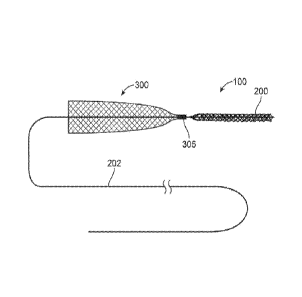
retrieval structure 200 can
move relative to the cover 300 so that the cover 300 everts over the proximal
end 206 of the
structure 200 when the cover 300 is expanded within a vessel and as the
structure 200 is
withdrawn into the distal end 302 of the cover 300, This mechanism is
discussed in detail
below.
100631 Figs. 2B and 2C illustrate alternative variations of a coverable
retrieval device 100.
As shown in Fig. 2B and 2C, the distal end 302 of the cover 300 can be spaced
from the
proximal end 206 of the retrieval structure 200. Alternatively, the distal end
302 of the cover
300 can extend over a portion of the retrieval structure 200. In some
variations, at least a
section of the cover 300 expands to a greater diameter than a diameter of the
retrieval structure
200. This allows the cover 300 to expand to a vessel wall where the vessel
holds the cover
stationary while the device is pulled proximally through the cover to evert
the cover. In
alternate variations, the cover 300 expands to the same or lesser diameter
than the retrieval
structure 200 or other device.
11
CA 2874586 2018-08-28
CA 02874586 2014-11-24
WO 2012/162437 PCT/US2012/039216
100641 Fig. 2D shows a retrieval device 100 with a catheter112 (usually a
microcatheter).
The retrieval device 100 can comprise a single unitary device of a cover 300
and retrieval
structure 200 (in this case the retrieval structure is an elongated stent
structure). One benefit
of a unitary device is that additional devices complicates the procedure and
can increase the
duration of what is ordinarily a time sensitive procedure. The retrieval
device .100 can be
positioned through the catheter 112 that includes a hub 114. As a result, the
physician only
needs to manipulate the unitary retrieval device 100 and the
catheter/microcatheter 112. The
retrieval device .100 is loaded into the catheter 112 for placement at the
target site. In addition,
the retrieval device can be reloaded if the procedure must be repeated. The
cover 300 and
retrieval structure 200 described 'herein can comprise any construction
described herein or as
known by those skilled in the art.
100651 Fig. 2E shows a retrieval device 100 with a cover 300 and retrieval
device 200 with a
radiopaque marker 305 therebetween. As shown, variations of the device 100 do
not require a
catheter or microcatheter.
100661 Fig. 2F illustrates an eversible cover 300 located on a sheath 330
having a lumen 332
extending thereth rough. A separable retrieval device 200 can be coupled to
the cover 300 and
sheath 330 by inserting the wire 202 of the cover retrieval device 200 through
the lumen 332
of the sheath 330. In this variation, the eversible cover 300 can be used with
any number of
different interventional tools. The separate devices can be assembled prior to
delivery into the
= patient. Alternatively, the devices can be positioned within the body and
subsequently joined
once the retrieval device 200 engages the target area.
[0067] Fig. 3A illustrates an example of a coverable retrieval device 100
where the cover 300
is in the process of everting about the retrieval structure 200. As shown,
arrow 50 illustrates a
force applied on the wire 202 in a proximal direction. Arrows 52 illustrate a
resistance force
applied by the friction of the expanded cover 300 against a vessel or similar
wall. This
friction force 52 prevents or resists proximal movement of the free end 304 of
the cover 300
while the fixed end 302 moves in a proximal direction with the proximal end
206 of the
retrieval structure 200. This action causes a wall 306 of the cover 300 to
even over the
retrieval structure 200. 'Ultimately, and as shown in Fig. 3:B, the free end
304 of the cover 300
ends up distally over the fixed end 302. As shown, the wall of the everted
cover 300 provides
a safety type cover for the retrieval device 200. In additional variations,
the fixed end 302 of
the cover can actually be slidable or moveable along the delivery wire 202.
.However, the
similar principle as discussed above shall apply to cause everting of the
cover 300 over the
retrieval structure 200.
12
100681 = Fig. 3C illustrates another variation of a coverable retrieval
device 100 after the cover
300 is everted about the retrieval structure 200. In this variation, the free
end 304 of the cover
300 ends up distally of the fixed end 302 and tapers or collapses towards the
free end 304.
The cover 300 can be shape set so that prior to eversion the cover is as shown
above where the
forces acting on the cover wall 306 expand outwards, but after eversion the
forces on the cover
wall 306 cause the tapering or collapsing as shown in Fig. 3C.
100691 In accordance with the illustrations discussed above, the cover
300 can be made so
that the cover wall 306 is atraumatic when dragged across a lumen wall. The
cover can be
manufactured from any number of materials including a fabric, a reinforced
fabric, a braid,
weave, or any such material that allows for expansion against a wall of the
body lumen or
vessel as well as to allow everting of a wall 306 of the cover over the
retrieval device 200.
The cover wall 306 can also comprise combinations of these materials such as
braids of
polymer material with metal fibers, soft braids with coil reinforcements or
various other
combinations.
100701 The cover wall can comprise a mesh that can include any medically
acceptable
materials such as a Nitinol braid. Furthermore, the mesh allows for flow
through the vessel or
lumen while expanded. However, additional variations of the device can include
a solid layer
of material substituted for the mesh. Moreover the cover can comprise any
number of
configurations. For example, the cover can comprise a single layer wall or a
multi layer wall,
the open end of the cover could be made to have terminated ends such as by
using continuous
wire loops formed during the braiding process. Alternatively, the ends can be
cut and then
terminated by encasing in polymer, laser welds, or by folding inward for a
discreet length and
then terminating
[00711 In one example, the cover 300 comprises a continuous wire
construction as described
in earlier commonly assigned patent applications. In one
variation
the cover 300 comprises a finely braided wire, such as 48-96 wires of .0005"
to 0.002"
diameter fine Nitinol wire or similar. Additionally, the wire can comprise
cobalt chromium,
stainless steel, or similar, or drawn filled tube (dft) with platinum core. In
additional
variations, a flat wire or oval wire can be used. The wire does not need to be
uniform.
Instead, a number of different types of wires can be used. Some of the
individual wires could
be platinum alloys for added radiopacity.
100721 Fig. 4A to 41 illustrates an example where an improved retrieval
device 100 with
passive protection retrieves a clot 2 from tortuous anatomy. Fig. 4A
illustrates a clot 2 that
obstructs blood flow in a vessel 6. As noted herein, the vessel 6 can comprise
any vessel in
13
CA 2874586 2018-08-28
cerebral vasculature, coronary or peripheral vasculature. Alternatively, the
device and
methods for use are not limited to use in the vasculature . Variations of the
principles,
concepts, method and devices described herein can be applicable wherever a
retrieval device
can be used. Fig. 4A also illustrates a guide sheath or access catheter 108
that is advanced
within the vessel. During a procedure, the physician will advance the access
catheter 108 as
far distally as possible. However, due to the size of the access catheter 108,
a physician
typically positions it a distance away from the obstruction 2. As shown, there
can be any
number of bifurcations 8 in the vessel 6 located between the access catheter
108 and the
obstruction 2. As discussed herein, in some variations, the access catheter
108 can be used to
remove the obstruction 2 from the body once the obstruction is captured by a
retrieval device.
However, the greater the distance between the initial location of the
obstruction 2 and the
location of the access catheter 108, the greater the risk that the obstruction
2 can break free
from the retrieval device or become dislodged clue to anatomic or
environmental features,
including but not limited to bifurcations, the wall of the lumen, the
tortuousity of the anatomy,
vessel wall plaque, etc.
100731 Fig. 4B illustrates an optional catheter 112 that advances from
the access catheter 108
to the site of the obstruction 2. Once at the site, the catheter 112 can
deploy a retrieval device
(not shown in Fig. 4B) so that the retrieval device can engage the clot 2.
Alternatively, the
catheter 112 can traverse the obstruction 2 as shown in Fig. 4C and deploy a
portion of the
retrieval device 100 distally to the obstruction 2. The physician then
manipulates the retrieval
device 100 to secure the obstruction 2. For example, the physician can deploy
the retrieval
structure 200 distally to the obstruction 6 and withdraw the retrieval
structure 200 proximally
to secure the obstruction 2. In another variation, the physician can position
the retrieval
structure 200 within the catheter 2 while the catheter 112 is through or
adjacent to the
obstruction 2. Then, the physician can withdraw the catheter 112 to expose the
retrieval
structure 200 so that it secures to the obstruction 2 after expansion. In the
illustrated example,
the retrieval structure 200 comprises an elongated stent type structure that
expands (or is
expanded) to enmesh or secure to the obstruction. Although not illustrated,
the system can
include a distal capture filter or basket as described in any of the commonly
assigned
applications.
100741 Next, as shown in Fig. 4E, the physician can further withdraw the
catheter 112 to
expose a cover 300 as described above. In many cases, the physician exposes
the cove 300
once the retrieval structure 200 is engaged with the obstruction 2. This
sequential process
allows for easier repositioning of the retrieval structure 200 if necessary.
Alternatively, the
14
CA 2874586 2018-08-28
CA 02874586 2014-11-24
WO 2012/162437 PCMJS2012/039216
cover 300 can be deployed prior to engaging the retrieval structure 200 with
the obstruction 2.
If necessary, the physician can apply a proximal force on the delivery wire
202 while
withdrawing the catheter 112 to prevent inadvertent movement of the
obstruction 2 and
retrieval device 200.
100751 Fig. 4.17 illustrates the stage with a fully exposed the cover 300
and a catheter 112
moved closer towards the access sheath 108. As shown, the free end 304 oldie
cover 300 is
proximal to fixed end 302 of the cover 300. As also noted above, the cover 300
can be a
shape memory alloy that expands against the walls of the vessel 6 upon
reaching body
temperature. Alternatively, the cover 300 can be self expanding upon
deployment into the
vessel 6. In some variations, the cover wall 306 comprises a porous material
or construction
that allows blood to continue to flow through the cover 300. =
100761 In addition, some variations of the retrieval device 100 include a
cover 300 that has at
least a section that expands to a greater diameter or dimension than the
retrieval structure 200.
This allows for expansion of the cover 300 against the wall of the vessel 6.
In most variation,
expansion of the cover 300 provides sufficient friction against the walls of
the vessel to
overcome column strength of the cover walls 306 allowing for everting of the
cover walls 306
over the retrieval structure 200 and obstruction 2 as discussed -herein. As
noted above, in
alternate variations the cover 300 can expand a diameter or dimension that is
equal to or less
than the retrieval structure 200.
100771 Figs. 4G illustrates proximal movement of the delivery wire 202,
which causes
proximal translation of the obstruction 2 and retrieval structure 200. Because
the cover 300 is
expanded against the walls of the vessel 6 the .free end 304 of the cover 300
does not move or
moves less than the fixed end 306 of the cover 300. The fixed end 306 moves
with the
obstruction 2 and retrieval structure 200 in a proximal direction causing the
cover walls 306 to
evert over the obstruction 2 and retrieval structure 200. Unlike a
conventional funnel, the
everting cover functions similar to a conveyor belt type movement as the
obstruction and
retrieval structure move together. This action allows for a passive type of
protection since
cover 300 does not need to be actuated over the obstruction 2 and retrieval
structure 200 and
can be performed in a quick manner by simply withdrawing the deployed
retrieval device 100.
100781 Fig. 411 illustrates a stage where the fixed end 306 of the cover
300 is now proximal to
the free end 304. As shown, the everted cover 300 forms a protective sheath or
cover over the
obstruction 2 and the retrieval structure 200. Fig. 4H also illustrates how
the cover 300
.protects the obstruction 2 and retrieval structure 200 as they are pulled
along the vessel and
navigate the tortuous anatomy, walls oldie vessel, as well as bifurcations 8.
'The cover 300
CA 02874586 2014-11-24
WO 2012/162437 PCT/US2012/039216
and cover wall 306 also protects the vasculature from the surface of the
retrieval structure 200
and obstruction 2.
100791 Fig. 41 shows the obstruction 2 and retrieval structure 200
protected by the cover 300
as the retrieval device 100 is positioned against or within the access
catheter 108 in
preparation for removal from the body. The retrieval device 100 can remain
outside of the
access catheter 108 as the physician removes both devices from the body.
Alternatively, the
cover 300 can assist in pulling the retrieval device .100 and obstruction 2
into the access
catheter 108 by compressing the obstruction 2 as it is pulled into the access
catheter 108.
100801 Figs. 4J and 4K illustrate examples of an obstruction or other
material 2 captured
within a retrieval device 2 with a cover 300 further protecting the loaded
retrieval device 200.
100811 Figs. 5A to 5K show a variety of cover configurations. =Fig. 5A
illustrates a retrieval
device 100 having a retrieval structure 200 adjacent to a double layer cover
300 with an
exterior wall 306 and an interior wall 308.
100821 Fig. 5B shows a cover 300 with a free end 304 that tapers down about
the delivery
wire 202 where the cover 300 will eventually form a double wall configuration
when the cover
300 everts over the retrieval structure 200. The tapered free end 304 limits
the cover 304 from
-moving once the retrieval structure 200 reaches the free end 304 thereby
forming double wall
protection over the retrieval structure 200.
100831 Figs. 5C and 5D show how a fixed end 302 of a cover 300 can be pre-
shaped to
reduce the force required to evert the cover wall 306 or to lower the
threshold to trigger
passive covering of the retrieval structure by the cover.
100841 Fig. 5E shows alternate variation of a passive cover 300 integrated
into a retrieval
device 1.00. In this variation, the retrieval device 100 includes a control
shaft or wire 202 to
manipulate the working end of the retrieval device 100. The cover 300 floats
along the shaft
202 between two fixed anchors or nodes 220, 222. The cover 300 can float or
slide between
the fixed nodes 220, 222. The nodes 220, 222can comprise radiopaque marker
bands, glue
joints, or any other mechanical obstructions capable of stopping the
translation of cover 300.
When the device .100 advances through a microcatheter, the rear or proximal
node 220 limits
rearward movement of the cover 300. When positioned appropriately, the
microcatheter can
be withdrawn to expose the retrieval device 200 and cover 300 as described
herein. When the
retrieval structure 200 engages the obstruction (not shown) the retrieval
device 100 can be
withdrawn by pulling on the delivery shaft 202. While this occurs, the cover
300, being
expanded against the vessel remains stationary (or moves at a slower rate than
the obstruction
and retrieval structure 200 due to the friction against the vessel wall). The
retrieval structure
16
CA 02874586 2014-11-24
WO 2012/162437 PCT/US2012/039216
200 and clot enter the cover 300, causing the distal node 222 to make contact
with the near
end 320 of the cover 300. This contact causes the retrieval structure 200 and
cover 300 to
translate as an integrated unit. It should be appreciated that the cover could
be a single layer
or double layer cover, and could have any of the wire design variables and
termination
variables described herein.
100851 Fig. 5F illustrates a cover having a pre-set :flattened cover wall
304 at a fixed end 302
that is spaced from a proximal end of the retrieval structure 200. Figs. 5G to
51 illustrate
various layered covers 300. The layered covers allow for shortening the axial
length of the
cover and therefore shortens the required translation length. Layering of the
cover wall 306
allows for a shortened deployed length of the cover 300 when deployed in the
vessel or body
structure. As the cover 300 evens over the retrieval structure 200 the layered
wall 306
extends. As a result, shortening the length reduces the length that the cover
300 extends into
the proximal vessels and reduces the length of that the retrieval structure
200 must travelrto
'become protected by the cover 300. This also helps shorten the distance
required to move the
device 100 to complete eversion of the cover 300.
100861 Fig. Si shows a cover 300 that is constructed directly onto the
retrieval structure 200
:rather than the delivery shaft 202. This construction also assists in
reducing the distance
necessary to complete passive protection of the retrieval structure by the
cover.
100871 Fig. 5K show a variation of a cover 300 that is mounted in a distal
direction over the
retrieval device 200 and then everted in a proximal direction over the wires
or shall 202 as
shown by arrows 230. Once evened, as shown by Fig. 5L, the device 100 is ready
:for
deployment as discussed herein.
100881 Figs. 6A to 613 illustrate a variation of a cover 350 for use as
describe herein.
Additionally, the cover 350 can be used with any obstruction retrieval device
not limited to the
retrieval baskets and stents described herein. The covers 350 disclosed herein
can be used
where the physician desires to shield the obstruction being removed from the
frictional effects
of the arteries or from the local anatomy (e.g., 'branching vessels, tortuous
anatomy, or other
substances on the vessel walls). in use, the covers can be sized for use with
guide catheters,
micro-catheters, and/or distal access catheters. The covers can include any
number of
radiopaque marker bands to allow non-invasive imaging of the device (see
marker 390 affixed
between cover 350 and shaft 212 in Fig. 7B as one example). In any case, once
the retrieval
device captures a clot or obstruction, as described above, the device and clot
are protected by
the cover so that the cover eliminates or reduces direct contact between the
interior of the wall
of the vessel and the clot.
17
CA 02874586 2014-11-24
WO 2012/162437 PCMJS2012/039216
100891 Figs. 6A to 6C show a variation in which a cover is created from
one or more mesh
tubes 372. Fig. 6B illustrates inversion of the tube 372 so that a first end
374 is drawn over
the tube 372 towards a second end 376. As shown in Fig. 6C, this creates a
double walled
cover having an exterior va II 378 separated from an interior wall 380. In one
example, such a
spacing or gap could range between 0.001 inches to 0.100 inches. However, any
range is
contemplated within alternative variations of the device. In some variations
the inverted cover
350 is heat set to maintain a separation between layers or walls 378 380 of
the cover 350.
Typically, if the cover 350 is not created from a radiopaque material, a
marker band will be
= placed on the proximal end 376 and adjacent to a shaft- or catheter to
which the cover 350 is
attached. In some variations the construction of the mesh material is
compliant to allow for
movement of a -first part of the mesh relative to a second part of the mesh
through compression
and expansion of the mesh material. In such a case, the individual strands
forming the mesh
are moveable relative to one another to cause the mesh to be naturally
compliant.
Accordingly, this construction permits the inner wall 380 to move or deflect
with the retrieval
device and/or obstruction as the device is withdrawn into the cover 350. In
some variations,
both ends of the mesh 374 and 376 are affixed to the catheter,shaft or wire.
100901 In many variations, the cover mesh is selected to minimize friction
when the interior
layer 380 moves against the exterior layer 378. For example, the braid
pattern, wire, wire
diameter, angle of the braid and or other features can be selected to reduce
friction between
the outer layer 378 and inner layer 380. This permits the inner layer 380 to
move proximally
with a retrieval device while the outer layer remains stationary. Again, as
discussed above,
the construction of the mesh permits compression and expansion of the mesh
layer to permit
movement of the inner layer while the outer layer remains affixed when engaged
against the
vessel wall. in certain variations, the cover is heat set so that the inner
layer has cushioning
and the ability to deflect to assist in movement of the inner layer. Fig. 5C
also illustrates a
coer 350 having a tapered design.
100911 Figs. 6D to 6L illustrate additional variations of cover
construction to produce covers
having more than two walls. For example, a mesh tube 372 is everted or drawn
over a second
end 376 in the direction 420. As shown in Fig. 6E this produces a dual layer
cover having a
open ends 422 and 424 and a folded end 426. The dual layer tube is then folded
over again in
the direction 420. This creates a cover construction with an exterior layer
378 and an interior
layer 380 as well as a first intermediate layer 381 and a second intermediate
layer 383. As
shown in Fig. 6F, the cover can be set to assume the tapered shape having an
opening at the
18
CA 02874586 2014-11-24
WO 2012/162437 PCMJS2012/039216
first end 374 that is flared with the ends of the mesh at the second end 372,
which are
ultimately affixed to a shaft, wire or other catheter device as described
herein.
100921 Fig. 6G illustrates another example of a cover construction. As
shown, a first mesh
tube 372 is placed coaxially with a second tube 372. The concentric tubes are
then everted in
direction 420 to produce a four layer cover. As shown in Fig. 6.11, the cover
can comprise an
interior mesh layer 380, and exterior mesh layer 378 as well as any number of
intermediate
layers 381, 383 depending on the number of tubes that are initially used. The
second end 372
of the cover 350 includes four unconnected ends of the mesh tubes that can be
affixed to a
shaft or tube as discussed herein, while the first end 374 of the cover 350
can be shape set to
taper from the opening.
100931 Figs. 61 to 6L illustrate another example of the construction of a
multi-wall cover. As
shown in Fig. 61, a first end 374 of a mesh tube 372 is everted over and
beyond a second end
376 in direction 420 to produce the configuration of Fig. 6J. 'Next, the first
end 374 is everted
or folded back in direction 420 to produce the configuration of Fig. 6K.
Finally, the first end
374 is folded again in direction 420 so that the ends 374 and 376 are even to
produce the cover
configuration shown in Fig. 6K. Again, one end of the cover 350 can be set to
form the
tapered shape while the other respective end can be affixed to a catheter or
shaft.
100941 Although the covers of the present disclosure are presented without
additional
structures, it should be noted that these covers are coupled with a shaft or
other member so
that the cover can be advanced within the target anatomy to assist in removal
of a device,
structure, or debris from the site.
100951 Figs. 7A to 7C show addition variations of covers 350. Fig. 7A
illustrates a cover in
which the cover wall as defined by the inner layer 380 and outer-layer 378 is
set in a shape
that varies along a length of the cover. For example, the end adjacent to the
cover opening
382 can be set to a bulbous shape. Such a configuration assists in maintaining
separation of
layers 378 and 380, which aids in re-entry of the retrieval device. Additional
configurations of
cover walls that vary in thickness are within the scope of this disclosure.
100961 One of the benefits of using a cover 350 as described herein is that
the cover reduces
flow through the vessel When deployed so that the retrieval device can remove
the obstruction
without the full force of the flow of blood opposing the obstruction.
Typically, conventional
devices relied upon the use of an inflated balloon to obstruct flow. However,
use of a cover
eliminates the need for total occlusion of blood flow. Fig. 7B illustrates a
further
improvement on a cover 350 that aids in flow reduction. As shown, the cover
350 includes a
dense region 386 and a relatively less dense region 384. This configuration
permits greater
19
CA 02874586 2014-11-24
WO 2012/162437 PCMJS2012/039216
blood flow through the region 385 while region 386 reduces or prevents blood
flow.
Furthermore, the distal section of the cover is more flexible and conformable.
Additional
mesh layers can be added to any of the cover designs to alter flow
characteristics or even
provide reinforcement to the cover. Alternatively, or in combination, the
braid density can be
altered to adjust the porosity of the braid at different sections.
'Furthermore, additional braid
layers can also be used to affect porosity of portions of the cover or even
the entire cover.
Deployment of a cover can reduce blood flow by 30% to 40%. Adding additional
layers or
coatings can additionally reduce flow.
100971 'Fig. 7C shows another variation of a cover 350 in which the mesh
partially or totally
is obscured using a polymeric coating 388 that reduces the permeability of the
mesh design.
Furthermore, drugs or other substances can be placed within the cover wall of
any of the
covers or can be deposited on the cover using the polymeric coatings. In some
examples, the
covers described herein can range from a length of 10 mm up to 50 mm. The OD
at the
opening of the cover can range from 7 mm and could range between 4 mm to 10
mm. Again,
any range of dimensions is contemplated within the disclosure.
100981 The covers described herein can further be stacked on a device. For
example, two or
more covers can be placed on a device to provide added protection.
100991 The cover/rentry devices described herein can be constructed of any
material currently
used in vascular applications, including those discussed above. Furthermore,
fabrication of
the cover from a DFT material can provide additional benefits as the entire
cover remains
radiopaque and can be imaged non-invasively. Furthermore, the covers can be
provided with
any type of medicament or bioactive substance either in a polymer that coats
the mesh or in a
delivery agent within the mesh or between layers. Such substances include tpa,
urokinase,
1.1b/1.1.1a inhibitors, and other clot disruptors or inhibitors.
101001 Fig. 8 illustrates another variation of a retrieval device 400
including a distal capture
portion 426 coupled to one or more leading wires in the form of a main bundle
402. The main
bundle extends through a sheath 106 that includes a proximal capture portion
460. The
configuration of the retrieval device 400 can incorporate the proximal and
distal capture
portions discussed herein as well as various other configurations discussed in
the commonly
assigned patent applications noted above.
101011 An end 464 of the proximal capture portion 460 is affixed to a
distal end of the sheath
106. However, as noted above, other variations are within the scope of the
disclosure. The
main bundle 402 can optionally terminate at a handle 442. As noted above, in
certain
variations, the main bundle is joined to a stiffer wire or stiffer bundle of
wires. This allows
CA 02874586 2014-11-24
WO 2012/162437 PCMJS2012/039216
the device 400 to have a very flexible distal section with a relatively
stiffer proximal section.
The device 400 can have a proximal bundle 403 that comprises either the
exposed wires or a
covering/tube over the wires. In certain variations, the bundle or wire 402,
403 can be
encapsulated with a coating. The device also includes a cover 300 adjacent to
the retrieval
device.
101021 The proximal end of the sheath 106 includes a sheath handle 444. As
discussed
herein, axial movement of the bundle 402 or proximal bundle 403 (typically at
the handle 442)
results in movement 126, or translation of the bundle within the sheath 106.
This action
moves the distal capture portion 426 (as shown by arrows 126). In certain
variations, the
device 400 is loaded into a microcatheter (not shown but discussed above) that
is delivered to
the site of the obstruction and crosses the obstruction.
101031 .In some variations, the sheath hub 444 includes one or more
locking hubs 446. Where
actuation (either axial or rotational) of the locking hub 446 locks the main
bundle 402 relative
to the sheath handle 444 and sheath 106. It follows that such locking action
also locks the
distal capture portion 426 relative to the proximal capture portion 460. A
variety of methods
can be employed to increase a frictional interference between the locking hub
446 and the
proximal bundle 403. As a result, when a physician determines a length of an
obstruction, the
physician can set a spacing between the capturing portions 426 460 by locking
the proximal
bundle 403 relative to the sheath hub 444. Accordingly, the proximal bundle
403 can include
any type of incremental markings to allow the physician to readily determine a
spacing of the
capturing portions. As illustrated, the sheath hub 444 can include additional
injection ports to
deliver fluid or other substances through the sheath 106.
101041 As noted above, the device 400 can be used with a micro-catheter.
In those variations
it is important that the device 400 is loaded without damaging the distal
bundle 402, capture
portions 426 460, and/or sheath 106. As a result, the device 400 can include
an optional cover
486 that reduces the proximal capture portion 460 (and /or the distal capture
portion 426) for
loading within the microcatheter and/or Sheath 106.
101051 Another variation of the device 400 includes an insertion tool 480
slidably affixed to
the sheath 480. Because variations of the device 400 can be extremely
flexible, the insertion
tool 480 can be used to provide column strength to the sheath 106, bundle 402
or other
components as the device 400 is pushed into the microcatheter. The insertion
tool comprises a
rigid section 482 and a frictional coupler 484. The rigid section 282 has a
column strength
that supports the device 400 to prevent buckling. The frictional coupler 484
can be a flexible
material that allows an operator to squeeze or grip the coupler 484 to create
a temporary
21
CA 02874586 2014-11-24
WO 2012/162437 PCT/US2012/039216
frictional interface between the loading tool 480 and the device 400
(typically the sheath 106).
Such an action allows axial advancement of the device 400 as the loading tool
480 is advanced
into the microcatheter. Once the rigid section 482 is fully inserted into the
microcatheter, the
operator releases the frictional coupler 484 and can withdraw the loading tool
480 from the
catheter without withdrawing the device 400. The insertion tool 480 can also
include an
optional loading tube 486 slidably coupled to the rigid section 482. When
used, the cover 486
can withdraw the proximal and distal capturing portion 226 260 within the
loading tube 486.
The loading tube 486 then couples to a microcatheter allowing the capturing
portions to
advance therein as the rigid section 482 and frictional coupler 484 advance
the device 400
relative to the loading tube 486.
[0.1.06) Figs. 9A to 9C show cross sectional views taken along the line 9A-
9A in Fig. 2A. As
shown, the wire form construction described herein allows for a number of
configurations
depending on the particular application. For example, the individual wires 254
(as discussed
'herein) may themselves comprise a bundle of smaller wires or filaments. In
addition, the
wires can be selected from materials such as stainless steel, titanium,
platinum, gold, iridium,
tantalum, Nitinol, alloys, and/or polymeric strands. In addition, the wires
used in a device
may comprise a heterogeneous structure by using combinations of wires of
different materials
to produce a device having the particular desired properties. For example, one
or more wires
in the device may comprise a shape memory or superelastic alloy to impart
predetermined
shapes or resiliency to the device. In some variations, the mechanical
properties of select
wires can be altered. In such a case, the select wires can be treated to alter
properties
including: brittleness, ductility, elasticity, hardness, malleability,
plasticity, strength, and
toughness.
101071 The device may include a number of radiopaque wires, such as gold
and platinum for
improved visibility under fluoroscopic imaging. in other words, any
combination of materials
may be incorporated into the device. In addition to the materials, the size of
the wires may
vary as needed. For example, the diameters of the wires may be the same or may
vary as
needed.
10108.1 In Addition, the individual wires may have cross-sectional shapes
ranging from
circular, oval, d-shaped, rectangular shape, etc. 'Fig. 9A illustrates one
possible variation in
which a number of circular wires 254 are included around another larger wire
256. Moreover,
the device is not limited to having wires having the same cross-sectional
shape or size.
Instead, the device can have wires having different cross-sectional shapes.
For example, as
shown in Fig. 98. one or more wires 256 can have a different cross-sectional
shape or size
22
CA 02874586 2014-11-24
WO 2012/162437 PCMJS2012/039216
than a reminder of the wires 254. Clearly, any number of Variations is within
the scope of this
disclosure. This construction can apply to the retrieval portion, capturing
portion and/or the
covering portion of the device.
10.109.1 To illustrate one such example, a device can have 8-12 wires made
of .003" round
superelastic material (e.g., Nitinol). The device may additionally have 2-4
wires made from
.002" platinum for fluoroscopy. Of the 8-12 'Nitinol wires, 1-4 of these wires
can be made of a
larger diameter or different cross-section to increase the overall strength of
the device.
Finally, a couple of polymer fibers can be added where the fibers have a
desired surface
property for clot adherence, etc. Such a combination of wires provides a
composite device
with properties not conventionally possible in view of other formation means
(such as laser
cutting or etching the shape from a tube or joining materials with welds,
etc.). Clearly, any
number of permutations is possible given the principles of the invention.
[MN In another example, the device may be fabricated from wires formed
from a polymeric
material or composite blend of polymeric materials. The .polymeric composite
can be selected.
such that it is very floppy until it is exposed to either the body fluids and
or some other
delivered activator that causes the polymer to further polymerize or stiffen
for strength.
Various coatings could protect the polymer from further polymerizing before
the device is
properly placed. The coatings could provide a specific duration for placement
(e.g., 5 minutes)
after which the covering degrades or is activated with an agent (that doesn't
affect the
surrounding tissues) allowing the device to increase in stiffness so that it
doesn't stretch as the
thrombus .is pulled out. For example, shape memory polymers would allow the
device to
increase in stiffness.
101111 In another variation, one or more of the wires used in the device
may comprise a
Drawn Filled Tube (DFT) such as those provided by .Fort Wayne Metals, .Fort
Wayne, 'Indiana.
As shown in Fig. 9C, such a DFT wire 252 comprises a first material or shell
258 over a
second material 260 having properties different from the outer shell. While a
variety of
materials can be .used, one variation under the present devices includes a DFT
wire having a
superelastic (e.g., Nitinol) outer tube with a radiopaque material within the
super-elastic outer
shell. For example, the radiopaque material can include any commercially used
radiopaque
material, including but not limited to platinum, iridium, gold, tantalum, or
similar alloy. One
benefit of making a capturing portion from the DFT wire noted above, is that
rather than
having one or more markers over the capturing portion, the entire capturing
portion can be
.fabricated from a super-elastic material while, at the same time, the super-
elastic capturing
portion is made radiopaque given the core of radiopaque material within the
super-elastic
23
CA 02874586 2014-11-24
WO 2012/162437 PCT/US2012/039216
shell. Clearly, any composite DFT wire 252 can be incorporated into the system
and capturing
portions described herein.
101121 Another aspect applicable to all variations of the devices is to
configure the devices or
portions thereof that engage the obstruction to improve adherence to the
obstruction. One
such mode includes the use of coatings that bond to certain clots (or other
materials causing
the obstruction.) For example, the wires may be coated with a hydrogel or
adhesive that
bonds to a thrombus. Accordingly, as the device secures about a clot, the
combination of the
additive and the mechanical structure of the device may improve the
effectiveness of the
device in removing the obstruction. Coatings may also be combined with the
capturing
portions or catheter to improve the ability of the device to encapsulate and
remove the
obstruction (e.g., a hydrophilic coating).
101131 Such improvements may also be mechanical or structural. Any portion
of the
capturing portion can have hooks, fibers, or barbs that grip into the
obstruction as the device
surrounds the obstruction. The hooks, fibers, or barbs 370 can be incorporated
into any
portion of the device. However, it will be important that such features do not
hinder the
ability of the practitioner to remove the device from the body.
101141 -hi addition to additives, the device can be coupled to an R17 or
other power source
(such as 14 or 16 in Fig. I), to allow current, ultrasound or -RF energy to
transmit through the
device and induce clotting or cause additional coagulation of a clot or other
the obstruction.
101151 Figs. 10A to 10E illustrate additional variations of covers 300 for
use as described
above. For example, as show in Fig. 10A, a cover 300 can comprise a single
wire, coil, or
laser cut tube 350. Alternatively, as shown in .Fig. 10B, the cover 300 can
comprises two or
more 350, 352 wires or coils. Fig. IOC shows a cover 300 comprising a coil 350
inside a
mesh structure 354. A variation of the device shown in Fig. :IOC can include a
compliant
atraumatic mesh 354 that is radially supported by the coil (whether interior
or exterior to the
mesh). The coil 350 provides the outward force against the vessel. Fig. 10D
illustrates a
polymeric film or membrane 356 coupled to a coil 350. The polymeric film 356
can be
permeable to fluid flow or impermeable. Fig. 10E illustrates a dual layer
braid construction
having an inner braid 358 and an outer braid 360. The braids can be
constructed to have
unique properties. For example, the inner braid 358 can be composed of fewer
wires or larger
diameter wires, such that it provides an expansion force against the vessel
wall. The outer
braid 360 can comprise a softer construction and increased compliance.
Accordingly, it can
be comprised of a 'number of smaller diameter wires having a denser pattern to
provide
increased surface area to protect the obstruction as it is removed from the
body. Alternatively,
24
CA 02874586 2014-11-24
WO 2012/162437 PCMJS2012/039216
these two constructional elements (e.g., braids of varying diameters) can be
combined into a
single layer or even multiple layers for the cover.
101161 'Fig. I IA illustrates yet another variation of a device 100
having a retrieval structure
200 and cover 300 where the cover is simply fabricated from the same material
as the retrieval
structure so long as it functions as described herein. The variation can
optionally include one
or more barbs 370 to increase resistance against a vessel wall.
10.1171 Fig. 1.1B and IIC illustrate a variation where the cover 300
comprises a balloon
material. Fig. 1113 illustrates the balloon cover 370 prior to deployment.
Fig. I IC illustrates
the balloon cover 370 once deployed.
[01.181 The retrieval devices described herein can optionally comprise
elongated sterns 400 as
Shown in Figs. 12A to 12E. These steins 400 can include any number of features
to better
assist the stent 400 in becoming enmeshed into the obstruction. For example,
Fig. I 2A
illustrates a variation o=fa stein 400 affixed to a shaft 412. As noted
herein, the shaft 412 can
include a lumen extending therethrough. Alternatively, the shaft 412 can
include a solid
member with the stent 400 affixed to a distal end thereof. The variation shown
in Fig. 12A
includes a stein where a distal end 414 that is "closed off' by intersecting
elements or wires
402 403. Accordingly, any of the variations of the stents disclosed 'herein
can include an open
lumen type stein or a closed lumen type stent as shown in Fig. 12A. As noted
herein, the
wires forming the stem 400 can comprise a single wire that is wound from a
first direction
(e.g., from proximal to distal) and then wound back in a second direction
(e.g., from distal to
proximal).
101191 Fig. 12A also illustrates a stem 400 comprised of twisted wires
402 or elements. For
example, Fig. 1213 Shows a magnified view of the section 12E3 in Fig. 12A. As
illustrated, the
elements 402 403 are twisted to increase the surface area at the exterior
perimeter of the stent
400. The twisting or spiraling of the elements 402 403 creates additional
surface area to
increase the ability of the stent 400 to capture debris, thrombus, foreign
body, etc. as the stem
is expanded against the debris. The twisting elements 402 403 can twist along
the entire
length of the stem 400 or along one or more portions of the stent. In certain
variations, the
= twisting of the elements 402 403 is sufficiently loose such that as the
stent expands into a clot
= or obstruction, the twisted pairs slightly separate to allow material to
become trapped between
the elements making up the pairs. The construction shown in Figs. 12A and 12B
also provide
an additional benefit to a retrieval stem. In the illustrated variation, the
twisted or spiraling
elements interlock with crossing elements to form intersections 405 that
provided added radial
expansive force. As shown, a first twisted element 407 passes in between
elements 402 403 of
=
CA 02874586 2014-11-24
WO 2012/162437 PCT/US2012/039216
an intersecting element 409. When in an expanded state, the element on the
interior of the
intersection 405 (in this case element 403) provides an added outward radial
force against the
intersection 405. 'However, since the elements are not affixed but instead are
slidable at the
intersection 405, the force required to linearlize and compress the stent 400
is reduced due to
the fact that the intersections are not affixed but slidable over the adjacent
elements. This
reduced linearization force allows the stent to be compressed to a small
diameter .for
positioning within a microcatheter but allow for a significant .radial
expansive force once
removed from the microcatheter. This design allows for a reduction in radial
force of the stent
against the vessel wall when the stern is pulled and removed from the vessel.
However, this
design also provides a high degree of radial force due to the interweaving of
elements when
the stern is deployed in the vessel prior to withdrawal of the stent.
10.1201 Figs. 12C to I21 illustrate another variation of types of stents
400 that have an
irregular surface at an exterior of the stent 400 that is formed by an
intersection of elements
402 403. The intersection or crossing of the elements forms a type of barb or
knuckle 416 that
creates an irregular surface on the exterior of the stent 400. Fig. 12C
illustrates a variation of
a stent 400 having a plurality of knuckles 52 that are radially spaced about
an axis 390 of the
stem 400. Fig. 12E shows another variation of a stent 400 with knuckles 416
aligned with an
axis 390 of the stent 400 as shown in Fig. 12D. Although the figures show the
axial and radial
aligned knuckles 416 on separate devices, both types of knuckles 416 can be
incorporated into
a single stent structure. Varying the alignment of knuckles can permit
increased radial force
as the stent expands into the obstruction or increased flexibility as the
stent navigates through
tortuous anatomy.
101211 Fig. 12G illustrates a proximal end of the stent structure 400 as
Shown, a plurality of
elements 402 and 403 extend along the shaft 412 and diverge to form the fluid
permeable
closed proximal end of the stent structure 400. The elements 402 and 403 that
extend along
the shaft 412 can be covered by a sheath, tube, spiral cut tube, or any
structure 418 that
prevents separation of the elements 402 403. A variation of the stem structure
402 includes a
construction where the elements 402 403 are not glued, welded, or have any
similar type of
joint in the distal portion 420 of the shaft 4-12. Instead, the joint 411 is
located proximal to the
distal section of the shall 412 in an intermediate section 422. 1Because
joints or other similar
features reduce flexibility of the joined structure, positioning the joints
411 in a proximal area
allows the the distal portion 420 of the shaft to remain flexible.
101221 The methods described herein may also include treating the
obstruction prior to
attempting to remove the obstruction. Such a treatment can include applying a
chemical or
26
CA 02874586 2014-11-24
WO 2012/162437 PCMJS2012/039216
pharmaceutical agent with the goal of making the occlusion shrink or to make
it more rigid for
easier removal. Such agents include, but are not limited to chemotherapy
drugs, or solutions,
a mild formalin, or aldehyde solution.
101231 As for other details of the present invention, materials and
manufacturing techniques
may be employed as within the level of those with skill in the relevant art.
The same may
hold true with respect to method-based aspects of the invention in terms of
additional acts that
are commonly or logically employed. In addition, though the invention has been
described in
reference to several examples, optionally incorporating various features, the
invention is not to
be limited to that which is described or indicated as contemplated with
respect to each
variation of the invention.
101241 'Various changes may be made to the invention described and
equivalents (whether
recited herein or not included for the sake of some brevity) may be
substituted without
departing from the true spirit and scope of the invention. Also, any optional
feature of the
inventive variations may be set forth and claimed independently, or in
combination with any
one or more of the features described herein. Accordingly, the invention
contemplates
combinations of various aspects of the embodiments or combinations of the
embodiments
themselves, where possible. Reference to a singular item, includes the
possibility that there
are plural of the same items present. More specifically, as used herein and in
the appended
claims, the singular forms "a," "and," "said," and "the" include plural
references unless the
context clearly dictates otherwise.
101251 It is important to note that where possible, aspects of the various
described
embodiments, or the embodiments themselves can be combined. Where such
combinations
are intended to be within the scope of this disclosure.
27