Note: Descriptions are shown in the official language in which they were submitted.
CA 03124779 2021-06-23
DESCRIPTION
METHOD FOR INDUCING MUSCULAR CELLS USING CELLS IN SPOT URINE
[0001]
Technical Field
The present invention relates to a method and a kit for preparing myotubes
from urine-
derived cells. Also, the present invention relates to a method for testing an
agent used for exon
skipping therapy of muscular dystrophy using the myotubes.
[0002]
Background Art
Duchenne muscular dystrophy (DMD) is a serious hereditary muscular disease
caused by dystrophin deficiency. For the treatment for DMD, practical
application of exon
skipping therapy using an antisense oligonucleotide (AON) has been expected.
The exon
skipping therapy is based on skipping an exon in the vicinity of a gene
mutation by targeting
an mRNA precursor with the use of AON (i.e., modification of abnormal
splicing), modifying
a frame-shift mutation to in-frame, and restoring the expression of a
shortened dystrophin
protein. As such agent used for exon skipping therapy, the inventors had
developed the
antisense oligonucleotide, NS-065/NCNP-01, that allows skipping of exon 53 of
the dystrophin
gene to restore dystrophin protein expression and completed the doctor-
initiated early
explorative test (Non-Patent Literature 1). At present, the next-phase testing
is in progress.
From now on, development of a novel exon skipping agent targeting an exon
associated with a
large number of target patients is expected.
[0003]
It is also known that therapeutic effects cannot be always predicted at
dystrophin
mRNA and protein levels based on genomic DNA mutation patterns. Even when the
exon
skipping therapy is performed based on a particular genomic DNA mutation
pattern, differences
are sometimes observed in preferable dystrophin protein expression levels. In
order to
accelerate the development of an exon skipping agent for DMD treatment, select
a subject on
which therapeutic effects of such agent can be expected, and provide effective
treatment to the
1
Date Recue/Date Received 2021-06-23
CA 03124779 2021-06-23
subject, accordingly, it would be important to examine the effects of
therapeutic agents using
muscle cells derived from the subject in vitro prior to the initiation of the
actual treatment.
[0004]
Prior Art Literature
Non-Patent Literature
Non-Patent Literature 1: Komaki, H. et al., Science translational medicine.
vol. 10, eaan0713,
2018
Non-Patent Literature 2: Saito, T. et al, Plos One. vol. 5, e12239, 2010
Non-Patent Literature 3: Kim, E.Y. et al., Skeletal Muscle vol.6: 32, 2016
[0005]
Summary of the Invention
Problems to be Solved by the Invention
In the past, the inventors had established the in vitro testing system
comprising
transforming the fibroblasts derived from the patient's skin into the myotubes
via introduction
of a muscle regulatory factor (i.e., MY0D1), and, after myotube
differentiation, examining the
effects of the exon skipping agent (Non-Patent Literature 2). However, a
technique involving
the use of dermal fibroblasts suffer from difficulties, such as the need for
invasive skin biopsy
and the need for performance of flow cytometry, which requires special
equipment and
techniques, in order to sort MY0D1-positive cells. In order to overcome such
difficulties,
development of an in vitro testing system of a therapeutic agent that can be
performed in a non-
invasive and simple manner is desired.
[0006]
While a method of introducing MY0D1 into urine-derived cells to perform direct
reprogramming into the myotubes has been reported (Non-Patent Literature 3),
such technique
had problems, such that cells with particular morphology were selected from
among urine-
derived cells in advance and it would take 4 to 5 weeks after the induction of
differentiation in
order to induce the myotubes.
[0007]
Means for Solving the Problems
2
Date Recue/Date Received 2021-06-23
CA 03124779 2021-06-23
The present inventors had focused on the urine-derived cells from the
viewpoint of
non-invasive testing and attempted the induction of the urine-derived cells
into the myotubes
by introducing the MY0D1 gene into the urine-derived cells as described in Non-
Patent
Literature 3. However, Myogenin, which is a muscle regulatory factor located
downstream of
MY0D1, was not substantially expressed, and myotubes could not be sufficiently
induced. In
order to overcome such problems, the present inventors had searched for the
conditions in
which the myotubes could be induced and succeeded in effective induction of
the myotubes by
exposing the urine-derived cells transduced with the MY0D1 gene to an
epigenetic regulatory
compound, such as a histone methyltransferase inhibitor (HMTI). We had also
succeeded in
testing the effects of an agent used for exon skipping therapy, which is a
therapeutic agent for
muscular dystrophy, with the use of the induced myotubes. The present
invention had been
completed based on such findings.
[0008]
Specifically, the present invention encompasses the following aspects.
(1) A method for preparing myotubes from urine-derived cells comprising:
a step of introducing the MY0D1 gene into urine-derived cells; and
a step of exposing the urine-derived cells to at least one of epigenetic
regulatory
compounds.
(2) The method according to (1), wherein, after the introducing step and
the exposing
step, the urine-derived cells comprise at least one selected from the group
consisting of
myoblasts and myotubes.
(3) The method according to (1) or (2), wherein the epigenetic regulatory
compound
comprises at least one selected from the group consisting of a histone
methyltransferase
inhibitor, a histone demethylase inhibitor, a histone deacetylase inhibitor, a
SIRT2 inhibitor,
and a PARP inhibitor.
(4) The method according to (3), wherein the histone methyltransferase
inhibitor
comprises at least one selected from the group consisting of 3-deazaneplanocin
A, 3-
deazaneplanocin A hydrochloride (DZNep), G51(343, 5GC707, furamidine
dihydrochloride,
UNC2327, E7438, and MI-2 (menin-MLL inhibitor).
3
Date Recue/Date Received 2021-06-23
CA 03124779 2021-06-23
(5) The method according to (3), wherein the histone demethylase inhibitor
comprises at
least one selected from the group consisting of IOX 1 and GSK-J1.
(6) The method according to (3), wherein the histone deacetylase inhibitor
comprises at
least one selected from the group consisting of LMK-235, CAY10603, BRD73954,
and
VORINOSTAT.
(7) The method according to (3), wherein the SIRT2 inhibitor comprises
SirReal 2.
(8) The method according to (3), wherein the PARP inhibitor comprises EB47.
(9) The method according to any of (1) to (8), wherein the MY0D1 gene is
introduced
by introduction of an expression vector comprising the MY0D1 gene under the
control of an
inducible promoter.
(10) The method according to (9), wherein the expression vector further
comprises a
selection marker gene.
(11) The method according to any of (1) to (10), wherein the urine-derived
cells are
derived from a patient with a muscular disease or a patient with muscular
dystrophy.
(12) A kit for preparing myotubes from urine-derived cells comprising:
a means for introducing the MY0D1 gene into urine-derived cells; and
at least one of epigenetic regulatory compound.
(13) The kit according to (12), wherein the introducing means is an
expression vector used
for introducing the MY0D1 gene into urine-derived cells.
(14) The kit according to (12) or (13), wherein the epigenetic regulatory
compound
comprises at least one selected from the group consisting of a histone
methyltransferase
inhibitor, a histone demethylase inhibitor, a histone deacetylase inhibitor, a
SIRT2 inhibitor,
and a PARP inhibitor.
(15) The kit according to any of (12) to (14), wherein the epigenetic
regulatory compound
comprises at least one selected from the group consisting of 3-deazaneplanocin
A, 3-
deazaneplanocin A hydrochloride (DZNep), GSK343, SGC707, furamidine
dihydrochloride,
UNC2327, E7438, MI-2 (menin-MLL inhibitor), IOX 1, GSK-J1, LMK-235, CAY10603,
BRD73954, VORINOSTAT, SirReal 2, and EB47.
(16) A method for testing an agent used for exon skipping therapy for a
patient with
muscular dystrophy comprising:
4
Date Recue/Date Received 2021-06-23
CA 03124779 2021-06-23
a step of preparing myotubes from urine-derived cells obtained from a patient
with
muscular dystrophy by the method according to any of (1) to (11);
a step of applying the agent used for exon skipping therapy to the myotubes;
and
a step of detecting recovery of the dystrophin mRNA and/or protein in the
myotubes.
(17) The method according to (16), wherein, in the detecting step, recovery
of the
dystrophin mRNA and/or protein is detected by at least one method selected
from the group
consisting of RT-PCR, Western blotting, and immunocytochemistry.
(18) The method according to (16) or (17), wherein the agent used for exon
skipping
therapy comprises at least one selected from the group consisting of an exon-
44-skipping agent,
an exon-45-skipping agent, an exon-50-skipping agent, an exon-51-skipping
agent, and an
exon-53-skipping agent.
(19) A method for screening for a candidate therapeutic agent or preventive
agent of a
condition of inducing skeletal muscle damage comprising:
a step of preparing myotubes from urine-derived cells obtained from a patient
with a
condition of inducing skeletal muscle damage by the method according to any of
(1) to (11);
a step of applying a test substance or factor to the myotubes; and
a step of identifying the test substance or factor as the candidate
therapeutic agent or
preventive agent by monitoring a change in the myotubes after the applying
step.
[0009]
Effects of the Invention
The method and the kit according to the present invention enable induction of
myotubes from urine-derived cells in a non-invasive and efficient manner. With
the use of the
induced myotubes, the progress of fundamental studies involving the use of
human-derived
disease model muscle cells and the progress of personalized medicine provided
for each patient
developing myopathy, including muscular diseases and skeletal muscle damages,
can be
accelerated. Therefore, the present invention may be useful in the medical and
drug discovery
fields.
[0010]
Brief Description of the Drawings
Date Recue/Date Received 2021-06-23
CA 03124779 2021-06-23
Fig. 1 shows an image of urine-derived cells that had formed colonies via
urine
culture obtained by phase contrast microscopy. The image was obtained 7 days
after the
initiation of primary culture.
Fig. 2 shows a retrovirus vector used to introduce the MY0D1 gene into urine-
derived cells.
Fig. 3 shows a graph demonstrating a degree of muscular differentiation
evaluated
based on the expression level of the myosin heavy chain protein by
immunocytochemistry. A
horizontal axis represents a type of a compound added, and a vertical axis
represents an area of
a myosin heavy chain-positive region determined by immunocytochemistry (Fig.
3A: 1 [EM
low-molecular compound; Fig. 3B: 10 [EM low-molecular compound).
Fig. 4 shows images demonstrating the effects of 3-deazaneplanocin A
hydrochloride
(DZNep) on promoting muscular differentiation analyzed by immunocytochemistry.
Red
represents a myosin heavy chain, and blue represents nuclear staining.
Fig. 5A shows blots and Fig. 5B shows graphs demonstrating the effects of 3-
deazaneplanocin A hydrochloride (DZNep) on promoting muscular differentiation
analyzed by
Western blotting.
Fig. 6 shows the results of testing (RT-PCR) the effects of exon skipping
therapy
using the myotubes induced from urine-derived cells obtained from a DMD
patient (the urine-
cell derived myotubes). Fig. 6A shows dystrophin gene expression analyzed by
RT-PCR, and
Fig. 6B shows a graph demonstrating the exon skipping efficiency determined
based on the
results shown in Fig. 6A.
Fig. 7 shows the results of testing the effects of exon skipping therapy using
the
myotubes derived from urine-cell obtained from a DMD patient. Fig. 7A shows
dystrophin
gene expression analyzed by Western blotting, and Fig. 7B shows a graph
prepared based on
the results shown in Fig. 7A.
Fig. 8 shows the results of testing (immunocytochemistry) the effects of exon
skipping therapy using the myotubes derived from urine-cell obtained from a
DMD patient.
Red represents a dystrophin protein, and blue represents nuclear staining.
Fig. 9 shows the results of the test system for selecting a sequence of an
optimal agent
used for exon skipping therapy. Fig. 9A shows dystrophin protein expression
analyzed by
6
Date Recue/Date Received 2021-06-23
CA 03124779 2021-06-23
immunocytochemistry, Fig. 9B shows a heat map for semi-quantitative analysis
of
fluorescence-positive regions based on Fig 9A, and Fig. 9C shows a graph
prepared based on
Fig. 9B.
[0011]
Embodiments of the Invention
Hereafter, the present invention is described in detail.
The present invention relates to a method and a kit for preparing a urine-cell
derived
myotubes from urine-derived cells in a non-invasive and efficient manner and
use of such urine-
cell derived myotubes.
[0012]
An aspect of the present invention relates to a method for preparing myotubes
from
urine-derived cells, and such method comprises: a step of introducing the
MY0D1 gene into
urine-derived cells; and a step of exposing the urine-derived cells to at
least one of epigenetic
regulatory compound. According to the method, induction of urine-derived cells
into myotubes
may be promoted by the introducing step and the exposing step.
[0013]
In the present invention, a "myotube" means that expresses MY0D1 and is
composed
of a plurality of myoblasts fused to each other. Whether or not cells of
interest are the myotubes
can be evaluated in accordance with a method known in the art. For example,
multinucleated
cellular morphology may be observed, or the expression level of a muscle
regulatory factor
(e.g., MY0D1 or Myogenin), myosin, or dystrophin may be measured. Thus,
whether or not
cells of interest are the myotubes can be evaluated.
[0014]
In the present invention, the term "urine-derived cells" is also referred to
as "cells in
spot urine" or "UDCs (urine-derived cells)," and the term refers to a cell
population obtained
by urine culture. While a urine sample before culture contains cells with
various morphologies,
such as renal epithelial cells or urothelial cells, as a result of cell
proliferation through culture
a relatively homogeneous cell population can be obtained (Zhou, T. et al.,
Generation of human
induced pluripotent stem cells from urine samples, Nature protocols 7, 2080-
2089, 2012).
[0015]
7
Date Recue/Date Received 2021-06-23
CA 03124779 2021-06-23
The method of the present invention involves the use of urine-derived cells
obtained
by urine culture. While a urine source from which urine-derived cells are
derived may vary
depending on the purpose and the application after the myotube induction, a
urine sample can
be obtained from an animal, and preferably mammalian animals, such as a human,
laboratory
animal (e.g., mouse, rat, dog, or rabbit), or domestic animal (e.g., cattle or
pig). According to
a preferable embodiment, a urine source may be a human, and more preferably a
human with a
muscular disease caused by gene defect (e.g., muscular dystrophy).
[0016]
Urine-derived cells can be obtained by a method known in the art (e.g., Zhou,
T. et
al., Nature protocols, vol. 7, pp. 2080-2089, 2012), and a method is not
particularly limited.
For example, a urine sample may be centrifuged to remove a supernatant, cell
pellets may be
mixed with the initial medium, incubated at approximately 37 C and cultured in
a growth
medium, and cell colonies formed several days to about 2 weeks after the
initiation of culture
may then be selected. The cells thus obtained can be stable cell lines that
can maintain similar
properties after a plurality of times of passage culture.
[0017]
According to the method of the present invention, the MY0D1 gene may be
introduced into urine-derived cells. The MY0D1 gene is one of muscle
regulatory factors and
belongs to the MYOD family. When the MY0D1 gene is introduced into fibroblasts
or the
like, the cell can be induced to differentiate into myotubes. The MY0D1 gene
and a method
for introducing the gene into a cell have been well known in the art and are
not particularly
limited. Preferably, the MY0D1 gene of an animal from which urine-derived
cells are derived,
such as a human, may be used. The MY0D1 gene sequence, such as the human MY0D1
gene
sequence, is registered to GenBank under Accession Number NM 002478.4.
[0018]
The MY0D1 gene can be introduced into urine-derived cells by a method known in
the art. Thus, the introducing step is performed. For example, the MY0D1 gene
may be cloned
and inserted into an appropriate expression vector (e.g., a retrovirus
vector). In addition to the
MY0D1 gene, a promoter, an enhancer, a selection marker gene, or the like may
be inserted
into an expression vector. A promoter can be appropriately selected in
accordance with the
8
Date Recue/Date Received 2021-06-23
CA 03124779 2021-06-23
origin of the urine-derived cells (e.g., a human origin), and use of an
inducible promoter may
be preferable. Upon MY0D1 expression, urine-derived cells initiate muscular
differentiation,
and the growth ability is decreased to a significant extent. Thus, it may be
preferable that cell
growth and differentiation into the myotubes be regulated with the use of an
inducible promoter.
Specifically, after introducing the MY0D1 gene into urine-derived cells using
TRE3GS
promoter as the inducible promoter, the urine-derived cells transduced with
the MY0D1 gene
may be allowed to grow, then the promoter may be activated with the addition
of doxycycline
(Dox) to the medium, and then the MY0D1 gene may be expressed such that the
cells may be
induced to differentiate into the myotubes. While a selection marker gene is
not essential, it
enables easy selection of the urine-derived cells transduced with the MY0D1
gene. Thus, a
selection marker gene may preferably be inserted into an expression vector.
Examples of
selection marker genes include puromycin resistance gene, neomycin resistance
gene, zeocin
resistance gene, hygromycin resistance gene, and blasticidin resistance gene.
Such expression
vector may be introduced into urine-derived cells by a method known in the art
with the use of
a commercially available transfection reagent or the like. The cells
containing the expression
vector introduced thereinto can be selected in accordance with a method known
in the art. When
the puromycin resistance gene is inserted into an expression vector, for
example, a cell having
resistance to puromycin is to be selected.
[0019]
According to the method of the present invention, urine-derived cells may be
exposed
to an epigenetic regulatory compound(s). Thus, the exposing step is performed.
Specifically,
urine-derived cells may be cultured in the presence of an epigenetic
regulatory compound(s).
According to an embodiment, the introducing step is followed by the exposing
step.
Alternatively, the introducing step may be performed simultaneously with or
after the exposing
step. Specifically, the MY0D1 gene may be introduced while or after urine-
derived cells are
cultured in the presence of an epigenetic regulatory compound(s) for a given
period of time.
[0020]
The term "epigenetic regulation" refers to regulation of gene expression via
chromosome modification without modification of the nucleotide sequence of
DNA. Examples
of chromosome modification include chemical modification such as methylation
of DNA in the
9
Date Recue/Date Received 2021-06-23
CA 03124779 2021-06-23
nucleosome and acetylation and methylation of histone, and such chemical
modification of
DNA and histone regulates gene expression. Thus, the epigenetic regulatory
compound(s) may
include an inhibitor of an enzyme associated with such epigenetic regulation,
such as an
inhibitor of histone methyltransferase (HMT), histone demethylase, histone
deacetylase
(HDAC), SIRT2 (Sirtuin 2), or PARP (poly-ADP ribose polymerase).
[0021]
The histone methyltransferase inhibitor is also referred to as "histone
methyltransferase inhibitor" or "HMTI," and it is a compound that inhibits
histone methylation.
Examples of appropriate histone methyltransferase inhibitors include 3-
deazaneplanocin A, 3-
deazaneplanocin A hydrochloride (DZNep), GSK343, SGC707, furamidine
dihydrochloride,
UNC2327, E7438, and MI-2 (menin-MLL inhibitor), with 3-deazaneplanocin A
hydrochloride
(DZNep), GSK343, furamidine dihydrochloride, UNC2327, and E7438 being
preferable and 3-
deazaneplanocin A hydrochloride (DZNep) being more preferable. Derivatives of
such
compounds having histone methyltransferase inhibitory activity can also be
used.
[0022]
A histone demethylase inhibitor is a compound that inhibits histone
demethylation.
Examples of appropriate histone demethylase inhibitors include IOX 1 and GSK-
J1.
Derivatives of such compounds having histone demethylase inhibitory activity
can also be used.
[0023]
A histone deacetylase inhibitor is also referred to as a histone deacetylase
inhibitor
or HDAC inhibitor, and it is a compound that inhibits histone deacetylation.
Examples of
appropriate histone deacetylase inhibitors include LMK-235, CAY10603,
BRD73954, and
VORINOSTAT, with LMK-235, CAY10603, and BRD73954 being preferable. Derivatives
of
such compounds having histone deacetylase inhibitory activity can also be
used.
[0024]
A SIRT2 (Sirtuin 2) inhibitor is a compound that inhibits SIRT2, and an
example
thereof includes SirReal 2. Derivatives of such compound having SIRT2
inhibitory activity
can also be used.
[0025]
Date Recue/Date Received 2021-06-23
CA 03124779 2021-06-23
A PARP (poly-ADP ribose polymerase) inhibitor is a compound that inhibits
PARP,
and an example thereof includes EB47. Derivatives of such compound having PARP
inhibitory
activity can also be used.
[0026]
A single type of epigenetic regulatory compound may be used, or 2 or more
types of
compounds may be used in combination, for example, simultaneously or
successively.
[0027]
In the exposing step, the exposure conditions can be appropriately determined
in
accordance with a type of an epigenetic regulatory compound(s) used.
Specifically, a medium,
temperature, and the environment suitable for culture of urine-derived cells
may be determined,
the epigenetic regulatory compound(s) may be added to the medium, and urine-
derived cells
may be cultured therein. Examples of media that can be used may include, but
are not limited
to, a growth medium comprising the REGM Bullet Kit (Lonza; CC-3190) mixed with
an
equivalent amount of high-glucose DMEM, tetracyclin-free 15% fetal bovine
serum, 0.5%
Glutamax (Thermo Fisher Scientific; 35050-061), 0.5% non-essential amino acid
(Thermo
Fisher Scientific; 11140-050), 2.5 ng/ml fibroblast growth factor-basic (bFGF)
(Sigma, St
Louis, U.S.A.; F0291), PDGF-AB (Peprotech, Rocky Hill, NJ; 100-00AB), EGF
(Peprotech;
AF-100-15), 1% penicillin/streptomycin, and 0.5 p.g/m1 amphotericin B; and a
differentiation
medium comprising high-glucose-containing DMEM with GlutaMAX-I (Thermo Fisher
Scientific; 10569-010), 5% horse serum, ITS Liquid Media Supplement (Sigma;
13146), and 1
jig/ml doxycycline. Such medium may be supplemented with an epigenetic
regulatory
compound(s) at appropriate concentration, such as a final concentration of
0.01 p.M to 100 p.M.
A person skilled in the art can appropriately determine the condition of the
epigenetic regulatory
compound(s) in consideration of the myotube-inducing ability or cytotoxicity.
Culture can be
conducted at temperature suitable for mammalian animal cell culture, such as
30 C to 40 C,
and preferably approximately 37 C and at around neutral pH. A culture period
can be 1 hour
to 4 weeks and preferably about 1 day to 2 weeks.
[0028]
As a result of the introducing step and the exposing step, induction of urine-
derived
cells to differentiate into the myotubes may be promoted. Myotube induction
can be confirmed
11
Date Recue/Date Received 2021-06-23
CA 03124779 2021-06-23
by evaluating as to whether or not the cultured cell is the myotubes by, for
example, measuring
the expression level of the muscle regulatory factor (e.g., MY0D1 or
Myogenin), myosin, or
dystrophin and so on.
[0029]
As described above, urine-derived cells may be sampled from the subject's
urine, and
myotubes can be prepared from the urine-derived cells. According to the method
of the present
invention, the myotubes can be prepared in a non-invasive and efficient
manner. In this respect,
accordingly, the method of the present invention is advantageous over
conventional techniques.
[0030]
The method described above can be performed in an easy and simple manner with
the use of a kit. Specifically, another aspect of the present invention
relates to a kit for preparing
myotubes from urine-derived cells. This kit comprises a means for introducing
the MY0D1
gene into urine-derived cells and at least one epigenetic regulatory compound.
The introducing
means may be, for example, the expression vector used for introducing the
MY0D1 gene into
urine-derived cells as described above. The epigenetic regulatory compound(s)
may be
provided together with a medium suitable for induction of differentiation into
the myotubes.
The kit may comprise, as components, the introducing means and an epigenetic
regulatory
compound(s), and the components may further comprise instructions describing
the procedure
and the protocol for implementing the method described above.
[0031]
Components of the kit may be individually and separately provided, or
components
may be accommodated in a single container and provided in that state.
Preferably, the kit
comprises all components necessary to perform the method described above at
adjusted
concentration, so that the kit can be used immediately.
[0032]
The myotubes prepared by the method or by the use of the kit described above
can
be used to evaluate the effects of a therapeutic agent for a condition of
inducing skeletal muscle
damage. For example, the myotubes prepared by the method or by the use of the
kit described
above can be used for evaluation of the effects of an agent used for exon
skipping therapy for
12
Date Recue/Date Received 2021-06-23
CA 03124779 2021-06-23
a patient with muscular dystrophy and/or screening of a candidate therapeutic
agent or
preventive agent for the condition of inducing skeletal muscle damage.
[0033]
Specifically, another aspect of the present invention relates to a method for
testing a
therapeutic agent for the condition of inducing skeletal muscle damage. The
method for testing
comprises:
a step of preparing myotubes from urine-derived cells obtained from a patient
with a
condition of inducing skeletal muscle damage by the method described above;
a step of applying a therapeutic agent to the myotubes; and
a step of detecting an improvement in the condition of skeletal muscle damage
in the
myotubes. More specifically, the present invention relates to a method for
testing an agent used
for exon skipping therapy for a patient with muscular dystrophy comprising:
a step of preparing myotubes from urine-derived cells obtained from a patient
with
muscular dystrophy by the method described above;
a step of applying the agent used for exon skipping therapy to the myotubes;
and
a step of detecting recovery of the dystrophin mRNA and/or protein in the
myotubes
after the applying step.
[0034]
According to the method for testing, the term a "condition of inducing
skeletal muscle
damage" is a generic term indicating a condition in which various symptoms are
developed
upon myogenic or neurogenic damage of the muscle. Examples thereof may include
congenital
muscular dystrophies, such as Duchenne muscular dystrophy, Becker muscular
dystrophy,
Fukuyama congenital muscular dystrophy, merosin-deficient congenital muscular
dystrophy,
and Ullrich congenital muscular dystrophy; neuromuscular junction disorders,
such as
myopathy, inflammatory muscular disease, and myasthenic syndrome;
neurodegenerative
disorders, such as amyotrophic lateral sclerosis; peripheral nerve disorders,
such as myelopathic
muscular atrophy; diseases that induce disuse atrophy including after effects
of cerebral stroke;
sarcopenia; and cancer cachexia.
[0035]
13
Date Recue/Date Received 2021-06-23
CA 03124779 2021-06-23
The method for testing according to the present invention comprises preparing
myotubes from urine-derived cells obtained from a patient with a condition of
inducing skeletal
muscle damage. Thus, the preparing step is performed. A patient with a
condition of inducing
skeletal muscle damage may be a human patient actually having the skeletal
muscle damage or
a condition of inducing skeletal muscle damage, and such patient may
preferably be a candidate
human patient to which the test therapeutic agent is to be administered.
According to the
method described above, a urine sample may be obtained from a patient with a
condition of
inducing skeletal muscle damage, urine-derived cells may be obtained
therefrom, and the
myotubes derived from the patient may be prepared.
[0036]
Subsequently, the test therapeutic agent may be applied to the myotubes
prepared
above. Thus, the applying step is performed. A therapeutic agent may not be
particularly
limited, provided that it is used for treatment of the skeletal muscle damage
or a condition of
inducing skeletal muscle damage. For example, the use of an agent used for
exon skipping
therapy, a read-through therapeutic agent, and gene therapy with a virus
vector have been
known as the therapy for muscular dystrophy. An agent used for exon skipping
therapy is a
therapeutic agent that recovers the expression of shortened dystrophin protein
by skipping an
exon in the vicinity of a genetic mutation by targeting a dystrophin mRNA
precursor using an
antisense oligonucleotide (AON) and modifying a frame-shift mutation into in-
frame. For
example, an exon-44-skipping agent, an exon-45-skipping agent, an exon-50-
skipping agent,
an exon-51-skipping agent, and an exon-53-skipping agent are known, and the
AON sequences
thereof are also known (see, for example, Wilton, S. D. et al., Mol. Ther.,
15, 1288-1296, 2007
for the exon-44-skipping agent, the exon-45-skipping agent, and the exon-53-
skipping agent;
Wu, B. et al., PLoS One 6, e19906, 2011 for the exon-50-skipping agent, and
eteplirsen (AVI-
4658) for the exon-51-skipping agent). In the method for testing, a single
therapeutic agent
may be tested, or a plurality of therapeutic agents may be simultaneously
tested to compare the
effects of the therapeutic agents.
[0037]
A person skilled in the art can readily determine the conditions in which the
therapeutic agent is applied. For example, the myotubes may be cultured in a
medium
14
Date Recue/Date Received 2021-06-23
CA 03124779 2021-06-23
supplemented with the therapeutic agent for a given period of time, such as
for 1 hour to 5 days.
The effects and the efficacy of the therapeutic agent can be tested under
several conditions.
Examples of conditions include the time at which the therapeutic agent is
applied, the amount
of the therapeutic agent to be applied, and the number of times the
therapeutic agent is applied.
[0038]
Subsequently, an improvement in the condition of skeletal muscle damage in the
myotubes may be detected. Thus, the detecting step is performed. The condition
to be detected
varies depending on a type of skeletal muscle damage or a condition of
inducing skeletal muscle
damage. In the case of muscular dystrophy having a deficiency in the
dystrophin protein
expression in muscle cells, for example, recovery of the dystrophin mRNA
and/or protein in
the myotubes may be detected. Recovery of dystrophin can be detected by a
method known in
the art. Specifically, recovery can be detected at the mRNA level (e.g., by RT-
PCR) or at the
protein level (e.g., by Western blotting or immunocytochemistry). For
comparison, the effects
of the therapeutic agents may be compared with the use of, for example, the
myotubes to which
no therapeutic agent has been applied or the myotubes derived from a healthy
subject (such
myotubes may preferably be induced from urine-derived cells by the same
technique).
[0039]
According to the method for testing, the effects of the therapeutic agents on
a
condition of inducing skeletal muscle damage, and, in particular, on muscular
dystrophy, can
be evaluated. More specifically, a therapeutic agent for a patient with
particular skeletal muscle
damage or a condition of inducing skeletal muscle damage (a patient with
muscular dystrophy)
can be tested, and a therapeutic agent that is predicted to be highly
effective can be selected.
[0040]
Another aspect of the present invention relates to a method for screening for
a
candidate therapeutic agent or preventive agent for a condition of inducing
skeletal muscle
damage.
The method for screening according to the present invention comprises:
a step of preparing myotubes from urine-derived cells obtained from a patient
with a
condition of inducing skeletal muscle damage by the method described above;
a step of applying a test substance or factor to the myotubes; and
Date Recue/Date Received 2021-06-23
CA 03124779 2021-06-23
a step of identifying the test substance or factor as the candidate
therapeutic agent or
preventive agent by monitoring a change in the myotubes after the applying
step.
[0041]
According to the method for screening, the term a "condition of inducing
skeletal
muscle damage" is a generic term indicating a condition in which various
symptoms are
developed upon myogenic or neurogenic damage of the muscle. Examples thereof
may include
congenital muscular dystrophies, such as Duchenne muscular dystrophy, Becker
muscular
dystrophy, Fukuyama congenital muscular dystrophy, merosin-deficient
congenital muscular
dystrophy, and Ullrich congenital muscular dystrophy; neuromuscular junction
disorders, such
as myopathy, inflammatory muscular disease, and myasthenic syndrome;
neurodegenerative
disorders, such as amyotrophic lateral sclerosis; peripheral nerve disorders,
such as myelopathic
muscular atrophy; diseases that induce disuse atrophy including after effects
of cerebral stroke;
sarcopenia; and cancer cachexia.
[0042]
The method for screening comprises preparing myotubes from urine-derived cells
obtained from a patient with a condition of inducing skeletal muscle damage. A
patient with a
condition of inducing skeletal muscle damage may be a human patient actually
having the
condition of inducing skeletal muscle damage or an animal model of the
condition of inducing
skeletal muscle damage. For example, mouse models of muscular dystrophy (mdx
mice), dog
models (GRMD and CXMDJ dogs), and cat models (HFMD cats) are known. A urine
sample
may be obtained from a patient with a condition of inducing skeletal muscle
damage or an
animal model thereof, urine-derived cells may be obtained therefrom, and the
myotubes derived
from the patient or animal model may be prepared. Thus, the preparing step is
performed.
[0043]
According to the method for screening, the target test substances or factors
are not
particularly limited. For example, test substances or factors may be any
substances. Specific
examples include: naturally-occurring molecules, such as amino acids,
peptides, oligopeptides,
polypeptides, proteins, nucleic acids, lipids, carbohydrates (e.g., sugar),
steroids, glycopeptides,
glycoproteins, and proteoglycans; synthetic analogs or derivatives of
naturally-occurring
molecules, such as peptide mimics and nucleic acid molecules (e.g., aptamers,
antisense nucleic
16
Date Recue/Date Received 2021-06-23
CA 03124779 2021-06-23
acids, an agent used for exon skipping therapy, and double-stranded RNA
(RNAi)); non-
naturally occurring molecules, such as low molecular organic compounds
prepared using a
combinatorial chemistry technique (e.g., inorganic and organic compound
libraries or
combinatorial libraries); and mixtures of any thereof. The test substance or
factor may be a
single substance, it may be a complex or composite of a plurality of
substances, or it may be
transcription factors or the like. In addition, factors may be environmental
factors, such as
radiation, ultraviolet, oxygen or carbon dioxide concentration, or
temperature.
[0044]
In the method for screening, the test substance or factor may be applied to
the
myotubes. A person skilled in the art can readily determine the conditions.
For example, the
myotubes may be cultured in a medium supplemented with the test substance, the
myotubes
may be soaked in a solution containing the test substance, the test substance
may be overlaid
on the myotubes, or the myotubes may be cultured in the presence of the test
factor. Thus, the
applying step is performed.
[0045]
The effects and the efficacy of the test substance or factor can be tested
under several
conditions. Examples of conditions include the time at which the test
substance or factor is
applied, the duration during which the test substance or factor is applied,
the amount of the test
substance or factor to be applied, and the number of times the test substance
or factor is applied.
For example, a dilution series of the test substance may be prepared to
determine a plurality of
doses. The duration for treatment with the test substance or factor can be
appropriately
determined. For example, such treatment can be performed over the period of 1
hour to several
days, several weeks, several months, or several years.
[0046]
When additive action, synergistic action, and other action of a plurality of
test
substances and/or factors are to be examined, in addition, test substances
and/or factors may be
used in combination.
[0047]
Subsequently, a change in the myotubes may be monitored. A change to be
monitored varies depending on conditions of inducing skeletal muscle damage.
In the case of
17
Date Recue/Date Received 2021-06-23
CA 03124779 2021-06-23
muscular dystrophy having a deficiency in the dystrophin protein expression in
muscle cells,
for example, expression of the dystrophin protein in the myotubes may be
monitored. After a
change in the myotubes is monitored, the results of monitoring may be compared
with the
results of the control samples, and the test substance or factor that can
improve the condition of
the skeletal muscle damage may then be selected as a candidate therapeutic
agent or preventive
agent. For comparison, the myotubes in the absence of the test substance or
factor or the
myotubes derived from a healthy subject (such myotubes may preferably be
induced from
urine-derived cells by the same technique) can be used. Thus, the identifying
step is performed.
[0048]
Upon screening for a candidate therapeutic agent or preventive agent, in
addition, the
selected test substance or factor may be administered to an animal model of
skeletal muscle
damage or a condition of inducing skeletal muscle damage (i.e., an animal that
developed
skeletal muscle damage or an animal that carries skeletal muscle damage) to
evaluate as to
whether or not the test substance or factor would influence the pathological
conditions of the
skeletal muscle damage in the animal model. Whether or not the test substance
or factor would
influence the pathological conditions of the skeletal muscle damage in the
animal model can be
evaluated depending on, for example, a skeletal muscle damage type, an animal
model type, a
pathological condition to be evaluated, or a causal factor. A person skilled
in the art can
appropriately evaluate the influence on the skeletal muscle damage. In the
case of muscular
dystrophy, for example, measurement of the muscle strength, measurement of the
serum
creatine kinase level, measurement of the tension of the isolated skeletal
muscle, histological
measurement of the maximal muscle diameter, or measurement of the frequency of
the central
nuclear fiber can be performed. In general, the efficacy of the test substance
or factor is first
verified in the animal model, and the efficacy is then evaluated via, for
example, clinical trial
in a human.
[0049]
As described above, the test substance or factor can be selected as a
candidate
therapeutic agent or preventive agent for skeletal muscle damage or a
condition of inducing
skeletal muscle damage when an improvement is observed in the condition of
inducing skeletal
muscle damage (e.g., an improvement in symptoms or delay in the development or
18
Date Recue/Date Received 2021-06-23
CA 03124779 2021-06-23
advancement of symptoms). For example, the test substance or factor that
improves muscular
dystrophy symptoms (e.g., lowered muscle strength, muscle atrophy, lowered
motor ability,
gait disturbance, and myocardial disease) or that delays the development or
advancement of
symptoms is to be selected.
[0050]
Examples
Hereafter, the present invention is described in further detail with reference
to the
examples and the drawings. It should be noted that the present invention is
not limited to the
examples described below.
[0051]
All the experiments described in the examples were performed upon receipt of
approval from the National Center of Neurology and Psychiatry (NCNP). Spot
urine samples
were obtained upon receipt of consent in writing from donors or proxies.
[0052]
[Example 11 Sampling and culture of urine-derived cells
Urine samples were obtained by having the subjects to urinate in sterilized
plastic
bottles (Corning Incorporated, NY, U.S.A.; 430281). The method of Zhou et al.
(Zhou, T. et
al., Nature protocols, vol. 7, pp. 2080-2089, 2012) was appropriately
modified, and urine
samples were subjected to the primary cell culture within several hours after
sampling.
[0053]
Briefly, the urine samples were aliquoted into a plurality of 50-ml conical
tubes, the
urine samples were centrifuged at 400x g at room temperature for 10 minutes,
and the
supernatant was then removed. Thereafter, the pellets were suspended in PBS
and collected in
a conical tube. A wash solution (10 ml, Ca2+- and Mg'-free PBS containing 1%
penicillin/streptomycin (Thermo Fisher Scientific, Waltham, MA; 15140-122) and
0.5 [tg/ml
amphotericin B (Sigma, St Louis, U.S.A.; A2942)) was added to the conical
tube, the resultant
was centrifuged at 200x g at room temperature for 10 minutes, and the
supernatant was then
removed. The pellets were suspended in 1.5 ml of the initial medium (high-
glucose DMEM
(GE Healthcare, Logan, UT; 5H30022.FS) was mixed with an equivalent amount of
Ham's F-
12 Nutrient Mix (Thermo Fisher Scientific; 11765-054), and REGM SingleQuots
(Lonza, Basel,
19
Date Recue/Date Received 2021-06-23
CA 03124779 2021-06-23
Switzerland; CC-4127), tetracyclin-free 10% fetal bovine serum (Clontech;
631106), 1%
penicillin/streptomycin, and 0.5 [tg/ml amphotericin B were added), and the
cell suspension
was cultured on a gelatin-coated 6-well plate (IWAKI, Shizuoka, Japan; 4810-
020) in an
incubator in the presence of 5% CO2 at 37 C. The initial medium was added in
an amount of
1.5 ml each every day, and the culture medium was substituted with 2 ml of the
growth medium
(the medium containing the REGM Bullet Kit (Lonza; CC-3190) mixed with an
equivalent
amount of high-glucose DMEM, tetracyclin-free 15% fetal bovine serum, 0.5%
Glutamax
(Thermo Fisher Scientific; 35050-061), 0.5% non-essential amino acid (Thermo
Fisher
Scientific; 11140-050), 2.5 ng/ml fibroblast growth factor-basic (bFGF)
(Sigma, St Louis,
U.S.A.; F0291), PDGF-AB (Peprotech, Rocky Hill, NJ; 100-00AB), EGF (Peprotech;
AF-100-
15), 1% penicillin/streptomycin, and 0.5 [tg/ml amphotericin B, provided that
amphotericin
B/gentamicin of the REGM Bullet Kit is excluded) 4 days after the initiation
of culture. The
urine-derived cells formed colonies several days to about 2 weeks after the
initiation of culture.
Fig. 1 shows an image obtained by phase contrast microscopy 7 days after the
initiation of
culture.
[0054]
[Example 21 Preparation of retrovirus vector
With the use of In-Fusion HD Cloning Plus (Clontech; 638909), the MY0D1
sequence (CCDS 7826.1) was inserted into the pRetroX-TetOne-Puro vector
(Clontech;
634307). GP2-293 cells (Clontech; 631458) were cultured on a collagen-coated
cell culture
plate in a DMEM medium containing 10% fetal bovine serum. With the use of
Xfect
transfection reagent (Clontech; 631317), the pVZV-G capsid vector and the
pRetroX-TetOne-
Puro vector containing MY0D1 inserted therein were transfected into the GP2-
293 cells. A
retrovirus vector produced in the GP2-293 cells (hereafter referred to as the
"MY0D1 virus
vector," Fig. 2) was recovered from the culture supernatant after 24 hours and
48 hours and
stored in a freezer at -80 C. In the retrovirus vector shown in Fig. 2, the
MY0D1 gene is under
the control of the TRE3GS promoter. Thus, the expression of MY0D1 gene can be
induced by
doxycycline (Dox). In addition, the vector contains the puromycin resistance
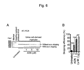
gene as a selection
marker.
[0055]
Date Recue/Date Received 2021-06-23
CA 03124779 2021-06-23
[Example 31 Introduction of MY0D1 into urine-derived cells
The urine-derived cells were plated onto a culture dish or plate (e.g., 3,000
to 5,000
cells/cm2), cultured in a growth medium, and infected with the MY0D1 virus
vector using
polybrene or the like (e.g., after 24 hours) to introduce MY0D1 into the urine-
derived cells.
Thus, the introducing step was performed. After a given period of infection,
puromycin was
added to the medium, culture was conducted for several days, and MY0D1-
positive urine-
derived cells were then selected.
[0056]
[Example 41 Promotion of induction of urine-cell derived myotubes via exposure
to low
molecular compound
The MY0D1-positive urine-derived cells were plated onto a collagen-coated
culture
dish or plate and cultured in a differentiation medium supplemented with
doxycycline (e.g., 1
[tg/m1) (the medium containing high-glucose-containing DMEM with GlutaMAX-I
(Thermo
Fisher Scientific; 10569-010), 5% horse serum, ITS Liquid Media Supplement
(Sigma; 13146),
and 1 [tg/ml doxycycline) to induce the myotubes. Whether or not muscular
differentiation
could be promoted with the addition of a low molecular compound in a compound
library
(Sigma; 5990043-EPI1) to the differentiation medium was examined. The low
molecular
compound was added at a final concentration of 0.1, 1, or 10 [EM. Myotube
induction was
evaluated by immunocytochemistry and Western blotting.
[0057]
For immunocytochemistry, cultured cells were washed in PBS, fixed in 4%
paraformaldehyde, and then incubated with the addition of 0.1% Triton-X at
room temperature
for 10 minutes. The anti-myosin heavy chain antibody (1:50, R&D, Minneapolis,
U.S.A.;
MAB4470) and the anti-dystrophin antibody (1:30, Novocastra, Newcastle, UK;
NCL-DYS1)
were used as primary antibodies, and Alexa Fluor 546 goat anti-mouse IgG (H+L)
(1:300,
Invitrogen; A11003) was used as a secondary antibody. Nuclear staining was
performed with
the use of Hoechst 33342. An image was obtained using a fluorescence
microscope (BZ-9000
or BZ-X800, KEYENCE, Osaka, Japan) and analyzed using the BZ-X Analyzer
(KEYENCE).
[0058]
21
Date Recue/Date Received 2021-06-23
CA 03124779 2021-06-23
As a result, it was founded that a degree of muscular differentiation
evaluated in
terms of the myosin heavy chain protein expression level by
immunocytochemistry was
enhanced to a significant extent with the addition of the epigenetic
regulatory compound(s) to
the differentiation medium (Figs. 3 and 4). In particular, effects of a
histone methyltransferase
inhibitor, 3-deazaneplanocin A hydrochloride (hereafter, referred to as
"DZNep"), were found
to be high. Also, effects of histone methyltransferase inhibitors (GSK343,
SGC707, furamidine
dihydrochloride, UNC2327, E7438, and MI-2 (menin-MLL inhibitor)), histone
demethylase
inhibitors (IOX 1 and GSK-J1), histone deacetylase (HDAC) inhibitors
(VORINOSTAT,
LMK-235, CAY10603, and BRD73954), SIRT2 inhibitor (SirReal 2), and PARP
inhibitor
(EB47) were observed. In Fig. 3, a horizontal axis represents a type of a
compound added, and
a vertical axis represents an area of a myosin heavy chain-positive region
determined by
immunocytochemistry. Fig. 3A shows the results obtained with the use of a 1 uM
low-
molecular compound, and Fig. 3B shows the results obtained with the use of a
10 uM low-
molecular compound. Statistical analysis was performed by a Kruskal-Wallis
test at a
significance level of p <0.05. "*," "**," and "***" indicate p < 0.05, p <
0.01, and p < 0.001,
respectively.
[0059]
The effects of DZNep on promoting myotube induction were also examined by
Western blotting. Specifically, Western blotting was performed in the manner
described below.
The cells were lysed in a RIPA buffer (Thermo Fisher Scientific; 89900)
containing a protease
inhibitor (Roche, Indianapolis, IN, U.S.A.; 04693116001), the cell lysate was
centrifuged at
4 C and 14,000x g for 15 minutes, and the supernatant was then recovered. The
total protein
concentration was measured using the BCA protein assay kit (Thermo Fisher
Scientific; 23227),
denaturation was performed using NuPAGE LDS Sample Buffer (Thermo Fisher
Scientific;
NP0007), SDS-PAGE was performed on 3% to 8% NuPAGE Novex Tris-Acetate Gel
(Invitrogen; EA03785B0X), and the resultant was transferred onto a PVDF
membrane
(Millipore, Billerica, MA, U.S.A.; IPVH304F0). The antibody reaction was
conducted by using,
as primary antibodies, rabbit anti-dystrophin antibody (1:500, Abeam,
Cambridge, UK;
ab15277), mouse anti-myosin heavy chain antibody (1:200, R&D, Minneapolis,
U.S.A.;
MAB4470), and mouse anti-a-tubulin antibody (1:1000, Sigma; T6199) and, as a
secondary
22
Date Recue/Date Received 2021-06-23
CA 03124779 2021-06-23
antibody, Histofine Simple Stain MAX-PO (1:100, NICHIREI BIOSCIENCE INC.,
Tokyo,
Japan; 424151). After the antibody reaction, a band of interest was detected
using the ECL
Prime Western Blotting Detection Reagent (GE Healthcare, UK; RPN2232).
[0060]
Fig. 5A shows the results of Western blotting, and Fig. 5B shows graphs
showing
relative intensities of band signals. As shown in Fig. 5, both the myosin
heavy chain and
dystrophin were found to be expressed at high levels and the myotubes were
found to have been
induced in the presence of DZNep by Western blotting performed with the use of
the myotubes
induced from urine-derived cells obtained from 4 healthy subjects. Thus, an
epigenetic
regulatory compound containing DZNep was found to have effects on promoting
induction of
the myotubes from the urine-derived cells transduced with the MY0D1. Thus, the
exposing
step was performed, and the preparing step was completed.
[0061]
[Example 51 In vitro test of an agent used for exon skipping therapy using the
myotubes induced
from urine-derived cells obtained from a DMD patient
The myotubes induced from the urine-derived cells obtained from a DMD patient
(i.e., the urine-cell derived myotubes) was subjected to the experiment
described below in order
to examine as to whether or not the therapeutic effects of an agent used for
exon skipping
therapy; i.e., an antisense oligonucleotide (AON), could be tested. A urine
sample was obtained
from a DMD patient with exon 45 deletion in the DMD gene, and the myotubes
were induced
from the urine-derived cells in the manner described in Examples 1 to 3. Seven
days after the
induction of muscular differentiation, the culture medium was replaced with a
differentiation
medium containing an agent used for exon skipping therapy (AON) and a 6 uM
endo-porter
(Gene Tools, Philomath, OR, U.S.A.). In addition, the medium was replaced with
a medium
consisting of a differentiation medium 3 days thereafter, and cells were
recovered 14 days after
the induction of muscular differentiation. The AON described in detail in
Wilton, S. D. et al.,
Mol. Ther., 15, 1288-1296, 2007 was used herein. Thus, the applying step was
performed.
[0062]
The exon skipping efficiency was examined by RT-PCR in the manner described
below. At the outset, total RNA was recovered using the RNeasy kit (Qiagen,
Hilden,
23
Date Recue/Date Received 2021-06-23
CA 03124779 2021-06-23
Germany), 1 jig of total RNA was reverse-transcribed using the cDNA reverse
transcription
kits (Applied Biosystems, Warrington, UK), and RT-PCR was performed using 1
jil of cDNA
template, 14.9 jil of distilled water, 0.2 jil of a forward primer (10 iM),
0.2 [El of a reverse
primer (10 iM), 1.6 jil of 2.5 mM dNTPs, 2 jil of 10x Ex Taq Buffer, and 0.1
jil of Ex Taq HS
(Takara Bio, Shiga, Japan). The forward primer used was 5'-
GCTCAGGTCGGATTGACATT-
3' (SEQ ID NO: 1), and the reverse primer used was 5'-GGGCAACTCTTCCACCAGTA-3'
(SEQ ID NO: 2). The band of the PCR product was analyzed using MultiNA
(Shimadzu, Kyoto,
Japan) to determine the exon skipping efficiency.
[0063]
Dystrophin protein expression was analyzed by Western blotting in the same
manner
as described in Example 4. Also, the dystrophin protein was observed under a
fluorescence
microscope by immunocytochemistry as with the case of Example 4. Thus, the
detecting step
was performed.
[0064]
Figs. 6 to 8 each show the results of experiments concerning the exon skipping
efficiency. Fig. 6 shows dystrophin gene expression determined by RT-PCR, Fig.
6A shows a
band detected by RT-PCR, and Fig. 6B shows a graph demonstrating the exon
skipping
efficiency determined by quantification of the band shown in Fig. 6A. In Fig.
6A, a band
appearing in a sample obtained from a healthy subject indicates a full-length
dystrophin gene.
In the case "untreated," a band indicating a dystrophin gene with exon 45
deletion indicated by
an arrow with the term "without exon skipping" is observed. In the presence of
an agent used
for exon skipping therapy (AON), expression of the dystrophin gene shorter
than the full-length
is indicated by an arrow with the term "with exon skipping."
[0065]
The exon skipping efficiency was determined in accordance with the following
equation.
Exon skipping efficiency = with exon skipping/ (without exon skipping + with
exon
skipping)
The graph shown in Fig. 6B shows the exon skipping efficiency in terms of the
mean
+ standard error, "***" indicates P <0.001, and "****" indicates P < 0.0001.
24
Date Recue/Date Received 2021-06-23
CA 03124779 2021-06-23
[0066]
Fig. 7 and Fig. 8 show the dystrophin protein expression analyzed by Western
blotting and immunocytochemistry, respectively. Fig. 7A shows the results of
Western blotting,
and Fig. 7B shows the graphs of the dystrophin protein levels prepared based
on Fig. 7A. The
graph shown in Fig. 7B shows the dystrophin protein level relative to a-
tubulin in terms of the
mean + standard error, "*" indicates P < 0.01, "***" indicates P < 0.001, and
"****" indicates
P < 0.0001.
[0067]
Fig. 8 shows the results of immunocytochemistry performed on the urine-cell
derived
myotubes obtained from a DMD patient and a comparison of the untreated sample
and the
sample after exon skipping therapy. Compared with the untreated sample,
dystrophin protein
(red) expression is more clearly observed in the sample after exon skipping
therapy.
[0068]
Thus, the detecting step was performed. As a result, it was found that AON-
dose-
dependent effects of exon skipping therapy could be tested at mRNA and protein
levels.
[0069]
[Example 61 Establishment of a test system that selects a sequence of the
optimal agent used
for exon skipping therapy for particular DMD gene mutation
A urine sample was obtained from a DMD patient with exon 45-54 deletion in the
DMD gene, and the urine-cell derived myotubes were induced. Seven days after
the induction
of muscular differentiation, the culture medium was replaced with a
differentiation medium
containing each antisense oligonucleotides (AON) having different sequences
and a 6 p..M endo-
porter (Gene Tools, Philomath, OR, U.S.A.). In addition, the medium was
replaced with a
medium consisting of a differentiation medium 3 days thereafter, and, 14 days
after the
induction of muscular differentiation, dystrophin protein expression was semi-
quantified by
immunocytochemistry in the same manner as described in Example 4. The AON used
was the
exon-44-skipping agent, and the exon-45-skipping agent, the exon-50-skipping
agent, and the
exon-51-skipping agent were used for control. These AONs are described in
detail in Wilton,
S. D. et al., Mol. Ther., 15, 1288-1296, 2007 for the exon-44-skipping agent
and the exon-45-
Date Recue/Date Received 2021-06-23
CA 03124779 2021-06-23
skipping agent, Wu, B. et al., PLoS One 6, e19906, 2011 for the exon-50-
skipping agent, and
eteplirsen (AVI-4658) was used as the exon-51-skipping agent.
[0070]
Fig. 9 shows the results of experiment.
Fig. 9A shows the results of
immunocytochemistry, and Fig. 9B shows a heat map for semi-quantitative
analysis of
fluorescence-positive regions based on Fig. 9A. Fig. 9C shows the signal
intensity of the
dystrophin protein determined based on Fig. 9B in terms of mean standard
error. The 1-way
ANOVA test is performed (N = 4 to 5), and "****" indicates P < 0.0001.
[0071]
Fig. 9 demonstrates that a frame-shift mutation is modified to in-frame via
exon
skipping and a fluorescence signal of the exon-44-skipping agent, which is
deduced to express
the dystrophin protein, is significantly high. Thus, it is predicted that this
DMD patient would
have satisfactory effects by the treatment using the exon-44-skipping agent.
Thus, the
identifying step is performed.
[0072]
As described above, an agent used for exon skipping therapy can be tested with
the
use of the myotubes induced from the urine-derived cells before a particular
DMD patient is
subjected to actual treatment. This enables selection of a sequence of an
optimal agent used for
exon skipping therapy that is expected to be effective.
[0073]
Sequence Listing Free Text
SEQ ID NOs: 1 and 2: artificial (synthetic oligonucleotides)
26
Date Recue/Date Received 2021-06-23