Note: Descriptions are shown in the official language in which they were submitted.
5~
,~ ~., J
1--
METHOD AND APPARATUS FOR LASER SURGERY
BACKGROUND OF THE INVENTIO~
.
This invention relates to cutting and cauterizing
of body tissue by means of laser energy and in particular, to
a new and improved method and apparatus for lasex surgery
utilizing a YAG laser and an optical fiber for delivery of
the laser energy.
In the past, lasers have been utilized for surgi-
cal procedures. It is known to use the YAG lasex at a
wavelength of 1.06 microns to provide an output to an optical
fiber for surgical cauterizing. However, this device has not
been satisfactory for cutting of tissue. Also, it has been
known to use the CO2 laser in cutting procedures. However,
there are no commercially available fibers that operate
satisfactorily with the CO2 laser, and therefore surgical
uses of the CO2 laser are severely limited.
It has long been known that it is highly desirable
in surgical procedures for the surgeon to be able to easily
cauterize tissue and to make precise tissue cuts during a
surgical procedure in order to reduce bleeding and facilitate
further surgical procedures, as well as to enhance healing.
While this is true for external applications, it is especi-
ally so for surgery deep within the body. It is also desir-
able to be able to achieve these functions with a sinyle
surgical instrument so that the surgeon does not have to
change tools during a procedure.
t75,7
--2--
~ ccordingly, it is an object of the present
invention to provide a new and improved method and apparatus
for laser surgery w~ich permits use of laser energy both for
cutting and for cauterizing, with a single instrument with
the change in function under the direct control of the
surgeon, both outside the body and inside the body.
It is an additional object of the present inven-
tion to provide such a method and apparatus whi.ch can utilize
an optical fiber for delivery of the energy, with the optical
fiber having a small distal end which is readily manipulated
and which can be utilized through an endoscope or other
surgical instrumentat.ion, as desired.
It is an object of the present i.nvention to
provide such a method and apparatus which can utilize conven-
tional laser technology and conventional optical fibertechniques to provide a surgical instrument which is small,
rompact and easily manipulated and which is relatively
inexpensive, whil~ achieving the desired aims of substan-
tially instantaneous changing between cutting and cauteri~ing
functions.
Other objects, advantages, features and results
will more fully appear in the course of the following des-
cription.
SUMMARY OF THE INVENTION
A method of surgery using a YAG laser and an
optical fiber wherein energy from the laser at a wavelength
of 1.06 microns is directed through the optical fiber to the
tissue for cauteriæing of tissue, and energy at a wavelength
of 1.3 microns is directed through the fiber for cutting
tissue.
--3--
The invention also includes laser surgical appara-
tus with either a single YAG laser selectively opera~ing at1.06 and 1.3 microns wavelength or two lasers operating at
1.06 and 1.3 microns, respectively, with the laser output
directed through an optical fiber to the tissue to be
~reated. In its simplest form the apparatus of the invention
comprises a dual wavelength YAG laser selectively operable at
1.06 and 1.3 microns wavelength with means operable by the
surgeon for selecting the desired wavelength and means for
directing the output toward tissue, with or without an
optical fiber.
BRIEF ~ESCRIPTION OF THE DRAWINGS
Fig. 1 is a diagrammatic presentation of a multi-
mode or multiwavelength YAG laser and optical fiber combina-
tion, incorporating the presently preferred embodiment of theinvention;
Fig. 2 is a partial view similar to that of Fig. 1
showing a~ alternative configuration for the distal end of
the optical fiber; and
Fig. 3 is a view similar to that of Fig. 1 showing
an alternative embodiment incorporating two lasers.
DESCRIPTION OF T~IE PREFERRED EMBODIMENTS
.
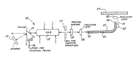
A laser is diagrammatically illustrated in Fig. 1,
more specifically a Nd:Y3A15O12 laser 10, which is usually
referred to as a YAG laser.
In the preferred embodiment illustrated, the
laser l0 includes a YAG rod 12, a hiyh reflecting mirror 13,
a partially reflecting mirror 1~, a pivotally mounted
prism 15 with a lever 16 for pivoting the prism, and an
iris 17 for varying the output aperture. The laser may be
conventional in construction and~ includes a housing with
pumping source, power supply and cooling source (not shown).
s~
--4--
The YAG laser operates at discrete spectral lines and will
function at a number of different wavelengths. The wave-
lengths of interes~ for the present inventlon are at about
1.06 and 1.3 microns. The operating wavelengths for the YAG
laser at room temperature are reported as including 1.0641~
1.319 and 1.338 microns. These are often referred to as
1.06, 1.32 and 1.34, respectively, and the figures 1.06 and
1.3 as used herein are intended to encompass these more
precise figures.
In operation, the prism 15 functions to disperse
radiation passing therethrough, in the conventional manner,
and hence can be used to tune the operation of the laser to a
particular wavelength by appropriately pivoting the prism.
In one alternatlve embodiment, the highly reflecting mirror
15 can be mounted directly on the face of the prism, and in
another alternative embodiment, a diffraction grating may be
utilized in place of the prism and mirror. The iris 17 may
be used for controlling the size of the output aperture, and
may be varied depending upon the operating wavelength
20 desired. It is pre~erred to reduce the aperature when
operating at 1.3 microns as this improves the single mode
I lasing and enhances beam concentration for cutting.
The surgical instrument also includes an optical
fiber 21, preferably a silica fiber, and preferably enclosed
in a protective sheath 22. While a single fiber is pre-
ferred, a bundle of fibers may be utilized, and~ the word"fiber" as used herein covers both the single fiber and the
fiber bundle. In the embodiment of Fig. 1, the laser lO and
the proximal end 23 of the fiber 21 are mounted so that the
output of the laser through the partial mirror 14 is directed
onto the proximal end 23, preferably with a focusing lens 2~.
The distal end 26 of the fiber 21 is designed to be readily
manipulatable for positioning at the tissue 27 to be treated.
In one embodiment, the surgeon may manually grasp the fiber
5 7
adjacen~ the distal end; in another embodiment, the fiber may
be mounted in an endoscope and be remotely manipulated, in
the conventional endoscope manner.
While the presently preferred embodiment of the
surgical instrument and the surgical process utilizes an
optical fiber as the element for delivering the laser output
to the worksite, the optical fiber may be omitted, with the
laser output being delivered by conventional output delivery
devices. Also the distal end of the instrument, with or
without the optical fiber, may be manipulated by an articu-
lated arm and guided by the surgeon using a surgical micro
scope for viewing the worksite.
Another focusing lens 30 may be mounted at the
distal end 26 for improved concentration of energy for the
cutting procedure. As shown in Fig. 1, the lens 30 may be
mounted on a shaft 31 which is carried in a bracket 32. The
shaft 30 may rotate in the bracket to move the lens from a
position in the optical path as shown in Fig. 1, to a posi-
tion out of the optical path as shown in Fig. 2. Alter-
natively, the shaft 31 may translate in the bracket 32,moving the lens 30 toward and away from the distal end 26 of
the fiber 210 When the lens 30 is incorporated in the
instrument, the lens will be used to focus the energy to a
small spot for the cutting operation, and will be moved to
have a larger spot or an unfocused spot for the cauterizing
procedure.
It has been found that YAG laser energy~ delivered
through an optical fiber at a wavelength of about 1.06
microns is especially suited for surgical cauterizing of
tissue because of its relatively deep penetration into tissue
(several Inm or more), but is not satisfactory for cutting at
tissue. Hence, the 1.06 wavelength laser has not been
suitable for many surgical procedures. Now it has been found
5~
--6--
that the same YAG laser wi~h optical fiber when operating at
a wavelength oE about 1.3 microns is especially suitable for
cutting tissue because its beam is absorbed in a fraction of
a mm of tissue, while no~ being satis~actory for cauterizing
tissue.
Hence, when in use for cauterizing tissue, the
instrument of the present invention is operated at 1.06
microns by appropriate adjustment of the prism lS, and of the
lens 30 when used t and the surgeon manipulates the distal end
of the fiber as desired. ~owever when a cutting procedure is
desired, the surgeon changes the operating wavelength of the
Y~G laser to 1.3 microns, typically by a foot operated lever
or manual switch, and immediately proceeds with the cutting
of tissue using the same instrument without requiring any
hand or head movements.
During use, the instrument is quickly switched
back and forth between the 1.06 and the 1.3 micron wave-
length, without requiring release of or movement of the
distal end, thereby greatly enhancing the ease and quickness
of the surgical procedure.
By using the process and apparatus of this inven-
tion for switching between coagulation or cauterizing and
cutting, the surgeon can approach true "bloodless" surgery.
The inven~ion also provides a no touch technique for both
cutting and coagulating. The instrument substantially
reduces the degree and amount of manual mechanical dexterity
required to complete the procedure, by using directed light
energy instead of an interposing mechanical device, i.e. a
scapel. Removal of a lesion or destruction of the lesion can
be accomplished through a tiny incision using a narxow beam
of light through a deep channel or hole too small for mechan-
ical devices or hands, to coagulate the lesion and then
remove it via converting the lesion to smoke, with the
procedure therefore being less invasive.
J5~
--7--
Fig. l shows the distal end 26 of the fiber 21
directed to the surface of a portion of tissue 27, for
cutting at the surface. Fig. 2 shows the distal end 26
positioned at a cut 33 in the tissue 27 for coagulation at
the cut.
An alternative embodiment of the invention is
shown in Fig. 3, which embodiment uses two YAG lasers 35, 36,
with the laser 35 operating at 1.06 microns and the laser 36
operating at 1.3 microns wavelength. In one configuration
the lasers 35, 36 are fixed in the instrument housing. The
output of one laser, here the laser 35, is directed to the
lens 24 through a beam splitter 37, and the output of the
other laser is reflected through the lens 24 by the beam
splitter 37. The respective lasers are turned on and off as
desired by a wavelength selecting switch 38 operable by the
surgeon.