Note: Descriptions are shown in the official language in which they were submitted.
~63447
--1--
NONINVASIVE METHOD FOR
MEASURING AND MONITORING
INTRAPLEURAL PRESSURE IN NEWBORNS
Technical Field
-
The present invention generally relates to a
method for noninvasively monitoring intrapleural pressure
of a newborn subject and, more particularly, to a method
for qualitatively and quantitatively measuring the
intrapleural pressure of a newborn in a noninvasive man-
ner.
The invention additionally relates to a method
for noninvasively detecting the presence of, and for dif-
10 ferentiating between, central and obstructive apneas andhypopneas in newborn subjects.
.~ . .
1263447
Background Art
Various techniques and apparatus are known for
measuring, and for detecting changes in, intrapleural
pressure of a human subject or other living organism.
Present techniques are invasive in requiring that at least
5 some portion of a device be inserted into the body as, for
example, directly into the pleural or adjacent esophageal
space. The most commonly used such device, the esophageal
balloon, is based upon the known close correspondence
between esophageal and intrapleural pressures. Although
10 the esophageal balloon is perhaps the least objectionable
of the available invasive devices with respect to subject
discomfort and acceptance, it cannot be used for
intrapleural pressure monitoring over extended periods of
time and it is particularly difficult to successfully
15 maintain in situ when dealing with newborn subjects. In
addition, it has recently been suggested that distortion
of the rib cage of preterm and term infants during
breathing invalidates the use of esophageal pressure as an
estimate of mean pleural pressure.
Maintaining a monitoring probe or device on or
about an infant's body is frequently difficult to achieve
and applicant is unaware of any prior art techique or
device that can be conveniently and noninvasively utilized
to continuously monitor inteapleural pressure in a new-
25 born.
Prior art methods and apparatus known for moni-
toring a newborn to detect the presence of apnea may be
designed to sense body movements, as by a detector under-
lying the subject's mattress during sleep'. This method
30 has inherent unreliability since any normal change in body
position during sleep can introduce substantial variations
into the signal generated by the sensor in response to
respiration-related body movements. In addition, such
techniques fail to provide a reliable means by which the
35 apnea can be readily differentiated as being either cen-
tral or obstructive in origin. Immediate differentiation
is important in that while central apnea is often treated
.''' ~
.. ', ~
12634A7
--3--
with drugs, obstructive apnea requires mechanical relief
of airway obstruction and, in either event, the appropri-
ate procedure or countermeasure must be introduced at once
to restore normal respication. Even a relatively short
5 delay required to separately diagnose the problem can
prove fatal to the newborn. External monitoring devices
worn around the rib cage and abdomen, such as
magnetimeters, respiratory inductive plethysmographs, and
impedance pneumographs may detect central apneas but if
10 respiratory efforts are minimal (i.e. where changes in
intrapleural pressure are small) then obstructive apneas
may not be diagnosed. Further, if external monitors such
as the impedance pneumograph are worn over only the rib
cage and abdomen, obstructive apneas will not be diaqnosed
15 if respiratory efforts are present. Finally, devices sens-
ing air flow at the nose, such as thermocouples, thermis-
tors and C02 analyzers will detect apneas but fail to dif-
ferentiate central from the obstructive types.
126~447
--4--
Disclosure of the 1 vention
The present invention is based upon my discovery
that the cranial bones of a newborn subject move relative
to each other during respiration as a result of a pressure
wave transmitted from the pleural space through the
cerebrospinal fluid and great veins to the cranial cavity.
Detection and monitoring of these movements produces a
waveform which closely resembles intrapleural pressure.
According to one aspect of the invention there is
provided a method of non-invasively monitoring and detect-
ing changes in intrapleural pressure of a newborn subject,
comprising: mounting an external means for detecting
movement across at least two adjacently-proximate cranial
bones of the subject to detect relative movement between
said bones; and generating a signal indicative of changes
in the relative positions of said cranial bones detected
by said means, changes in said signal being indicative of
changes in intrapleural pressure of the subject.
According to a first preferred form of the
invention, a surface inductive plethysmographic transducer
-- in the form of a length of wire formed in the shape of
a loop -- is secured on the newborn's head across at least
two adjacently-proximate cranial bones to detect relative
movement between the bones. Preferably, the transducer is
placed over the sagittal suture or the anterior or
occipital fontanels. Relative movement of the cranial
bones results in proportional movement of the portion of
the loop lying thereon, and correspondingly proportional
changes in the cross sectional area of the loop. This, in
turn, causes a proportional change in the self-inductance
of the loop. By incorporating the inductive loop as the
inductance element in a variable frequency LC oscillator,
changes in loop self-inductance result in proportional
changes in the oscillator output signal frequency, which
may then be converted to a corresponding voltage signal
suitable for display on an output device or further
conditioned as desired for the particular application.
:
- ,
1263447
-4a-
It is well known that the cranial bones of an
infant remain separated until at least nine months of age;
thus, the anterior fontanel of the newborn infant may
become effectively closed at any time from nine to
approximately eighteen months. Consequently, detection
and measurement of relative movement between adjacently-
proximate cranial bones in accordance with the invention
should be possible for at least approximately nine months
after birth.
Changes in the output signal of the oscillator
are correspondingly indicative of changes in intrapleural
pressure of the newborn subject. That signal can be
i263447
calibrated to pcovide a measucement of actual intcapleural
pcessure of the subject by momentarily manually occluding
the nose of the subject, measucing the subject's airway
pressure while the nose is occluded -- as by Placing a
5 catheter within the nose just distal to the obstruction
or in the mouth -- and adjustinq the signal to equal the
airway pressure measuced with the nose occluded. This
calibration technique makes use of the known fact that,
except ducing crying, newborns are obligatory nasal
10 bceathers. Alternatively, if the baby is intubated with
an endotracheal tube because of the need for mechanical
respiration assistance, the endotcacheal tube can be
momentarily occluded and the same calibration pcocedure
car r ied out.
One particularly significant application of the
method of the pcesent invention lies in the detection of
apnea in the newbocn subject, and in diffecentiating
between centcal and obstcuctive apnea. By simultaneously
monitocing both ccanial bone movement (as indicative of
20 changes in intcapleural pressure) and changes in the
velocity of air at the nostrils of the subject's nose (as
indicative of inhalation to and exhalation from the
lungs), apneas can be detected and differentiated as to
type oc ocigin. In a first preferred method of the apnea-detection and
25 differentiation invention, a surface inductive plethysmographic transducer
is secured on the newborn's head across at least two
adjacently-pcoximate cranial bones to detect relative
movement therebetween and to genecate a corresponding sig-
nal indicative of changes in intrapleural pcessure, and a
30 nasal oxygen cannula is secured at the subject's nose and
an output indicative of tidal breathing pressure is gener-
ated. By continuously monitoring these two genecated sig-
nals, central and obstructive apneas can be reliably
detected and differentiated. A substantial absence of
35 changes in both the signals is indicative of the presence
of central apnea, ~hereas a substantial absence of changes
in the signal generated from the nasal cannula, when
accompanied by continuing changes in the output genecated
~:.
.
~263447
from the transducer monitoring movements of the cranial
bones, is indicative o~ the presence of obstructive apnea.
The methods in accordance with the present
invention will be more fully apparent from the following
5 detailed description and annexed drawings of the presently
preferred embodiments thereof.
. .
1263447
-- 7 --
srief Description of the Drawings
In the drawings, wherein like reference numerals
denote similar elements throughout the several views:
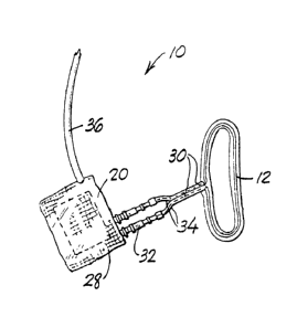
FIG. 1 is a perspective view showing a surface
inductive plethysmographic transducer for use in monitoring
intrapleural pressure in accordance with a first preferred
method of the invention;
FIG. 2 is a diagrammatic representation of pre-
ferred circuitry for measuring the inductance of the con-
ductive loop used in the first preferred method of theinvention;
FIG. 3 is a plan view of the skull of a newborn
human subject showing the various cranial bones and a pre-
ferred placement of the first preferred transducer of FIG. 1
on the skull;
FIG. 4 is a frontal view of a newborn subject
showing a nasal cannula in situ in accordance with the
inventive method for detecting and differentiating apneas;
FIG. 5 is a diagrammatic representation of a
system for automatically detecting and differentiating
apneas and hypopneas in accordance with the invention.
. . .
'
'
~2~i3447
--8--
Best ~ode For Carryinq Out the Invention
The present invention is based upon my discovery
that the cranial bones of a newborn subject move relative
to each other during respiration. This movement is a
5 result of a pressure wave transmitted from the pleural
space through the cerebrospinal fluid and great veins
through the cranial cavity. I have further found that
monitoring of these cranial bone movements and qeneration
of a signal corresponding to the bone movements produces a
10 waveform which closely resembles intrapleural pressure.
Thus, the relative movements of the cranial bones of a
newborn are a function of intrapleural pressure.
The spacing between and relative moveability of
adjacently-proximate cranial bones in the newborn exists
15 until at least three months and usually until approxi-
mately nine months of age after which the cranial bones
gradually beco~e fused to one another. The anterior fon~
tanel, for example, may become effectively closed between
nine and eighteen months of age. As a consequence, detec-
20 tion of cranial bone movement during respiration inaccordance with the invention will generally be attainable
until the newborn is at least nine months old.
The term "adjacently-proximate", as used in the
present descriptiion and disclosure, is intended to indi-
25cate a relationship between adjacently-disposed cranial
bones wherein these adjacent bones have con~ronting or
opposed edges. ~s the cranial bones move with respiration
of the newborn subject, their confronting or opposed or
"adjacently-proximate" edges correspondingly move relative
30 to each other and, as a consequence, a movement-sensing
transducer lying atop and across these opposed edges will
detect that movement. Moreover, the confronting edges may
be spaced apart by any amount normally present in the cra-
nial bone arrangement of a newborn and, so long as the
35 transducer overlies or is supported by at least two por-
tions of each of the adjacently-proximate bones, their
relative movements with respiration will be detectable in
accordance with the invention.
' :
1263447
The preferred apparatus utilized to pcactice the
method of the invention is shown in FIG. 1 and there
designated by the general reference numeral lO.
Transducer lO is a surface inductive pleth~smograph dis-
5 closed in my Canadian patent no. 1,216,635 issuedon January 13, 1987, and includes a pref-
erably insulated length of conductive wire 12 formed in
the shape of a loop. It is known that the self-inductance
of a conductive loop is proportional to the cross sec-
10 tional area enclosed by the loop. ~ccordingly, a changein the cross sectional area enclosed by the loop causes a
proportional change in the loop inductance.
In the practice of the invention, relative
movement of adjacently-proximate cranial bones is moni-
15 tored by disposing the conductive loop on the subject suchthat the loop lies on the surface of the subject's head
across at least two of the adjacently-proximate cranial
bones. The presently preferred placement of loop 12 over
the sagittal suture 14 is shown in the FIG. 3; other pre-
20 ferred locations for the loop include the anteriot fon-
tanel 16 and the occipital fontanel 1~. Nevertheless,
since all of the cranial bones have been found to move
relative to each other during respiration, it is within
the scope and contemplation of the invention that conduc-
25 tive loop 12 be operatively positioned across at least anytwo adjacently-proximate cranial bones of the newborn sub-
ject. The loop may be secured in place as by taping~ or
by employing an adhesive preparation such as a collodian
solution, althouqh care should be taken not to inhibit
30movement of the loop upon movement of the cranial bones
being monitored.
Relative movement of the ccanial bones causes
the loop portion lying atop the bones to move. This, in
turn, produces a change in the cross sectional area
35enclosed by the loop and hence in the self-inductance of
the loop. By monitoring these self-inductance changes in
the manner more fully explained below, an indication of
the extent of relative bone movement is pcovided.
lZ63447
Referring now to FIG. 2, a presently preferred circuit
for converting the self-inductance of loop 12 to a suitable
electrical signal is diagrammatically illustrated. As
shown, the circuit includes a variable frequency LC
oscillator circuit 20 connected to the ends of conductive
loop 12. The resonant frequency of oscillator circuit 20
is determined by an internal capacitor and the inductance
of loop 12. This frequency may, for example, be centered
about 400,000 Hz, and will vary as the cross sectional
area enclosed by the loop varies. Because the relative
cranial bone movements being measured are quite small, it
is essential that the oscillator circuit have sufficient
sensitivity and gain to measure these movements.
A suitable oscillator circuit 20 is disclosed in my
Canadian patent no. 1,216,635, and other appropriate
circuits will suggest themselves and be apparent to those
skilled in the art once this description is known.
The output signal from oscillator circuit 20 is
preferably converted to a suitable voltage signal by a
demodulating circuit 22. The output of demodulator 22 is
an analog voltage signal having an amplitude which varies
in response to changes in the frequency of oscillator 20.
An exemplary demodulator circuit 22 is disclosed in my
co-pending United States application Serial No. 317,418,
and other suitable circuits will be apparent to those
skilled in the art once this description is known.
The output signal from demodulator 22 may be displayed
on one or more suitable output devices, shown by way of
example in FIG. 2 as a CRT terminal 24 and strip chart
recorder 26.
As further seen in FIG 1, oscillator circuit 20 may be
incorporated in a module 28 for securement to the
subject's head adjacent conductive loop 12. A pair of
insulated wire leads 30 interconnect the oscillator module
28 to loop 12, the leads 30 preferably being joined
together in the vicinity of the loop. Connectors 32 in
wire leads 30 may be employed to accomodate separation of
1263447
loop 12 from the oscillatoc module 28. It will be
apparent that the inductance element of oscillatoc 20 is
determined not only by loop 12 but also by leads 30, and
that movement of the leads would thecefore be disadvanta-
5 geous as it would affect the oscillation fcequency ofoscillator 20. Accocdingly, leads 30 are preferably sub-
stantially rigid, oc ace secured against movement in some
other fashion. The leads 30 in FIG l are rendered rigid
by the combination of the substantially riqid wire sheaths
lO 34 and connectors 32. ~ cable 36 extending from module 28
connects the oscillator circuit 20 to the demodulator 22
and connected output devices 24 and 26.
As the subject exhales and inhales, changes in
intrapleural pressure cause correspondinq relative
15movements of the cranial bones. Thus, movements of the
cranial bones result in changes in the cross sectional
area enclosed by loop 12, and hence in the inductance of
the loop, as should be evident. Changes in the loop
inductance are monitored by the oscillatoc ciccuitcy and
20demodulatoc ciccuit 22, and are displayed on the CRT 24
and/or strip chart recorder 26. Consequently, the voltage
signal, as so displayed, is an analog wavefocm indicative
of the extent of relative movement of the cranial bones
over which loop 12 lies. Changes in the monitoced signal
25have been found to be a linear function of coccesponding
changes in intcapleucal pressure Oe the subject.
The signal wavefocm output fcom demodulatoc
circuit 22 may be calibcated ducing an initial calibcation
procedure, whereby subsequent ceadings will indicate actu-
30 al intcapleucal pcessuce of the newbocn subject. A pces-
ently pcefecced calibcation technique makes use of the
known fact that newbocns are obliqatory nasal breathecs.
In accordance with this pcoceduce, the subject's nose is
manually occluded, as by pinching the nose to momentarily
35 close the nasal passages. Since in a closed respiratocy
system, changes of airway (nasal) pressuce equal changes
of intcapleural pressuce, the subject's airway pressure is
then measured by any conventional means while the nose is
1263447
occluded. ~he output signal from demodulatoc circuit 22
is next adjusted to equal the airway pressure measured
during the occlusion maneuver, following which the occlu-
sion of the nose is cemoved to enable the resumption of
5 natural breathing. The output of demodulator circuit 22
will thereafter remain calibrated to intrapleural pressure
for natural breathing o~ the subject.
~ lthough the preferred focm Oe the invention
utilizes the disclosed surface inductive plethysmographic
10 transdùcer 10, it should be recognized and unde~stood that
monitoring of the respiration-caused movements of the cra-
nial bones may alternatively be carried out with any
transducer sensitive enough to detect the relatively small
displacements involved. For example, the cranial bone
lS movements can be m~nitoced by an inductive
plethysmographic ban~ oc a mercucy in silastic strain
gauge placed encirclingly about the skull. Other devices,
placed over the ccanial bones, such as linear displacement
transducecs, pneumatic pcessuce transducecs and optical
20 tcansducecs, by way of example, can also be employed in
accocdance with the method of the invention. Use of the
disclosed surface inductive plethysmographic transducer
10, however, is particularly advantageous in being rela-
tively small and light weiqht, and in its consequent abil-
25 ity to be maintained in situ secured on the infant's headdurinq extended periods Oe time -- as ducing sleep.
Furthermore, operative use of transducec 10 in no mannec
interferes with cespiration or with normal body movement
of the subject.
The disclosed inventive method for
non-invasively monitoring intrapleural pressure in a new-
born subject by detection of cranial bone movements finds
pacticulac application in the detection and differ-
entiation of central and obstructive apneas. Apneas are
35considered to be a majoc cause Oe sudden infant death syn-
drome which most often occucs ducing the first thcee
months of liee. The suc~ace inductive plethysmoqcaphic
tcansducer 10 is ideally suited as a celiable and easily
~ ,
.. :., ,. ,, _
1263447
-13-
applied device which may be readily maintained in situ on
the infant's head during extended sleep periods to contin-
uously monitor intrapleural pressure changes. In
accordance with this particular application of the disclo-
5 sed invention, the subject's nasal tidal volume is moni-
tored concurrently with the use of an airflow transducer
10 as will hereina~ter be understood.
Central apnea is commonly defined as the cessa-
tion of neural impulses from the respiratory center of the
10 brain whereby the respiratory muscles fail to contract; in
essence, the subject "forgets" how to breath. This con-
dition is accordingly characterized by a lack of fluc-
tuations in intrapleural pressure and, since the respira-
tory muscles are rendered inoperative so that no inspira-
15 tion or expiration occurs, tidal volume is essentiallyzero.
In obstructive apnea, the respiratory muscles
are instructed and continue to regularly contract.
However, an obstruction of the upper airways (the
20 oro-nasal-pharyngeal region) prevents ventilation of the
lungs. Under these circumstances, tidal volume is again
zero but, in contrast to central apnea, wide fluctuations
in intraesophageal and intrapleural pressure occur as
respiratory efforts from muscle contractions continue to
25 take place.
Thus, by monitoring both changes in intrapleural
pressure and changes in tidal (breathing) volume, the
presence of apneas can be detected and di~ferentiated as
to type or origin. Early recognition of the onset of
30 apnea is essential so that an effective treatment or cor-
rective plan can be instituted as rapidly as possible.
- Since central apnea is most often treated with drugs,
whereas obstructive apnea requires physical removal of the
obstruction as by an operation or the like, early and
35 immediate diffeeentiation as to the origin of the apnea
present is likewise critical.
The inventive technique herein disclosed for
identifying the presence and origin of apnea is based at
~.,~. .
. ,, . .. ., .............................................................. _
~63~4~
-14-
least in part on known observations that the newborn is an
obligatory nasal breather, except when crying, and that
even during episodes of crying a portion of the bceath
passes through the nose. Changes in tidal volume are
accordingly monitored with a device that noninvasively
detects tidal flow at the infant's nostrils. Although any
conventionally known device for such purpose can be uti-
lized -- such, ~or example, as a thermistor, a thermocou-
ple or a CO2 sensor (as by mass spectrometry or infrared
lO analyzer techniques) -- it is presently preferred that a
pediatric nasal oxygen cannula be employed.
Referring now to FIG. 4, a conventional nasal
cannula 38 is shown in situ on the newborn subject.
Cannula 38 includes a pair of probes 40 which partially
15 project into the subject's respective nostrils. If
desired, an alternative cannula configuration (not shown)
having but a single nostril projecting probe can be uti-
lized to minimize possible infant discomfort or as the
medical condition of the subject might warrant.
Cannula 38 is secured to the patient as, for
example, by the use of members 42 that hook about the ears
and a cooperating elastic band 44 that encircles the read
portion of the head. Alternative methods of securement
for mounting cannula 38, as by taping or utilizing an
25 adhesive collodian solution or the like, are also within
the contemplation of the invention.
Nasal cannula 38 monitors the infant's breathing
by qualitatively measuring pressure changes at the
nostrils. A pressure transducer 46 receives the output of
30 cannula 38 and converts the pressure changes to tidal vol-
ume changes by integrating the square root of the measured
pressuees as well known in the art. Transducer 46 may
conveniently generate a voltage signal, the amplitude of
which varies coerespondingly with changes in the pressure
35 detected by this arrangement. Standard output devices
such as CRT terminal 48 and strip chart recorder 50
receive the signal output of transducer 46 and display a
waveform corresponding to tidal volume.
.,_
~;263~
-15-
The inventive method for detecting and
differentiating central and obstructive apneas should now
be understood. The output of movement trans~ucer 10 (in
conjunction with demodulator 22) -- which directly indi-
cates relative movement Oe the cranial bones with respira-
tion -- is a varying waveform at least qualitatively re-
presentative of the subject's intrapleural pressure. l~
desired, that output can be calibrated to quantitatively
correspond to actual intrapleural pressure, although cali-
10 bration is not essential in utilizing the apnea detectionand differentiation technique herein disclosed.
Concurrent with the monitoring of cranial bone
movements by transducer 10, changes in nasal tidal volume
are detected utilizing cannula 38 and associated trans-
15 ducer 46. The output signal displayed on the devices 48,50 is a waveform at least qualitatively representative of
changes in the subject's tidal volume with respiration.
If desired, the output Oe cannula 38 can be calibrated by
any known method -- as, for example, by the technique dis-
20 closed by Guyatt et al (American Review of Respiratory
Disease 1982, Volume 126, pp.434-438) -- although once
again, a quantitative measurement of tidal volume is not
essential to effective use Oe the inventive apnea detec-
tion and differentiation method.
By observing the output of each o~ the moni-
toring devices -- i.e. transducer 10 and cannula 38 --
during natural or normal respiration, a control or stand-
ard value of each of the signals is next obtained. These
control values are defined as the average di~ferences
30 between the qualitative trough-to-peak values of each of
the waveforms over a period of perhaps ten to twenty
respiratory cycles or breaths. Put another way, the con-
trol value of the output signal erom each detector is the
average qualitative change in signal level during normal
35 or natural respiration.
Monitoring of the two output signals or wave-
forms can be readily interpreted to indicate the onset and
origin of apnea present. An absence of changes in the
~.
~263~47
-16-
outputs of both cranial bone movement transducer 10 and
nasal cannula 38 is indicative of the presence of central
apnea. On the other hand, a sudden absence of changes in
the output from nasal cannula 38, when accompanied by con-
tinuing changes in the signal generated by transducer 10,is indicative of the presence of obstructive apnea. The
rapidity with which central and obstructive apnea can be
reliably diagnosed in accordance with the disclosed method
enables appropriate effective countermeasures to be
10 immediately carried out with corresponding life saving
benefits to the newborn subject.
Those skilled in the art will recognize that
initial establishment of control values for the output
signals generated from transducer 10 and nasal cannula 38
15 are not essential to the detection of central and
obstructive apnea in accordance with the invention. In
the former instance, both output waveforms become substan-
tially flat, while in the latter the signal of transducer
10 continues to vary while the output generated from can-
20nula 38 is substantially flat; the control values areunnecessary to each determination. ~onetheless,
establishment of control values eor the output signals
enables intermediate conditions -- such as central and
obstructive hypopneas -- to be diagnosed as well. Central
25 hypopnea is characterized by a proportional diminution or
decrease in both intrapleural pressure and tidal volume.
Thus, an observation of predetermined partial decreases in
the output signals generated from transducer lO and can-
nula 38 is suggestive of the presence of central hypopnea.
30 Obstructive hypopnea, or partial upper airway obstruction,
might correspondingly be suspected if nasal tidal volume
predeterminately decreases from its control value while
the amplitude of cranial bone movements persists or
increases. Additional advantageous uses for the developed
35control values during continuous monitoring of
intrapleural pressure and tidal volume in accordance with
the invention will suggest themselves to those skilled in
the relevant art.
~, ,
lZfi3447
-17-
FIG. 5 diagrammatically illustrates an auto~ated
system for detecting and differentiating centcal and
obstructive apneas and hypopneas. Automated system 52
incoeporates a microprocessor-based controller 54 into
which the output signals from each transducer are input.
As shown, the out~ut signal generated by the combination
of cranial bone movement transducer 10 and demodulator 22,
and the output signal generated by the combination of
nasal cannula 38 and pressure transducer 46, are inter-
lO preted automatically by controller 54 which includes aplurality of alarm indicators 56, 58, 60 and 62. If
desired, modulator 22 and/or pressure transducer 46 may be
incorporated within controller 54, or they may be
externally provided as depicted in FIG. 5. Similarly,
15 additional visual alarm indicators, as well as supplemen-
tal auditory alarms, may be incorporated in controller 54.
The waveforms input to the controller may also be dis-
played on suitable output devices such as CRT 54 and/or
strip chart recorder 66.
The structural details and construction of con-
troller 54 are deemed to be within the skill of an
individual technically competent in the relevant art once
this description is known and understood. As such, no spe-
cific details are herein disc1Osed and any suitable con-
25 troller arrangement for carrying out the apnea detection
and differentiation technique of the invention may be
employed.
Preferably, controller 54 incorporates a plural-
ity of visual and/or auditory alarms, each corresponding
30 to the diagnosed presence of a particular apnea or hypop-
nea condition. Thus, by way of example, alarms 56 and 58
may correspond to conditions indicative of central and
obstructive apnea, respectively, while indicators 60 and
62 may respectively signal the possible presence of cen-
35 tral and obstructive hypopnea. It is also contemplatedthat controller 54 may include provisions for
user-adjustment of the maximum and/or minimum relative
signal levels at which each alarm will be activated
substantially by the apparatus.
.... .
~263447
-18-
It should also be recognized and understood that
although the methods of the present invention have been
disclosed and described herein for use with a newborn
human subject, they are equally applicable for use with
5 any newborn animal or organism having initial separated
cranial bones. Thus, the foregoing description is meant
to be by way of example only, and not as a limitation of
- the scope of the inventive methods and techniques.
There has accordingly been disclosed herein a
lO novel method for measuring intrapleural pressure in new-
born subjects, and an application of that method to a
novel technique for detecting and differentiating the
presence of central and obstructive apneas and hypopneas.
While there have been shown and described and pointed out
15 fundamental novel features of the invention as applied to
preferred embodiments thereof, it will be understood that
various omissions and substitutions and changes in the
details of the disclosed methods, and in the form and
details and operation of the disclosed devices, may be
20 made by those skilled in the art without departing from
the spirit of the invention. It is the intention, there-
foce, to be limited only as indicated by the scope of the
claims appended hereto.
.
,