Note: Descriptions are shown in the official language in which they were submitted.
: ~J3~3'73L~
MEDIC~L IASER PRCBE WITEI ~'EEDBAC~C MONIr~)R
PIELD OF INVENTION
This invention pertains to the use of laserQ in medicine
and, more particularly, to the controllable firing of medical
lasers when performing surgery.
DISCUSSION OF PRIO~ ART
Currently, medical and surgical laser output is guided
visually by the o~erator. The eye, or an optical vlewing
device, i~ used to identify the ~reatment area and ~ire the
laser. A major problem is that imperfect vi~ualization of
the treatment are3 lead~ to poor aim of the laser and con~e-
quently ~o damage of healthy tissue adjacent ~o the treatment
area~ When the laser device is accurately almed, it ~ ~till
di~flcult for the operator to know precisely the amount of
laser energy to be delivered to destroy the treat~ent area
without damaging the underlying tissue. Oversupply of laser
enexgy may lead to an irreversible destruction of healthy tis~ue
around the treatment site. This destruction can lead to side
effects and compllcations from the procedure. Undersupply of
laser energy may lead to inadequate destruction of the treatment
--1--
3~ 7~ ~
1 area and a therapeutic failure. Furthenmore, the problem is
complicated because of the diversity of tissue types that are
potential lesions
U S. patent 4,438,765 teaches the use of a laser surgical
device wherein the controlling of the ~iring of the laser Ls
by a motion detector to ensure that there is no movement of,
say, the eyeball when the laser is fire~ for retinal fusion.
U. S patent 4,316,467 teaches the use of a laser in
removing naturally pigmented tissue from the skin. The firing
of the laser i~s controlled by the color of the treatment area
sensed by a photodetector. Both of these patents are basically
concerned with the use of a laser in the surglcal treatment
of external body surfaces.
In order to enhance the visualization of the treatment
area, there have been developed certaln dyes which can
selectlvely stain the diseased tissue. The difference in the
optical property of the stained tissue and the unstained
healthy tissue lmproves the visuallzation of the treatment area.
U. S. patent 4,336,809 is typical of the teaching of a photo-
radiation method for tumor enhancement with hematoporphyrln dye,
wherein the dyed leslon site ls bathed wlth radlatlon of a
particular wavelength to cause it to fluoresce
When dealing with lesion sites withln the body cavity,
it is necessary to deliver the laser energy internally to the
leslon slte. U S patent 3,858,577 and U. S. patent 4,273,109
--2-
3~ 37 ~
1 are typlcal of fiberoptic light del:Lvery systems.
In splte of all of this existing technology, there is
still not available a laser surgical ~system which is capable
of performing laser surgery wlthin the body cavity such that
the laser effec~s are automatically monitored to control the
output of the laser and to termin~te its operation before
there ls a destruction of healthy tissue flround the treatment
site.
BRIEF SUMMARY OF THE INVENTION
It is, accordingly, a general object of the invention to
provide an improved method of delivering laser energy for the
treatment of an area within a body cavity.
It is a more specific ob~ect of the invention to provide
such laser energy only as long as the treatment area shows the
need for such laser energy and to terminate the application
of the laser energy when the malignant portion of the treatment
has been destroyed
Accordingly, with this aspect of the invention, there is
provided a method for radiatinga treatment area within a body
cavl~y by introducing an elongated flexible radiation transfer
conduit into the body cavity until the distal en(l thereof is
operatively opposite the treatment area.
There ls a particular optical characteristic of the
treatment area which is photoelectrically sensed and as long
" t
,
1 as this optlcal characteristic is sensed, laser pulses are
periodically transmLtted into the proximal end of the conduit
for transfer to the distal end and the treatment site
In accordance with a feature of the inventLon, if the
treatment area has no inherent optical properties which are
sufficiently different from the surrounding healthy tlssue,
then before the treatment begins there is introduced into the
treatment area a reagent which will cause the treatment area
to be characterlstlcally stained so that when the photoelectric
sensing takes place the optical properties of the character-
istic stainlng will be sensed,
According to a speclfic feature of the invention, there
is contemplated a method of destroying atheromatous plaque
within an artery of a patient comprising the steps of initially
sdministering to the patient a non-toxic atheroma-enhancing
reagent which causes plaque to have a characteristic optical
property when illuminated with a given radiation. Thereaf~er,
a catheter system including fiberoptical cable means is intro-
duced into the artery such that the distal end thereof is
operatively opposite the plaque site. There is then introduced
into the proximal end of the fiberoptical cable the given
radiation, When plaque is iiluminated with the given radiation,
a characteristic optical property is sense~ at the proximal end.
~ 7~
..
' '
l There is then fed via the cable means from the proxlmal end
to the distal end periodically occurring laser pulses until the
characteristic optical property is no longer sensed.
In order to implement the method of the invention, there
is contemplated a laser system having a ~iberoptlcal bundle
with a central optLcal diagnostic means, a receiving fiber-
optical array means annularly disposed about the dlagnostic
means and a treatment fiber optical arra~ means annularly
disposed about the receiving fiberoptical array means. A
treatment laser source is connected to one end of the treatment
fiberoptlcal array means, a diagnostic light source is con-
nected to a corresponding end of the central fiberoptical
diagnostic means, and a radiation detector is connected to the
corresponding end of the receiving optical fiber means.
Another implementation of the method of the invention
contemplates the use of a single optical fiber which transmits
time multiplexed radiation A further implementa~ion contem-
plates two fibers, one handling diagnostlc :-adiation and the
other multiplexed treatment and sensed radiation
BRIEF DESCRIPTION OF THE DRAWINGS
_
Other ob~ects, the features and advantages of the
inventlon will be apparent from the follow1ng detailed des-
cription when read with the accompanying drawir.g in which:
~ 7 ~
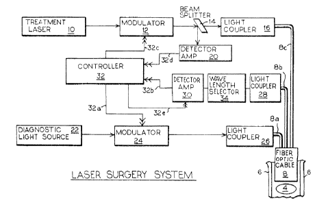
1 Figure 1 is a block diagram of a laser system utilizing
the invention;
Figure 2 is a schematic longitudinal section of the flber
optic cable of the system of Fig. l;
Figure 3 is a cross-sectlonal view of said cable along
the lines III-III of Fig 2;
FLgure 4 is a block dlagram of a portion of the laser
system of Figure 1 ~tilizing a single opt:ical fiber; and
Figure 5 is a block diagram of a portion of the laser
system of Figure 1 utillzing two optlcal fibers.
DETAILED DESCRIPTION OF THE PREFERRED EMBODIMENTS OF THE
INVENTION
The invention will be described utilzin~ the example of
the destruction of an atheromatous plaque 4 from the artery 6
of Fig. l. Initially, the patient is administered a dose of
a dye to enhance the contrast be~ween the treatment site
(plsque) and the healthy surrounding tissue. A ~ypical dye
is tetracycline which has the property of fluorescing when
radiated wlth an ultraviolet light This dye has a special
property of accumulating within the plaque relative to normal
he~lthy tissue. Therefore, a predetermined time after the
administration of the dye, the fiberoptic cable 8 is inserted
into the artery with the distal end thereof opposite ~he
. J ~
1 treatment site. The optlcal cable 8 ln a first embodiment
(see also Figs. 2 and 3) includes a central optical fiber 8a
coupled to the output of light coupler 26, an annular array
of optical fibers 8b surrounding the central fiber 8a con-
nected to the input of light coupler 28, and an outer annular
array of cables of fibers 8c is coupled to the output of
light coupler 16. Light coupler 26, of conventional design,
receives light from the diagnostic light source 22 via the
optical modulator 24. Diagnostic light source 22 for the
present example would be a source of ultraviolet light. If
other dyes were used, then appropriate light sources for those
dyes would be selected. The llght indicated by the single
arrowhead line feeds the input of modulator 24 whose output
is fed to the input of coupler 26. The modulator 24 can be
of a conventional opto-accoustic modulator or an electro-
mechanical shutter which passes or blocks the light in response
to an electrical signal from controller 32 (note all electrical
signal lines show double arrowheads). Thus, the presence or
absence of a signal on line 32a from the controller 32 can
close or open the light path between diagnostic light source
22 and light coupler 26.
The proxi~al end of array 8b feeds light into the light
coupler 28 whose output is fed into the wavelength selector
34 which selects light corresponding to the predetermined
characteristic wavelength to be detected. In turn, the selected
--7--
i,37~
.
1 light feeds detector-amplifier 30 whose output is fed via signal
lead 32b to the controller 32. The detector-amplif~er 30 can
be the combination of, for example, a photodiode wh~ch drives
a transistor amplifier, a photomultiplier, or in and of itself
can be a phototransistor. Thus, whenever light is received
from array 8b, a slgnal will be transmitted to controller 32
The detector/amplifier 30 is controlled by signals on line
32e from controller 32.
Treatment laser 10 will transmit light to modulator 12
which is controlled by signals on line 32c from controller 32.
The controlled light from modulator 12 i.s fed to a conventional
beam spliter 14 with a portion of the light being deflected
to detector/amplifier 20 and the remaining light passing to the
input of light coupler 16. The output o~ light coupler 16 is
fed to the proximal end of optical fiber array 8c. Beam
splitter 14 also feds part of the beam to detector/amplifier
20 which in turn feeds a signal on line 32d to controller 32
which provides feedback sensing of the laser output to ensure
constancy of ~he amplitude of the laser output over time.
In operation, after the dye has been inserted and the
cable 8 is in place, the diagnostic light source 22 passes
light via modulator 24, coupler 26 and array 8a to the treatment
site 4. The plaque in the treatment site will fluoresce and
the fluorescence will be picked up by the array 8b and fed to
the light coupler 28 and, thence, to the wavelength selector 34.
--8--
3 ~.~ 3
! .
l The output from the wavelength selector 34, corresponding to
the characteristic fluorescent emission of tetracycline,
is fed to the detector/amplifier 30 which in response will emit
a signal on line 32b to controller 32. The controller 32 in
response thereto will send a slgnal on line 32c to open the
modulator 12 to emit laser energy of a pred~termined power and
wavelength for a set time interval. Accordingly, a pulse of
light from treatment laser lO wLll be fed via the beam splitter,
11ght coupler 16 and the array 8c to the treatment area 4.
Because light reflected from the treatment area can be very
great during the time of the laser pulse, controller 32 via
line 32e feeds a signal to detector/amplifier 30 to turn off
the detector for a pre~etermined time interval. This signal
can also be fed to modulator 24 to prevent the radiation of
ultraviolet light during the laser pulse. Controller 32 then
switches signals on lines 32c and 32e at the pre~etermined timing
delays such that the laser output is blocked and the fluorescent
light can again be sensed from the treatment site. If ~he
fluorescence is then detected indicating that plaque is still
present, the detector 30 will send a signal to controller 32
which, again, switches the signals on the lines 32e and 32c,
lnitiating another laser pulse~ This sequence continues until
no fluorescence is detected indicating that all plaque has been
de~troyed. At that time, no signal is fed to controller 32
and no further laser pulse ls ~enerate~. In this way, us~n~ the
~ 3~ ~
1 probe-and-fire technique of the invention, the possibility of
destroying healthy tlssue is minimlzed. The controller 32 in
its simplest form can dispense with the use of detector-
amplifler 20 and can merely be a monostable ~evice whlch is
momentarily triggered on a pulse from line 32b and then reverts
to its rest state. The paraphase output of thls device can be
connected via appropriate amplifiers to lines 32a, 32b and 32e
To facilitate the positioning of the laser catheter within
narrow tortuous pathways a single flexible optical fiber 8'
(or small diameter bundle)is used(See Fig. 4~ instead of the
multibundle cable 8 of Fig 1 More particularly, the light
couplers 26,28 and 16 connected to their assoc~ ted bundles
8a, 8b and 8c are replaced by a slngle multiple-wavelength couple
MWFC which optlcally couples multiple-wavelength beam splitter
MWBS to single optical fiber 8'. Multi.ple-wavelength beam
splitter MWBS recelves laser light from beam spli~ter 14(Fig
1) along a given incident angle path and diagnostic light from
dulator 24(Fig 1) along another given incident angle path
and transmits such received llght via a port along a common
transmlt-receive pa~h to multiple-wavelength light coupler MWLC
Furthermore, radiation from the treatment site 4 is fed from
multiple-wavelength coupler MWLC via the common transmlt-
. receive path into the port of multiple-wavelength beam splitter
MWBS This light is emitted therefrom to wavelength fitter 34
: via a further path having an angle different from the two glven
-10 -
~ ~ ~;;37~1~
.~
1 lincident path angles. Because of the nature of the multiple-
wavelength beam splitter ~WBS it may be possible to delete fitter
34 and feed detector/amplifier 30 directly from the beam splitter.
The so-modified system operates in the same manner as
the system of Fig. 1. In Fig. 5 the fiber optical configuration
is modified to a dual fiber configuration. This configuration
may put less demands on the multiple-wavelength beam splitter
~S and may permit more diagnostic light to reach the treatment
site 4. In this embodiment a single fiber or bundle 8a'
is connected to light coupler 26(Fig. 1). The fiber 8" or
narrow diameter cable is connected to multiple-wavelength light
coupler M~C which is optically-coupled via a common transmit-
receive path to the ports of the multiple-wavelength beam
splitter M~BS. Laser light is received along a given incident
angle path from beam splitter 14(Fig. 1) and fluorescent light
from coupler ~IFC is fed from multiple-wavelength beam splitter
~WBS via an output optical path having a different angle to wave-
length selector 34(Fig. 1). As with the embodiment of Fig. 4
fitter 34 may be omitted.
Operation of the system utilizing the embodiment of Fig.
5 is the same as the other embodiments.
In addition to tetracycline it has been found that the dye
Nile Red(9-di-ethylamino-5h~benzo[et] phenoxazine-5-one) shows
l excellent results with plaque.
I
1l While only a limited number of embodiments of the invention
~has been shown and described in detail, there will now be
obvious to those skilled in the art many modifications and
variations satisfying many or all of the objects and features
~of the invention without departing from the spirit thereof.
l l
~, 1 - 1 1 -
1 For example, while only the treatment of plaque has been described
the invention can be used as a treatment of other diseases such
as tumors(cancer), stones ln urinary tract and gall bladder as
well as prostate obstructions. In addi~ion, depending on the
nature of the treatment site, the appropriate dye is selected
to enhance the contrast between normal tissue and malignant
tissue. When the treatment site is a tumor, one can successfully
use hematoporphyrin or its derivatives. In some cases, inherent
differences in optical properties between the treatment site
and the surrounding healthy tissue may eliminate the need for a
dye Again, depending on the treatment site and the dyes
involved, the diagnostic llght can be ultraviolet, infrared,
white light, etc~ Furtherm~re, again depending on the treatment
site, the laser source can take many forms such as argon, Nd-yag,
carbon dioxide, tunable dye, and exclmer lasers with pulse or
continuous output. The choice of the diagnostic light source
is predicated on the optical characteristlcs of the dye and/or
the treatment site. However, the choice of the coherent light
source for the treatment laser does not have to match the absor-
ption peak of the dye. The treatment laser can be any wavelength
that destroys the dlseased treatment site Normally, there is
a risk that this light will also destroy healthy tissue.
However~ the possibility does not exist since once the diseased
treatmen~ site is removed, the means for triggering the laser
pulse is also removed.
-12-
.
1 The fiberoptic cable can be coupled with catheter
designs which include, but are not limlted to, such features
as endoscopy, balloon devices, steerable guiding systems7
multiple lumens for lnfus~on and suctloning, ultrasonlc guidance,
monitoring or ablation, pressure and temperature monitoring
and catheter centerlng devices.
-13-
