Note: Descriptions are shown in the official language in which they were submitted.
5a~
The invention relates to closed system urinary
drainage bags. More ~peci~ically, the invention rela~es to a
device placed in the outlet tube for dispensing an agent for
controlling and preventing the retrograde migration of
pathogenic microorganisms into the urinary drainage bag.
BACRGROUND OF THE INVENTIO~
Urine drainage bags are routinely used by
post-opeeative patients as well as those with urological
disorders. Because of injury to the Gpinal cord, paraplegic
patients a~e unable to control bladder activity and
consequently must continuously use a catheter.
In pLac~ice, the pa~ient is ca~heterized and the
catheter then connected to the drainage bag through a length of
plastic tubing. The bag i8 normally supported below the level
o~ the pa~ient either from a bed rail or other support and the
urine drains by gravity from the patient through the catheter,
the tubing and then into a bag via a drip chamber. The bag may
be emptied from time to time by means of an outlet tube which
is normally closed to prevent leakage. The tube may discharge
its contents into any convenient receptacle and ~hen the outlet
tube is clamped and the bag reused for the same patient.
The catheterized urinary track i6 one of the most
common sites of hospital-acquired infection and in fact
accounts for almost thirty percent of such infections.
5~6
Signi~icant imp~ovements in the p~evention of catheter
associated infection has been by use o~ wha~ are known as
closed ste~ile drainage systems. Despite the~e advance~, still
ove~ twenty ~e~cent of pati.ent~ with indwelling catheters
continue ~o acquire urinary infections. See C7aribaldi et al,
New England J. Med., 29l: 215-219, 1974. Urine collection bags
must be emptied at frequent intervals usually at least once
every shift and the ~emoval of bacterially contamina-ted urine
can lead to the sp~ead of urine infection. I~ is even possible
for a patient in the same ward or room shared with a
catheterized patient to acquire the infection. In order to
minimize cross-contamination, the collected urine must be
maintained in ste~ile condition during the collection period,
even when the ueine has a high bacte~ial count when it enters
the drainage bag.
Despite ~he use of the most careful aseptic
techniques, almost fifty percent of catheterized ~atients
develop an infection when the catheter is in place fo~
twenty-four hours and approximately ninety-eight ~ercent or
even more develop an infection of after four days of use of
such cathete~s. This of course i8 quite harmful to the patient
and subjects them to the risk of cystiti6 and life threatening
seeticemia. ~rch. Internal ~ed., Vol. 110: 703-711 (1962) and
Lancet, Vol. 1, 310-312 (1960). The above noted infections
occur due to many circum~tances. These include prolonged use
of indwelling Foley-type catheters which are often accompanied
-- 2
~2~ 3~
by absence of steeile insertion and maintenance ~echniques;
ha~ing the cathete~ connected to clean but not ~terilized
drainage collection container~; and other~. The p~esence of
urinary ~athogens in the containe~ which multi~ly and enter the
urinary trac~ through the ascending ca~heter which is a major
~athway of infection is quite important. Various attempts have
been made to reduce the mig~ation of bacteria through the
closed sy~tem including the bag, the drip chamber and the
tubing connected to the catheter.
The patent to Jinkens et al, No. 3,332,442 employs a
connector between a catheter and a urine drainage bag for
preventing movement of bacteria from the bag to the patient.
The three patents of Langston et al, No~. 4,236,517 4,193,403;
and 4,241,733 show a dispensing de~ice which releases
parafoLmaldehyae to control the multiplication of pathogens and
prevent migration in catheters. Shaffer U.S. Pa~ent No.
4,233,263, teaches adding of hydrogan peroxide solution
periodically to a urine bag for prevention of bacterial
growth. Attempts have been made to provide a one way inlet
valve into the urine bag to prevent upward migration. See, for
example, U.S. Patent No. 3,312,221 and 4,232,677.
Other attempt~ have been made to treat the catheter
itself with an microbicidal substance. Note U.S. Patent No.
3,598,127 and the Shepard et al Patent Nos. 3,566,~74 and
3,695,921 which relate l:o an antibiotic material in a
hydrophilic catheter coating.
~2~5~
U.S. Paten~ No. ~,417,~92 descLibe~ a method of
releasing an microbicidal gubstanca by means of a frangible
capsule which is inser~ed into the outlet drainage tube. The
capsule must be broken by a nurse or other medical pe~sonnel in
order to relea~e the active agent.
SUMMARY OF THE INV~NTION
It is well known that indwelling catheterization of
patients, can lead to serious infections. In nor~al use of the
conventional urinary drainage bag, transmission of infection
via the outlet d~ainage tube is of major concern.
In this invention, a microbicidal tube or plug is
inserted into a section of the outlet tube. The microbicidal
tube is usually made by one of three processes. 1) ~ porous
material, such as polypropylene is impregnated with at least
one microbicidal agent. It is then coated with a hydrophilic
polymer which in response to contact with urine swells, causing
the leaching out of the microbicidal substance. 2) A porous
material, such as high density polyethylene is impregnated with
a hydropholic polymer and at least one microbicidal agent.
3) The microbicidal tube is made by compounding and
co-extruding a polymer, such as silicone, with at least one
microbicidal agent, and then coated with a hydrophilic
polymer. By appropriate combination of active agants,
virtually all pathogens can be effectively eliminated and
-- 4 --
~evented flom ente~ing the urinaLy bag and further into the
catheter.
The present invention is superior in many way~ to -the
methods of prior a~t. It i~ a pas~ive dispensing systeTn, thus
eliminating the need for human pa~ticipation, ~uch a~ is
necessary, for example, in the breaking of an antibiotic
containing capsule. It is a self-activating system which
responds to the presence of body fluids, in this case, urine.
The microbicidal substances are released in a timed sequence
for an extended period of time, thus creating an effective
barrier against migration of infectlous organisms into the
catheter.
The microcidal tube is easy to prepare u~ing readily
available materials and microbicidal sub6tances. The prolonged
effectivene~s of the invention obviates the need for frequent
draining of bags, thus 6aving on nurse'6 time. The passive,
self-actuating relea6e, likewise i6 a labor-saving aspect of
the present invention. Since the need for human handling is
substantially reduced, there i6 le6s of a chance for infection
due to such contact.
BRIEF DESCRIPTION OF TH~ DRAWING~
These and other objects and advantage6 of the
invention will become apparent upon reading the following
detailed desc~iption and upon referring to the drawings in
~s~
which:
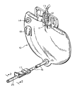
FIGURE L is a ~e~pec~ive view o~ a conventional urine
drainage bag and showing the outle~ tube in broken apart
fashion:
FIGURE 2 is an enlarged cross-section o~ the outlet
tube taken along the lines 2-2 of FIGURE 1: and
FIGURE 3 is a perspective view of a typical microcidal
tube used in the outlet tube.
While the invention will be described in conjunction
with an example embodiment, it will be understood that it i8
not intended to limit the invention to such embodiment. On the
contrary, it iB intended to cover all alternatives,
modifications and equivalents as may be included within the
spirit and scope of the invention as deined by the appended
claims.
DETAI ED DESC~IPTION OF THE INVENTI~N
In the following description, similar features in the
drawings have been given similar reference numerals.
Referring now to the drawings, a conventional closed
system urine drainage bag is shown generally at lO and is
formed by peripherally heat ~ealing or otherwise securing a
pair of flat vinyl or PVC ~heets. The bag is provided with an
inlet 11 adjacent the top thereof fo~ reception of a
conventional drip chamber 12 and its associated tubing 13 which
-- 6
n~
connect6 to a catheter which in turn i~ inserted in the
urethral canal of the patient. ~n air vent and bacterial
filter 14 is conventionally provided on one ace of the bag.
The bag al~o includes a drain 15 terminating in an
outlet tube or conduit 16 which may be for~ned of latex or any
other suitable material, and which may be clamped off when not
in use in a well known manner by means of the spring pinch
clamp o~ valve 17 which is received about the outlet tube. The
free end of the outlet is received in a protective housing 18
heat sealed to one face o~ the urine drainage bag.
Part of the outlet tube 16 houses a microcidal tube
25. The microcidal tube i8 typically 1 1/2 inches in length,
5.5 mm in internal diameter (I.D.), 8.5 mm in outer diameter
(O.D.) and i6 70% void. It is hollow in~ide in order to permit
unimpeded urine ~low. Obviously, the geometry and dimension of
the microcidal tube may be varied over wide limits yet still
function as a microbial barrier while permitting urine flow.
In general, the microbicidal tube i~ prepared by
impregnating porous polymeric material with at least one
microbicidal agent. Various kinds of polymeric materials can
be used, but they must be crystalline and have a high melting
point which will allow them to withstand exposure to body fluid
temperatures without ~oftening. U~ually the tube i8 made by
one of three methods. 1) ~ poLous material, such as
polypropylene is impregnated with at least one microbicidal
agent. It is then coated with a hydrophilic polymer which in
~54~36
reseonse to contact with urine ~wells, causing the leaching out
of ~he microbicidal substance. Z) A porous matecial, such as
high density polyethylene is impreynated with a hydro~holic
polymer and at least one microbicidal agent. 3) The
microbicidal tube is made by compounding and co-extruding a
polymer, 6uch as silicone, with at least one microbicidal
agent, and then coated with a hydrophilic polymer.
The solu~ion of ~he additives and the polymeric
material is allowed to react for a euitable length of time in
the presence of solvent or solven~s. The usual solvents in the
preparation of the microcidal tube are ethanol and dimethyl
sulfoxide. ~t the end o~ the reaction time, the microbicidal
tube is dried by conventional method and sterilized with
ethylene oxide (ET0).
Thç seecif ic antimicrobial substance to be used is
left to the choice of the manufacturer, however such sub6tance
must readily be compounded into polymers or adhere to the
porous polymeric material which in turn will ab60rb the
substance. The biocidal additives can be selected from a very
large group of commerically available antibiotics, drugs,
antiseetics, e~c.. Some exam~les of the common active agents
that can be incor~orated into the microcidal tube are:
penicillin, tetracycline, triclosan, nalidixic acid,
sulfamylon, amphotericin B, nonfloxacin, haloprogin,
gentamicin, chlorhexidine, clotrimazol, tolnaftate, polymyxin,
parachlorometaxylenol, pyrithione, hexachlorophene,
1%6S~
nitrofura20ne, nit~ofuLantoin, chloLixin and many other.
Microbicidal agents may be incorporated either singly or in
various combinations.
The microcidal ~ube may be inserted in~ide the outlet
~ube during normal manufacturing conditions and there will be
no loss of the biocidal activity ~ince the ~ubstance does not
become releasad until it comes in contact with urine. The
microcidal tube is ef~ective for at least two weeks. During
thi~ time, i~ continues to release in a timed sequence, the
microbicidal ~gents, thus creating an effective barrier against
upward movement and multiplication of organisms and the
subsequent infection of the urinary tract.
The amount of drug released will depend on a number of
factors, 6uch as for example, the specific biocidal agent u6ed,
the length oE time it is desired to release the drug, the
dosage that is to be administered in a specific time, etc
The dosage can be controlled by varying the concentrations (or
amounts~ of the drug(s) and hydrophlic polymer and physical
parameters such as pore si~e and shape of the support polymer
used.
There are a number of important advantages that the
microbicidal tube offers over the apparatus and method~ of
prior art.
Thus, the prolonged effectivene~s of ~he microbicidal
tube (two wee~s at least) saves on nurses' time, for the bag
need not be drained as often as it has to be, using prior art
~ g _
~2~X~
apparatus. The passive nature of the sustained drug release,
activated upon contact wi~h urine, likewise ~aves on the
nurse's time, fo~ the~e is no necessity for human
participa~ion. Moreover, this is likewise a more reliable and
certain method of controlling and preventing infection~ then
methods requlring periodic handling of apparatus. Such
periodic manipulation is frequently delayed or entirely
disregarded. ~dditionally, ~he less human handling that is
involved, the less i8 there a chance for contamination from the
outside, hence ~he decrease in an opportunity ~or an infection.
The following examples describe the manner and process
of making and using the invention and represent the best mode
contemplated by the inventor, but are not to be construed as
limiting.
The examples and procedures are to be regarded as
illustrative rather than restrictive. Variations and changes
may be made by those skilled in the art without departing ~rom
the spirit of the present invention.
PREPARATION OF MTCROCIDAL TUBE
ExamPle 1
Batchwise compound 85% polypropylene, 10%
hexachlorophene, 4% gentamicin A HCl and 1% clotrimaæole at
196C and, then, extrude it into a tube. A 1.0 inch segment of
-- 10 --
~2EiS~
the tube is dippsd into a solution o~ g6% ethanol and 4% D-3
~olyethylene glycol ~olyurethane (D~3) for 5 seconds, air-dLied
at RT for 10 minutes and oven-dried at 85C for 10 minutes.
Example 2
Batchwise compound B8% polypropylene, 6% triclosan, 4%
chloroxin, and 2% tolna~tate at 198C and then extrude it into
a tube (5.5 mm ID/~.5 mm O~ 1.5 inch seyment of the tube
is dip~ed into a solution containing 96.5% ethanol and 3.5% D-3
for 5 seconds, air-dried at RT for 10 minutes and oven-dried at
52C for 30 minutes.
Example 3
Batchwise compound 91% polythylene, 6% nalidixic acid
and 3% parachlorometaxylenol at 150C to a homogenous
dispersion and extrude it into a tube (6.5 mm ID/8.5 mm OD).
1.0 inch segment of the tube is dipped in a solution cont~ining
97% ethanol and 3% D-3 for 5 seconds, air-dried at RT for 10
min~tes and oven-dried at 78C for 10 minutes.
ExamPle 4
Batchwise compound 89.8% Polyethylene~ 8% zinc
pyrithione and 2.2~ tatracycline HCl at 152C to a homogenous
-- 11 --
1~:6i5~
dispe~sion and extrude it into a tube (6.5 mm I~/8.55 mm OD).
1.5 inch segment of it is ~ipped in a solution o 95% ethanol
and 5% D-3 foc 5 second~, air-dried at ~rr for 10 minutes and
oven-dried at 65C or 10 minutes.
Example 5
A segment of microporou~ poly6ul0ne tube i8 immersed
in a solution of 8B.8% ethanol, 7% gentamicin ~ HCl, 4% D-3
polyethylene glycol polyurethane and 0.2~ zinc pyrithione for
lO minute6. ~t i~ tnen air-dried at room temeera~ure (RT) for
10 minutes and oven-dried at 70C for 15 minutes. The concen-
tration of gentamicin ~ HCl i8 below its saturation point and
could be raised if wanted. Zinc pyrithione, an antifugal
agent, is approximately at it6 oetimum concentration. It could
be replaced by the more soluble ~odium pyrithione.
Ex mple 6
~ segment of microporous polysufone tube is immersed
in a solution of 79.5% ethanol, 11% tetracycline HCl, 5.3%
polymyxin B HCl, 3.5% D-3 polyethylene glycol polyurethane and
0.2% zinc pyrithione ~or lO minutes. It is air-dried at RT or
lO minute6 and oven-dried at 63C for 20 minutes. Both
tetracycline and polymyxin are below their 6aturation point~.
- 12 -
~;~65a~0~;
Example 7
~ segmen~ o~ mic~oporouR high den~ity polyethylene
tube (HDPE; pore ~ize: 50 microns) i8 immersed in a solution of
50% anhydrous acetone, 40% anhydrou~ ethanol, S% gentamicine
HCl, and 5~ paLachlorometaxylenol for 5 minutes. It is
air-dried at RT fo~ 15 minutes, dipped in 96.5% ethanol. 3.5%
D-3 polyethylene glycol polyurethane for 5 seconds, air-dried
at RT for 10 minute~ and oven-dried at 70C for 15 minute~.
Concen- trations o~ D 3, gentamicine and parachlorometaxylenol
could be increased if needed.
Example 8
A ~egment of microporous HDPE is immer~ed in a
solution of 73% ethanol, 10% tetracycline HCl, 10
earachlorometaxylenol, 3.8% D-3, 2~ dimethyl gulfoide (DMS0),
0.6% clotrimazol and 0.6% tolnaftate for 5 minutes. It is then
air-dried at RT for 10 minutes, oven-dried at 55C for 20
minutes and air-dried at RT for 18 hours.
Example 9
~ segment of HDPE tube i8 immerséd in a solution of
41.4~ DMS0, 44.6% acetone, 10% triclosano and 4.3% chloroxine
for 10 minutes. It is air-dried at RT for 15 minutes, dipeed
....
~2~S~6
into 96.5~ ethanol and 3.s% ~-3 for 5 seconds, air-dried at RT
Eor 10 minutes and oven-dried at 550C for 30 minutes. Concen-
tration6 of triclosan and chloroxine could be raised
substantially if needed.
Example 10
~ segment of microporoug polypropylene tube (5.5 mm
ID/8.5 mm 0~; 70% void) is immerged in a solution of 83.5%
anhydrous acetone, 8% chlorhexidine aceta~e, 5.5% triclosan and
3% Hypol 3000 polyurethane for 5 minutes. It is air-dLied at
RT for 5 minutes, ovan-dcied at 52C for 10 minutes and air-
dried at RT ~or 18 hou~s. Concentrationg of chlorhexidine and
triclosan can be increased if needed.
ExamPle 11
~ segment of microporous polypro~ylene tube ig
immersed in a solution containing 50% ethanol, 33~ chloroform,
11% hexachlorophene, 3.8% D-3, 1% nalidixic acid, 0.6% clotri-
mazole and 0.6% tolnaftate for 10 minutes. It is air-dried at
RT for 10 minutes and oven-dried at 72C for 25 minutes.
Concent~ations of clotrimazole and tolnaftate are substantially
below their saturation point6 in the 601vent sy6tem.
- 14 -
~65~
Example 12
A s~gment of microporous polypropylene tube is
immersed in a solution o~ ~6.2% ethanol, Z2% DMS0, 11%
parachlorome~a xylenol, 11% dchlorhexidine diacetate, 4.8%
water, 4.0% D-3 and 1.0% nitrofarazone for 10 minutes. It is
air-dried at RT for 10 minutes, oven-dried at 6BC for 10
minute~ and air-dried at RT for 18 hours. The concentration of
parachlorome~axylenol could be increased if needed.
Example 13
~ segment of microporous polypropylene tube is
immersed in a solution containing 84% ethanol, 7.5%
hyxachlorophane, 5% D-3, 2.5% DMS0, 0.7% H20, 0. 25~o
clotrimazole and 0.05% aminacrine foL 10 minute6. It i~
air-dried at RT for 10 minutes, oven~dried at 78C for 20
minutes and air-dried at RT for 18 hour6.
Example 14
A segment of microporous polypropylene tube is
immersed in a solu~ion of 50.5% ethanol, 31~ acetone, 10%
hexachlorophene, 4.3% D-3, 2.5% DMS0, 1% clotrimazole and 0~5%
H20 for 10 minutes. It is ai.r-dried at RT for 10 minutes,
oven-dried at 78C for 20 minutes, and air-dried at RT for 18
hours.
- 15 -
~2~5~06
Example 15
~ segment of microporous polypropylene ~ube i~
immersed in a solution containing ~.1% ethanol, 31 DMSO, 11%
~riclosan, 9% hexachlorophene, 3.4% D-3, 1.0% clotrimazole, and
0.5% nitrofurayon for 10 minutes. It is air-dried at RT for 15
minutes, oven-dried at 52C for 30 minutes and air-dried at RT
for 18 hours. Conçen~rations of triclosan, hexachloro~hene and
clotrimazole could be increased if needed.
The foregoing preparations of mic~obicidal tubes were
effective in maintaining sterility for 17 to 21 days upon
exposure to urine.
- 16 -