Note: Descriptions are shown in the official language in which they were submitted.
i9755
METHOD AND DEVICE FOR I~ VIVO WETTING DETERMINATIONS
l ~3~ Q~ h~
This invention relates to the testing and
evaluation of eye wetting, and more particularly to
the wetting of contact len~es worn on the eye of a
subject.
It is commonly assumed that the wettability
of a contact lens is dir0ctly related to its comfort,
optical qualities, and wearing qualities. The bare
eye is normally protected by a tear film having a
thickness in the range of S-7 microns. This tear
film is composed primarily of an aqueous saline
solution, with lesser components of lipid and of
mucin, an assortment of glycoproteins. The ~luid
also contains debris, such as dust and sloughed
ep;thelial cell~. The normal precorneal tear film on
the bare eye is quite stable. The aqueous component
may be calculated to thin out to the point of
break-up by pure evaporative processes ov~r a time
interval of 5-10 minutes. The film is refreshed upon
blinking, and the normal interblink interval is
substantially shorter than 5 minutes, so drying-out
is not normally encountered.
By contrast, when a contact lens is placed
on the eye, each blink of the lid refreshes a tear
film on the outer surface of the contact lens, called
the prelens tear film, which is typically less than 1
micron in thickness. Drying o the film to zero
thickness typically occurs in under 10 seconds. A
dry lens can be uncomfortable, sticks to the eye and
lids, and may be expected to display inferior optical
performance due to scattering from uncovered surface
imperfections. In addition, a lens which has been
~.
"' :
.
- ,. ~
~69755
1 subject to repeated drying cycles is likel~ to have
permanent solid deposits built up thereon, which
accelerate subsequent tear film breakup and promote
trauma or infection.
For these reasons, "wettabilîty" of contact
lenses is deemed desirable. Various wetting traits
have been taken by the industry as measures of
wettability. A common measure oE wettability is the
"contact angle" determined by the angle of the
tangential plane of the edge of a fluid drop with the
plane of the lens surface where they meet. This
measurement is generally made at a clean interface of
a solid with a given fluid, and the measurement must
be performed with some delicacy. It is not clear how
a contact angle measurement performed under
laboratory conditions correlates with the contact
angle of tear fluid on an in vivo contact lens in
which the lens has been repeatedly e~posed to mucins,
lipids and other tear film components. One
researcher has reported making in vivo contact angle
measurements using drops of distilled water placed on
the lens. Applicant is not aware of in y~y~ contact
angle measurements using actual tear fluid.
Other approaches to determining in vivo
wettability might involve adding a dye, such as
fluorescein, to the tear fluid to vi~ualize the thin
prelens tear film. However it is desirable to
develop a method or apparatus to measure lens
wettability which directly measures the thickness and
distribution of the prelens tear film, and which does
not introduce extraneous substances, such as dyes or
fluids which might alter the measurements.
' :'
~69755
-- 3 --
1 Accordingly, there is a need for a method
and apparatus for directly determining the
wettability of a contact lens in a manner which
meaningfully relates to the environment in which the
lenses are used.
Summary__nd Obiect of the Invention
It is an object of the invention to provide
a method for evaluating the in ~ ability of a
contact lens worn by a subject to become or remain
wet.
It is another object of the invention to
providP a method for evaluating the wettability in
vivo of different contact lens materials.
It is another object of the invention to
provide a method of evaluating the effect on
wettability of different lens cleaners or eye
treatment fluids.
It is another object of the invention to
provide a method for evaluating the effects of use or
age on the w~ttability of a contact lens.
It is another object of the invention to
provide a device for performing one or more of the
above methods.
These and other objects of the invention are
obtained in a method for evaluating the in vivo
ability of a contact lens to become or remain wet, in
which the method includes the steps of supporting the
subject's head to determine a fi~ed location of the
lens, illuminating the lens with coherent light,
imaging the pre-lens tear film in a manner to form an
interference pattern~ recording in a time sequence
manner the image formed thereby, and determining the
~IL2697~
-- 4
1 tear film thickness by correlating the interference
bands of the recorded image. The spacing of adjacent
bands givas a measure of the tear film thickness
distribution from which the classical contact an~le
may be derived. Other measures, such as the time
interval from blink until the onset of tear film
breakup, or the time until the film evaporates may be
determined, and the initial or average film thickness
may be derived. In addition, the nature and
evolution of localized dry spots may be directly
observed, clarifying the causes of tear film
breakdown on contact lensss. The invention also
contemplates a method for the evaluation of w~tting
characteristics as they are affected by lens
material, lens aging and lens soiling, as well as the
evaluation of solvents, additives and cleaners, for
their effects on wetting characteristics.
~n apparatus for performing the method
include a means for supporting the head so as to
determine a fixed location of the contact lens, a
coherent light source, and a camera focused on the
pre-lens film from a position that images the light
from the source that is specularly reflected from the
front and rear surfaces of the tear film. A film
motion analyzer is used to quickly obtain the
location and separation of interference bands. A
microprocessor computes the tear film thickness at
each band location, and provides a quantitative
measure of lens wetting characteristics.
. .. . . .
.
.: ~. - -
12~9755
-- 5 --
1 Brief Descril~iol~Qf th~ Prawinqs
These and other features will be understood
with reference to the figures, in which
Figure 1 is a block diagram of the steps
involved in the method according the present
invention;
Figure 2 is a diagram showing the theory of
interference band measurements;
Figure 3 is a schematic representation of
the optical apparatus of a preferred embodiment of a
device according to the invention for performing the
method of Figure l;
Figure 4 shows an alternative layout of
optical elements of another device according to the
invention;
Figure 5 shows a representative image of the
pre-lens tear film and interference patterns recorded
: in accordance with the invention;
Figure 5~ is a graph of film thickness; and
Figure 6 is a block diagram of a pr~ferred
apparatus.
Detailed Description
The present invention provides a method and
apparatus for directly measuring the thickness of the
prelens tear film distributed over the surface of a
contact lens and changing over the course of time.
. The invention further contemplates measuring the
layer as it is affected by solvents, cleaners,
treatment fluids, lens materials, and different
environmental or use factors which affect the lens
worn by the user. As applied to the method of the
;~
- ., .
-
; . - .: , . .
- -.. : -' ' : ~ -
:: , :-
-: : .. :. :
: ~ .
;97~5
-- 6 --
1 invention, the evaluation of such solvents,
components, or conditions are included under the
general term "wettability" or "wetting
characteristics", and accordingly as used in this
disclosure and the claims below it is the intention
to describe and claim the clinical or laboratory
practice of the methods and device of the invention
for evaluating all such solvents, lenses, products
and conditions affecting the wetting of a contact
lens.
Returning now to Figure 1 the method of the
invention includes the steps of ~1) illuminating the
lens with coherent light: (~) forming an image of the
prelens tear film by the light specularly reflected
from the pre-lens tear film, ~3) recording the image
including the interference patterns therein, in a
time sequence manner; and (4) determining the
thickness of the prelens tear film by correlating
bands of the recorded interference patterns.
It will be appreciated as a general matter,
that when light is specularly reflected from the
front and rear surfaces of a sufficiently thin film,
the images of the reflected light will form
interference patterns because the variable thickness
of the film is such that the front and rear reflected
light rays interfere constructively or destructively
at diEferent positions. In general, to provide a
meaningful interference pattern, a film must have a
thickness in the range of approximately lf4 to
several whole wavelengths of the incident light, and
must exist in an environment in which sufficient
specular reflections from its upper and lower
surfaces occur. As an aid to understanding the value
of the method used to visualize the dynamic
. .
~ ~ . ,,`' ~ :
"' . :, ',
. ' ' ,.
~ i975~
1 spreadin~, thinning and breakup of the prelens tear
film, basic principles of interference phenomena will
now be discussed in relation to Figure 2. Further
details of the optical theory may be found in optical
textbooks.
The changing colors of a soap bubble, or of
a thin film of oil floating on water, are each common
manifestations of light interference. This
phenomenon is observed when two or more bPams of
light from a common source arrive along different
paths at the same region of space. In each of the
two cases mentioned above, the interfering beams are
those reflected at the two surfaces of a thin film.
In the first case the film is a film of soapy water
in air; in the second case it is a film of oil
betwsen air and water. Under the proper conditions,
similar interference colors, or bands, can be
reflected from the tear film on the surface of the
cornea or a contact lens. As described below, in
accordance with the present invention, such
interference patterns are formed and recorded to give
precise and detailed information of the topography of
the tear (or lipid) layer overlying the cornea or a
contact lens.
The invention is best understood after a
brief discussion of interference phenomona, modeled
using the wave model of light. In general a light
wave is a transverse electromagnetic disturbance
propagated $hrough space. When two or more light
waves cross a point, the amplitude of the disturbance
at that point is the superposition of the amplitudes
of the individual waves. This superposition
principle follows from the linear character of
Maxwell's equations. Generally, the superposition of
' . ..-- .- .: ,,
'' ~ ' :~: ,
, - .. , : . .
6~755
-- 8 --
1 several light waves does not produce an interference
pattern. This is because most light sources used for
observations consist of a large number of
microscopic, uncorrelated sources, each of which is
active for certain short periods of time and
quiescent the rest of the time. Assuming, for the
sake of argument, that during their active periods
all of the sources emit trains of sinusoidal waves of
the same wavelength, the resultant optical
disturbance produced by the source as a whole would
be represented as a sinusoidal function of time,
whose phase and amplitude change whenever one of the
sources goes on or off. Thus the optical
disturbances produced by two such microscopic
sources, however similar they may be, will have a
phase difference that varies rapidly and irregularly
with time. Such a source is said to be incoherent.
The positions of the interference fringes will change
as the phase difference changes. At a given instant
the maximum intensity will occur at a point where a
minimum was present only a short time before, and
vice versa. Since most optical instruments cannot
resolve such rapid and irregular fluctuations of
intensity, the observable result will be a uniform
illumination. In order to observe interference
phenomena, it is necessary to use coherent light
sources, that is, sources whose phase difference
remains constant in time. The only manner in which
this can be practically accomplished is to use a
single light source and its optical image, or to use
two different images of the same source.
According to the present invention,
interference patterns are created by using two images
of the same source. Specifically two interfering
-, '' ' :'
. .
. .
: - .
~ ~i975~
1 beams are used: the reflected beam Erom the Eront
surface of the tear film and the reflected beam from
the rear surface of the tear film. The two beams
originate from the same single light source, and in
fact are two images of it, so the beams satisfy the
requirements of coherence. To observe specular
reflectance, the angle the incoming beam makes with
respect to a direction perpendicular to the tear film
surface must equal the angle of observation. This is
usually stated as "the angle of incidence equals the
angle of reflection.~' Any light seen at other angles
is due to light scattering rom the surface, and such
light does not maintain its uni orm phase
relationships with the incoming beam; thus
interference bands are not seen.
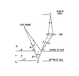
Figure 2 is a diagram of a representative
film 5 having front and rear surfaces 6, 7 and
illuminated by a source S. Light from source S falls
on the film 5, such as the tear layer, and light rays
are reflected by the film to a converging lens L
which forms an image of the film on a focal plane F.
Consider the ray SCD reflected from point C at the
upper surface of the film. Another ray SABCE passes
through the same point C after having been reflected
at point B of the lower film surface. The lens L
brings the two rays together again at point F which
forms an image of point C. To determine the phase
difference, note that the optical path lengths of the
two rays from S to C are Pl = nO SC, and
P2 = nO SA + n(AB ~ BC)
where n is the refractive index of the ilm and nO
that of the medium through which the incident light
ray is travelling (nO = 1 if this medium is air).
Therefore
P2 -Pl - -nO ~SC-SA)+n(AB -~ BC)o
,~
` :. . ` ~ : ',
' .~.. -' ' ~ '
9755
-- 10 --
1 Let t be the thickness of the Eilm, and
and ~' the angles of incidence and refraction of
the ray S~. According to Snell's law for refraction
at interfaces of difEerent reractive illdeX,
nO sin ~ = n sin ~'. Considering that the film
is very thin, CA is very small compared with SA, and
we obtain, to a good approximation,
nO ~SC-SA) = nO CA sin~
= 2tnO tan~ sin~
=2tn(sin2~'/cos~'~ and
n(AB+BC) = 2tnJcos~'.
Therefore,
P2 -Pl = 2tn( -sin2~'/cos~' ~ l/cos~) = 2tn cos~'.
The phase difference corresponding to this
difference in optical path length is
( 3(P2 Pl)/~o where ~0 is the
wavelength in vacuum (or air, in practical terms).
There is, however, an additional phase factor to
consider. When a light ray is reflected from a
surface such that the ray is incident from a media of
lower reractive index than the substrate reflecting
media, the situation is just as described above.
However, if the incident ray is travelling in a media
of higher refractive index than the reflecting
; 25 surface, there is a phase change of 180 degrees, or
radians, between the incident ray and its
reflection. The ray SCD is reflected at the upper
surface of the film, where the index of refraction
changes from nO to n, and since nO is less than n
(i.e., the refractive index of air is essentially
1.0, while that of the tear film is similar to that
of water, where n=1.33), there is no phase change Eor
the reflected ray. The ray SABCE is reflected at the
lower surface, where the index of refraction changes
~ .
'' ' .~ -
. ' -:
,. ` ~
~L~69755
-- 11 --
1 from n to nx, where nx is the refractive index o~
the medium beneath the film layer. If nx is less
than n, there will be a change of phase of ~ upon
reflectance; if nx is greater than n, the case will
be as with the surface ray, and no phase change will
occur. In the case where we are dealing with a tear
film layer (n=1.33~ on a contact lens, the lens will
always have a higher value (for nx) than 1.33. If
the lens is PMMA, nx will be about 1.49; if the
lens is a hydrogel, it will be less, but will still
be at least slightly greater than the tear film, so
no phase change need be considered.
; Thus, the two reflected rays meet at C and
then again at F with a phase difference a given by
a = 2~ (P2 -P~ o~ or
a = 2~ (2tn cos~ 0
In particular, if the lens is so located as
to collect rays that are reflected by the film in a
nearly perpendicular direction, cos ~' is very
close to unity, and the above equation reduces to
a = 2~ ~2tn/~0).
Interference of the two rays at F produces a
maximum of intensity if a is an odd multiple of
2~, that is, if the condition
2tn/~0 = k, k = 0, 1, 2,
is satisfied. Ths interference will produce a
minimum of intensity if a is an even multiple of
~, that is, if
2tn/~0 = k + 1/2, k = 0, 1, 2,
If we let ~ = ~0/n be the wavelength
in the film, we can rewrite these equations as
Interference maxima: t = k(~/2) Equation (A)
Interference minima: t = ~2k +1)~/4 Equation (B)
.
:
~L2~;~75~i
- 12 -
1 From this we can conclude that the
interference maxima occur where the tear film
thickness t is an even multiple of ~/4, and minima
(dark bands) occur where the tear film thickness t is
an odd multiple of ~/4.
With a tear film of varying thickness over
the field of view/ which is illuminated with a
monochromatic light source, a pattern of light and
dark bands, or interference fringes, will be seen.
The lines where the thickness of the tear fllm
satisfy Equation (A) will stand out as lines of
maximum brightness, and those where the thickness of
the film satisfy Equation (B) will appear as dark
lines.
Using a point source of light as
illumination, interference fringes are seen only on
that portion of the tear film that reflects the light
rays from the source into the observing (collectinqj
lens. This portion becomes smaller as the diameter
of the observing lens decreases. However, if the
diameter of the observing lens is sufficiently small
compared with the distance of the lens from the
surface of the tear film one can use a broad source
of light and observe interference patterns from a
wider portion of the tear film. This is because with
a small observing lens diameter, the angle of
reflection ~' is practically the same for all rays
reflected at a given point of the tear film into the
lens, and so, for all pairs or rays that can enter
the lens (L of Fig. 2) cos~' and t are constant and
the phase difference a also has a constant value.
: . .
' ~ ;~ ' :
755
- 13 -
l The "ideal" case shown in Fig. 2 represents
a plane for the reflecting surfaces. In the case of
a contact lens, the surfaces are, of course, sharply
curved. This limits the angles of re~lected rays
that are collected by the observing lens, so the
fiald of view over which the intarference patterns
are seen are much less than if plane surfaces were
involved. Nevertheless, by optimal use and placement
of the light source, subject, and collecting lens, a
field diameter of at least 4 to 5 mm can be observed
at one timeO This seems to be adequate for
evaluating the general characteristics and dynamics
of the tear film as it e~ists on the surface of the
contact lens.
Figure 3 shows a schematic optical set-up
for obs~rvlng interference fringes in the tear film
on a contact lens. This arrangement permits the
illumination and observation of the tear film to be
performed essentially along an a~is orthogonal to the
lens and aligned with the optical axis of the eye.
In this arrangement, the interference fringes remain
sharp over a large area of the contact lens since
cos~ changes quite slowly over the surface of the
lens in the region where ~ equals 0, that is when
the incident light is nearly coaxial with the center
of curvature of a contact lens radius line.
A prototype instrument according to Figure 3
was built using the optics from a conventional
keratometer, suitably modified. In this device a
light source 9 and collimating lens 12 direct light
onto a translucent diffusion screen lO. The image of
screen lO is reflected by mirror 15 having a central
aperture to a converging lens 16 which directs the
light in a cone onto contact lens 8. In this manner,
" ~lX6~:~755
- 14 -
1 contact lens 8 is illuminated essentially along its
central axis. Light reflected from the lens travels
back, essentially along the axis, through the
aperture of mirror 15 and is focused by lens 17 onto
an image plane 18. In one embodiment, the light
source g was a conventional photographic 35mm slide
projector. Heat absorbing glass was used to remove
the infrared light, and a narrow band filter 13
(shown in phantom) having a half band width of
approximately 20nm and centered at ~OOnm was used to
provide a source of known wavelength. The condensing
lens 16 and the focusing lens 17 were chosen to
provide a working distance of approximately 4 to 6
inches between the instrument and the contact lens.
Lens 17 had a focal length of approximately 4 inches,
a numerical aperture of approximately f/4, and was
set up at approximately a 1:1 image to object ratio.
The focal plane 18 was aligned with the focal plane
of a motion picture camera spaced approximately
fifteen inches from the eye. The apparatus of Figure
3 is representative, and variations in the design are
possible.
It will be observed that with the device in
Figure 3 having a diffuse light source, rays
originating from different points of the source will
generally form interference patterns which do not
overlap. The result is an essentially uniform level
of illumination across the plane of observation.
However, if the source is not too extended and if the
rays from the source strike the tear film in
directions not too far from the perpendicular to the
surface of the tear film, then in the plane of the
tear film itself the various systems of interference
fringes are almost exactly coincident. This will not
.' ~ ~' '
,
~9 7
- 15 -
1 happen in any other plane except the plane on which
the converging lens 17 forms a real image of the tear
film. The interference frinyes produced by the
variable thickness of the tear film under these
conditions are said to be localized in the plane of
the tear film. By placing camera 19 at the focal
plane 18, a picture of the interference pattern
representative of tear film thickness is obtained.
It is not necessary that the light souxce be
a diffuse source. One may, for example, use a laser
light source, with an appropriate scanning mechanism,
such as a rotating polygonal mirror. When scanning
with such a source one must be careful to arrange the
scanning optics so as to avoid "burning" caused by
the eye focusing the scanning laser at a fixed
interior point, e.g. on the fundus. When using a
laser it is also desirable to use a beam expander to
provide a beam of coherent light of sufficient
diameter. Neither is it necessary that the light
source be used in connection with the apertured
mirror observation system illustrated in Figure 3.
An alternate arrangement of optical elements
for recording interference patterns formed by the
prelens tear film is shown in Figure 4. This
arrangement includes a first mirror 21 directing the
coherent light beam 22 from source 9 to the contact
lens, and a second mirror 23 placed on the opposing
side of the central axis "0" for receiving the image
24 specularly reflected from the tear film and
directing it to a focussing lens 25 so as to form an
interference pattern in the film plane of camera 19.
The two-mirror arrangement permits illumination and
imaging of the film at the same angle from the
central axis, chosen to be small. Other arrangements
~ .
75 S
- 16 -
1 will occur to those skilled în the art. For
instance, the angles of the mirrors can be changed or
even rapidly tilted to allow a larger area of the
lens sur~ace to be observed. For such a device, when
the source light mirror directs the beam at a point x
on the lens at an angle ~ with respect to the
normal N at that point, the receiving light mirror 23
should be tilted to receive light reflected at such
angle ~. By providing mechanics for causing
mirrors 21, 23 to rotate in synchrony in this manner,
a larger portion of the lens may be imaged.
Figure 5 shows an example of the image 30
formed by lens L S17 of figure 3, or 25 of figure 4)
and recorded by the camera 19 as a single frame. A
representative image includes a diffuse, or
unfocussed image 32 including cornea 32a of the
subject's eye, and a sharper image 34 of the pre-lens
tear film. A central dark region 32b results from
the geometry of the light source and imaging optics
of Figure 3. The image of the pre-lens tear film
includes an interference band pattern 36a, 36b, etc.,
and also includes in sharply delineated focus, such
opaque physical debris as may be present in the tear
film. The dark interference bands 36a....each lie
along a contour of equal depth of the tear film, and
are formed at those contours where the depth of the
tear film is k~/4, where k is an odd integer, as
discussed above in relation to Figure 2.
Figure 5A shows a representative graph 39 of
tear film thickness along a line T', such as line T
of Figure 5, imposed on a recorded interference
pattern (shown in phantom). A dry edge is chosen as
the origin. Each point at which line T' crosses a
dark interference band is indicated by a transverse
line marked on the horizontal a~is. These
...
;: `' . "
~- ,
:ILX~755
- 17 -
1 coordinates may be directly ascertained by
measurement of the filmed image, since the scale is
known. Tear film thickness is indicated in
increments of ~/2, starting at ~/4, along the
vertical axis. Each successive dark band crossed by
line T~ indicates an increment or decrement of film
thickness by ~/2. For purposes of this example, it
is assumed that the bands adjacent to the edge are
representative of increasing thickness. It will be
seen that, with the T' and thickness axes calibrated
in identical units, the slope of the tangent line to
graph 39 is the tangent of the classical contact
angle.
The field of view with the apparatus shown
in figure 3, over which the focus of the image of the
curved prelens film is sharp enough to include
well-defined interference bands is approximately 4-6
mm diameter, and may be extended by appropriate
optics. Thus the device of figure 3 provides a
recorded image which is a topographic map of prelens
tear film depth over a substantial portion of the
lens. By themselves, the interference bands give
only relative, and not absolute, film thickness
information; the spacing of two dark bands gives a
measure of the slope of the tear film depth between
the two dark bands. In addition, certain patterns
such as local occurrences of concentric closely
spaced closed contours 38a, 38b may be recognized as
local peaks or valleys of the fluid film. During
recording of the frames 30, which is accomplished
with a fixed camera at constant magnification, a
millimeter rule is first placed in the object plane
and photographed. Thus, absolute spacing of the
bands shown in a frame 30 may be readily
.:. - . -
.
: . . ..
9t755
- 18 -
1 ascertained. Knowing the absolute spacing, the slope
of the film at a point is determined as ~/2 divided
by the band spacing. Thus, the band spacing
approaching a dry edge of the film gives a direct
numerical measure of the tangent of the contact
angle.
When one observes a time se~uence of frames
30, the bands 36a, 36b will be seen to move across
the field, change contour, and disappear. Most
commonly the "bull's eye" regions 38a, 38b will
exhibit behavior indicative that they are dry spots
of thickness t such that 0 ~ t~ ~/4. The bull's
eye will first appear at some time after the subject
blinks, and the rings of the bull's eye will migrate
radially outward as the film further dries. When a
spot D has been identified as dry by this criterion,
and its position noted, the absolute tsar film
thickness distribution over the entire field may then
be determined for each instant in time by merely
correlating the interference bands, starting at a dry
spot, in preceding frames 30. Thus for example the
first dark ring about D indicates a contour of film
thickness t = ~4. That ring, followed through
each preceding frame, is a contour indicating
locations over the lens having the same film depth.
The next contour out from the dry spot has a tear
film thickness ~2 greater.
Thus the invention includes a method, and
the apparatus for performing the method, for the
direct observation and computation of the actual tear
film thickness distribution, at each instant in time,
on a contact lens. In addition to film thickness,
other measurements of different types may readily be
defined and experimentally observed to determine both
' `" ' '
. .
,., ~ ` '
9~5~
-- 19 --
1 their appropriateness as criteria of wetting, and the
correlation of such measure with conventional
criteria.
By way of example, for a given lens material
or treatment fluid, a measure such as the time
interval until tear film breakup, or such as the
average t~ar film thickness following blinking may be
quickly ascertained. Dry spots may be correlated
with deposits ~uilt up from previous drying cycles,
and the efficac~ of cleaners on, or the resistance of
lens materials to, such deposits may be directly
measured.
Such measurements are performed in
accordance with the presently preferred embodim~nt of
the invention using the aspparatus shown in Figure
6. An interference pattern imaging and recording
apparatus 40, such as that shown in figure 3 records
a time sequence image of the interference pattern
localized in the tear film. The film is placed in a
film motion analy~er 50 which is us~d to deYelop
output signals indicative of the time, ~-position and
y-position of features selected by an operator. A
microprocessor 60 receives the output signals and,
operating according to an operator selected program,
calculates the desired measurement.
In the prototype embodiment the apparatus 40
recorded the îmages on 16mm black and white movie
film at 25 frames per second, so that seguential
frames had time coordinates differing by .04
seconds. The analyzer 50 was a Film Motion Analyzer
made by ~AC of Japan and marketed in the United
States by Instrumentation Marketing of Burbank,
California. The basic elements of analyzer 50
include a screen onto which the film is projected, a
- ",, ~
, : ,. ~ .~. : .
.. :. . . -, . :
. .
,,~. . .
. . ~ . . ~ . . -
.- .. , ~
:, ,:: -~. '
.... ..
;9755
_ 20 -
1 cooxdinate grid overlaid on the screen, and a
handpiece having cross-hairs and a sel~ctor button
thereon the actuation of which causes the analyzer to
output the frame number and the (x,~) coordinates of
the point on the screen under the cross-hairs. The
microprocessor 60 was an Apple computer, programmed
to carry out basic processing and storage of the data
from the analyzer 50, such as calculation of the film
thickness gradient, plotting of the thickness
distribution, or calculation of the film drying
time. Such a system 40, 50, 60 permits the ready
measurement of wetting characteristics of a lens worn
by a user, enabling the direct ~xperimental
evaluation of materials, cleaners and conditions
which were previously evaluated by theoretical
projections.
~ther measures of immediate clinical utility
I may be readily defined and reduced to elementary
computer-implemented programs. For example, the
drying half-time, defined as the time interval
following blink until one-half of the lens area is
dry, or the dry spot count, defined as the number of
discréte dry spots formed before a fi~ed time
following blinkJ may be ascertained.
The invention having been thus disclosed,
- diverse changes and variations in the apparatus and
method will occur to those skilled in the art, and
all such changes and modifications are intended to be
within the scope of the invention, as set forth in
the following claims.
.
. : ~ .
~,. .;
. . :~, .