Note: Descriptions are shown in the official language in which they were submitted.
27
--1--
FEMORAL SURFACE SHAPING GUIDE FOR KNEE IMPLANTS
This invention relates to a method and apparatus
for shaping the distal surface of a human femur employing a
novel adjustable shaping guide which is fixed to an
intramedullary alignment guide which aligns with the central
long axis of the femur.
In replacing a knee joint which has been damaged
due to disease or trauma, it is very important that the
prosthesis used to replace the damaqed portion of the joint
be properly aligned with respect to the bone to which t,he
prosthesis is fixed. To enable a surgeon to shape the distal
femur to receive a femoral component of a total knee joint
prosthesis, Leo A. Whiteside, one of the named inventors
herein, developed a method and apparatus for shaping a distal
femoral surface which is claimed in U.S. Patent No. 4,474,177
~issued lO/2/1984). That '177 patent teaches the use o~ an
intramedullary alignment guide which provides the surgeon
with a means for determining the central long axis of the
femur and a means by which the surgeon can shape the distal
femur relative to that axis by attaching distal femoral
shaping instruments to that alignment guide. The '177 patent
teaches the use o~ a number o shaping guides to accomplish
the shaping of the distal femoral surface. A more detailed
surgical procedure describing this method of shaping the
distal femur is described in Brochure No. L095-V201 9/85
entitled "Whiteside ORTHOLOC (TM) Total Knee System" from Dow
Corning Wright Corporation, Arlington, TN (1985). Speciic
examples of two such shaping guid~ instruments de~cribed in
that brochure (A~P Bevel Cutting Guide and Distal Cutting
Guide) are shown in Brochure No. L095-PN003 entitled "New
.~ '
3;~:~
Whiteside ORTHOLOC (TM) Total Knee Instruments" also by Dow
Corning Wright Corporation.
The shaping guide instruments described in the
above patent and ~rochure lock onto the handle o~ the
alignment guide and take their alignment from the position of
the alignment guide handle. The surgeon selects the size o
the prosthesis needed and a Standard, Large or Extra Large
A/P Bevel Cutting Guide is fi~ed to the handle and the distal
femoral surface is shaped using the guide surfaces on the
Cutting Guide. The Distal Cutting Guide is adjustable to
permit resection of the distal femoral condyles over a range
of 6-10 millimeters. However, once the cutting guide is
attached to the handle of the alignment yuide, the guide
surfaces on the cutting guide cannot be adjusted relative to
the handle and thus relative to the surfaces of the femur
being shaped. It is quite important that the anterior aspect
of the distal femoral condyles be shaped relatively even with
the anterior femoral cortical surface.
Brochure No. 86-038-5780-0525/16MA (1986) from
Zimmer, Inc., Warsaw, Indiana, entitled "ZIMMER (R)
Intramedullary Knee Instrumentation For the Miller Galante
Total Knee System" shows an Anterior Femoral Cutting Guide
Instrument No. 5785-018 which uses a locator to reference the
anterior femoral cortical surface and thus guide resection of
the anterior aspects of the femoral condyles relative to that
anterior cortical surace. That Guide Instrument is mounted
on a Femoral IM (intramedùllary) Alignment Guide No.
5785-012. However, the other distal femoral surfaces are
then shaped after the IM Alignment Guide is removed from the
femur and several different shaping guides are employed to
accomplish the shaping of the femur.
Androphy, in U.S. Patent No. 4,487,203 ~issued
12/11/84), teaches a triplanar knee resection method which
~ao3~7
employs a single cu-tting guide member which is used in
conjunction with L-shaped femur and tibia guide rods which
are placed in the intramedullary canal to accomplish the
shaping of the distal femur and the proximal tibia. The
anterior and posterior aspects of the femoral condyles axe
shaped using the cutting guide member loc~ed onto ~he
L-shaped guide rod while the distal femoral condyles are
shaped to a particular degree (based on a flexion gap
determination) using a slidably adjustable bar which moves
the cutting ~lide relative to the femoral guide rod.
Brochure No. 81-038-226-10~0/15MZ entitled "Knee
Replacement Using The INSALL/BURSTEIN Total Condylar Kn,~e
System" from Zimmer, Inc., Warsaw, IN (1981) teaches the use
of a femoral hole locator which references the anterior
femoral cortical surface to locate a point where a drill is
used to provide an opening for a rod on which a separate
femoral surface shaping guide is mounted. The shaping guide
itself is not adjustable relative to the rod once the guide
is mounted on the rod.
Lacey~ in U.S. Patent No. 4~502,483 (issued
3/5/1985), teaches a method and apparatus for shaping a
distal femoral surface which uses an external alignment guide
which has a main body which mounts on the surace of the
anterior femoral cortex and employs femoral surface shaping
guides whose shaping guide surfaces are adjusted relative to
certain points on the distal femoral surface using locator
pins. One such locator pin references the anterior surface
intercondylar notch to align the shaping guide surfaces for
shaping the anterior and posterior aspects of the distal
femoral condyles. No intramedullary~alignment guide is used
with this apparatus.
Other example~ of shaping guides which reference
the anterior femoral cortical surface to accomplish the
3327
shaping of the posterior and anterior aspects of the distal
femoral condyles can be found on pages 9-10 of Brochure No.
3246 Rev. 9-79 en~itled "R.M.C. Total Knee System -
Technique" from Richards Manufacturing Co., Inc., Memphis, TN
(1979) and on page 14 and 20-1~ of Brochure No. B-260-1
lOM778 entitled l'GE0-PATELLA (TM)/GE0-TIBIAL (TM) Total Knee
- Surgical Technique" from Zimmer, Inc., Warsaw, IN (1977).
Neither of these instruments employ intramedullary alignment
guides to provide a reference point for the shaping guides.
In all of the above methods, multiple s ts of
shaping guides are generally employed to shape the distal
femur.
One object of the present invention is to provide a
simple method and apparatus for shaping the distal femur to
receive the femoral component of a total knee prosthesis
using a distal femoral shaping guide which is designed to be
fixed to an external support means present on (~.g., the
handle of) a~ intramedullary alignment guide so that shaping
can be done relative to the central long axis of the femur
using one shaping guide to which all shaping guides are
attached or are present thereon. The primary object is to
minimize the number of shaping instruments and alignment
procedures necessary for knee joint replacement by providing
a surgeon with a shaping guide which has a main body that is
attached and aligned once during the surgical procedure and
on which all other ~haping guides are mounted; the main body
remains attached to the alignment guide handle throughout the
entire shaping procedure.
It is another object of the present invention to
provide a shaping guide which is adjustable with respect to --
the anterior femoral cortex surface to permit accurate
shaping of the anterior and posterior aspects of the distal
femoral condyles using a single e2ternal point of reference
-- .
.
3~
as well as the central long axis of the femur as points of
reference for all shaping operations. The inventiorl also
provides a stabiliziny means for fixedly securlny the cuktiny
guide to the sides of the distal femur to retain that
alignment.
The shapiny guide of the present invention is
adjustable with respect to the handle of the alignment guide
so that an anterior femoral cortical surface feeler ~auge
which is fixed to the surface of the main body comprising the
shaping guide can be used to raise or lower the cutting guide
surfaces to permit the anterior aspect of the femoral
condyles to be shaped relative to, and preferably even ~with,
the level of the antexior femoral cortical surface. The main
body of the shaping quide which attaches to the handle of the
alignment guide remains fixed to the handle after the main
body is adjusted relative to the anterior femoral cortex
during the entire shaping procedure. The main body furthar
contains a stabilizing means for fixing the main body to one
side of the distal femur after the main body is adjusted
relative to the anterior femoral cortex surface. The upper
surface of the main body which faces anteriorly relative to
the femur contains an attachment means to which the feeler
gauga and other shaping guides can be attached to accomplish
shaping of the distal femoral surface. Preferably, the main
bod~ contains at least one shaping guide surface formed as a
part of the main body. It is preferred that the main body
contain integrally formed shaping guide surfaces for shaping
the anterior and posterior aspects of the distal femoral
condyles and, more preferably, further contain bevel cutting
guide surfaces.
In an alternative embodiment, the main body
contains all shaping guide surfaces required to complete the
shaping of the distal femoral surfaces formed as an integral
~8~3~
part of the main body and no further shaping guides need be
attached to the main body during the proces~ of shaping the
distal femur.
The above and other objects, features, and
advantages of the present in~ention will become apparent to
those skilled in the axt upon an examination of the following
description and drawings which are illustrative of the
present invention.
The present invention, in one aspect,
resides in a distal femoral surface sh.aping ~uide for
fixation to an intramedullary alignment guide having (a) an
intramedullary alignment rod portion which is fixed within
the intramedullary canal of a femur in such a manner as to
have the central long axis of the rod concentric with the
central long axis of ~aid femur and (b) an external support
means attached to said rod portion in an aligning
relationship with respect to the central long axis of the rod
portion, said shaping guide comprising, in combination"
A~ a main body having an upper surface facing
anteriorly with respect to the distal femur when the main
body i~ fixed to the alignment guide, a means for
cooperatively engaging said external support means, an
adjustable means for fixing said main body in proper
alignment with respect to the central long axis of the
intramedullary rod portion, a stabilizing means for fixing
the main body to the distal femur, and an attachment means
fixed to said main body,
B) an anterior femoral cortical surface feeler
gauge which cooperatively engages said attachment means and
indicates when a shaping means ~uide surface for shaping the
anterior a~pect of the femoral condyles is properly aligned
with respect to the upper surface of the an~erior femoral
cortex, and
-6a- ~0327
C) a sufficient number of distal femoral surface
shaping guides having shaping means guide surfaces thereon
which guide~ cooperatively engage said attachment means to
ultimately permit complete ~haping of the di~tal femur in
~uch a manner that a preselected femoral knee pro~thesis can
be attached to the shaped distal femur, said main body
medial and lateral aspects of the distal femoral condyles
relative to tha central long axis of the femur.
In another aspect, the present in~enti-on
resides in a method of preparing a human femur ha~in~ a
distal femoral surface containing medial and lateral condyles
and an intramedullary canal located at the center of a
tubular shaft of hard compact bone to receive a distal
femoral knee prosthesis, said femur having ixed therein an
intramedullary alignment guide having (a) an intramedullary
alignment rod portion which i~ fixed within the
intramedullary canal of a femur in such a manner as to have
the central long axis of the rod concentric with the central
long axis of said femur and (b) an external support means
attached to said rod portion in an aligning relationship with
respect to the central long axis of the rod portion, a,id
method comprising the steps of
I) fixing to said external support means one
component of a distal femoral surface shaping guide, said
component being a main body having an upper surface facing
anteriorly with re~pect to the distal femur when the main
body i8 fixed to khe alignment guide, a means for
cooperatively eng~ging said external support means, an
adjustable means for fixing said main body in proper
alignment with respect to the central long axis of the
intramedullary rod portion, a means for fixing the main body
to the distal femur, and an attachment means fixsd to said
main body, said main body optionally containing at least one
emoral surface shaping means guide surface formed as an
integral part of the main body,
~280327
II) engagin~ to said attachment means an anterior
femoral cortical surface feeler gauge which cooperatively
engages said attachment means and indicates when a shaping
means guide surface for shaping the anterior aspect of the
femoral condyles i9 properly aligned with respect to the
upper surface of the anterior femoral cortex,
In the drawings appended to this specification:
EIG. 1 is a front view of main body 12 of the
femoral shaping guide 10 of the present invention.
FIG. 2 is a right side view of FIG. 1.
FIG. 3 is a plan view of main body 12 of FIG., 1
which further contains side stabili~ing members 30 and 35.
EIG. 4 is a right side view of guide 10 of FIG. 3
furt.her showing an anterior femoral cortical surface feeler
gauge 44 mounted over stud~ on upper surface 14 o main body
12.
FIG. 5 is a plan view of anterior femoral cortical
surface feeler gauge 44 and FIG. 5A shows a front view of
indicator 408.
FIG. 6 is a plan view of a distal femoral condyle
shaping guide 60 which also fits over the studs 16, 16 in
main body 12.
FIG. 7 is a left side view of FIG. 6.
FIG. 8 is a side perspective view showing
intramedullary alignment guide 82 being inserted into the
intramedullary canal of femur 80 using main body 12 as a
rotational alignment guide.
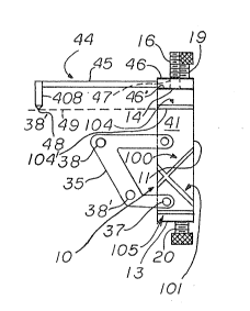
FIG. 9 shows main body 12 fixed to handle ~8 of
intramedullary alignment guide 82 and adjusted using feeler
gauge 44 so that the lower guide surface 104' of ~uide 104 is
level with anterior femoral cortical surface 81.
332~7
FIG. 10 is a partial fronk view taken in the
direction of arrows 10-10 of the adjustment markings 28
showing the alignment o~ main body 12 relative to the
intramedullary alignment guide 82 and to anterior femoral
cortical surface 81.
FIG. 11 is an enlarged partial side view of main
body 12 and feeler gauge 44 showing how a misalignment with
respect to the anterior femoral cortical surface 81 is
indicated.
EIGo 12 is a left side view showing main body 12
secured to the medial side of femur 80 by member 30 and ready
for shaping o femur 80 using the guide surfaces thereo~.
FIG. 13 shows distal femoral co~dyle shaping guide
60 fixed to studs 19 and 20 of main body 12 ready to
accomplish resection of the distal condyles after the other
portions of distal femur 80 have been shaped.
FIG. 14 is a side perspective view of shaped femur
80 with a distal femoral component 146 of a knee joint
prosthesis shown in relief over the shaped surface.
Referring to the Drawings, particularly FIGS. 1-5
and 8-10, the preferred embodiment of the apparatus of the
present invention is shown as dista~ femoral condyle shaping
guide 10 composed of main body 12 having upper surface 14
which faces in the direction of the anterior aspect of right
femur 80 twhile references will be made to the right femur,
the invention also applies to the same type of guide used for
the left fPmur) when guide 10 is fixed to handle 88 of
intramedullary alignment guide 82 as shown in FIGS. 8-13.
Upper surface 14 contains an attachment means in the form of
~two threaded studs, 15 and 16, which extend up away rom
surface 14. Main body 12 contains a means 18 by which main
body 12 can be rigidly fixed to handle 88 in the form of two
opposed knurled cap bolts l9 and 20 which serve to both grasp
~8~3~27
--8--
planar surfaces 88' and 88'' of handle 88 as a result of
contact with flat surfaces 24 and 25 of bolts 19 and 20,
respectively. Opening 22 in main body 12 permits viewiny of
the level at which main body 12 engages handle 88. Reference
marks indicated at 28 are inscribed on front face 11 o~ main
body 12 to better enable a surgeon to determine that level
and thus the level at which shaping means guide surfaces
associated with main body 12 are located relative ko guide
82. Studs 15 and 16 have reference marks 28' corresponding
to reference marks 28 to better permit the surgeon to
determine the proper setting of main body 12. In the
embodiment shown, the means by which the main body is fixed
to guide 82 and the means by which the level of main body 12
is adjusted relative to guide 82 are one and the same
although other embodiments may contain a separate means for
grasping handle and a separate level adjustment means such as
a sliding central portion permitting up and down (anterior
and posterior~ movement of the portion of the main body
containing or holding the shaping means guide surfaces.
Main body 12 contains two sets of shaping means
guide surfaces formed as a part of the main body as opposed
to a separately attachable shaping ~lide such as guide 60.
Thus, main body 12 contains anterior condyle shaping means
guide surfaces 104 an~ 106, posterior condyle shaping means
guide surfaces 105 and 107, and bevel condyle shaping means
guide surfaces 100, 101, 102 and 103.
Thraaded bores 310 and 320 on medial side surface
40 of main body 12 and corresponding threaded bores 360 and
370 on lateral side surface 41 engage knurled cap bolts 31,
32, 36 and 37 to permit members 30-and 35 to be fixed to each
respective side of main b~dy 12 when the bolts are passed
through bores in each member. Stabilizing membar 30 contains
two threaded, knurled cap bolts 33 and 33' each passing
03~2~
through a threaded bore in the end of member 30 opposite main
body 12 and each comes to a point 34 and 34', respectively.
Stabilizing member 35 contains the same type of bolts 38 and
38' coming to point 39 and 39', respectively. One of members
30 and 35 are employed to ~ix main body 12 rigidly to distal
femur 80 as will be described, infra.
FIGS. 4 and 5 show anterior cortical surfa~e feeler
gauge 44 which is composed of a base 46 having an L-shaped
pointer 45 extending away fxom base 46 and terminating in
blade-shaped indicator 408 having a several points 48. The
blade-like elongated configuration of indicator 408 with
several points 48 is preferred over a pointer with a single
point since handle 88 is often set at an angle with respect
to rod portion 84 to provide the desired valgus angle and
this may cause a feeler gauge with a single point to contact
cortical surface 81 at a point other than at the apex of the
anterior cortical surface 81. Because of its blade-like
shape, the lower edge of indicator 408 can push through soft
tissue overlying surface 81 and insure that actual contact
with the apex of surface 81 is made.
Base 46 contains two smooth bores 47 and 47 which
permit base 46 to be rigidly mounted over studs 15 and 16.
Base 44 is constructed such that when base 44 is mounted on
upper surface 14 of main body 12 and lower surface 46' of
base 44 contacts surface 14, point 48 lies on a line 49 which
is parallel with and at the same level as lower sur~ace 104
and 106', respectively, of shaping means guide surfaces 104
and 106.
FIGS. 6 and 7 show distal femoral surface shaping
means guide 60 which is composed of base 66 containing two
smooth bores 67 and 67 which permit base 46 to be rigidly
mounted over studs 15 and 16. A distal femoral surface
shaping guide means composed of guide 63 is formed by metal
.
3~7
--10--
guideplates 64 and 65 and ~uide 68 is formed by metal
guideplates 69 and 70. Guide plates 65 and 70 are attached
to base 66 and are separated rom ~lide plates 64 and 69 by
bar 62. Guideplates 65 and 70 as well as guideplates 64 and
69 can each be one continuous plate. The distance between
the center of centrally mounted studs 15 and 16 and the
surfaces of guides 63 and 68 on which the femoral surface
shaping means is to rest (e.g., an oscillating saw blade) is
selected to suit the amount of distal femoral condyle surface
necessary to be removed to properly fit a femoral component
of a knee prosthesis on femur 80. I~ desired, but less
preferable, separate guides for shaping9 for example, t~e
anterior and postPrior aspects of the distal femoral condyles
could also be made similar to that described or guide 60
using appropriately oriented cutting guide surfaces instead
of including such guide surfaces as part of the main body.
A shown in FIG. 13, the lower edges of guide
plates 64, 65, 69 and 70 come rather close to the bevelled
surface 142 of femur 80 to insure that a saw blade is
accurately guided in shaping the distal femoral condyles.
This configuration along with a removable shaping guide such
as guide 44 is preferred where the surgeon desires to shape
the distal femur to receive a prosthesis which i8 afixed to
the femur via bone ingrowth into a porous substrate such as
sintered metal beads as opposed to fixation by cementing the
prosthesi~ to the distal femur. This configuration provide~
the very accurate degree of shaping of the distal femur
needed for such implants and in such a case, it is preferred
that the distal femoral condyle shaping step be done last to
provide clearance for the lower edges of guide 44.
If the prosthesis to be implanted is to be cemented
to the femur, the layer of cement can fill in minor
inconsistencies in surface shaping. In that case, the distal
327
--11--
femoral shaping guide 60 can be made as a fixed part of main
body 12 (e.g., extend the length of studs 15 and 16, fix
guide 44 to the top surface 14, and modify ~eeler gauge 44 so
that points 48 line up with line 49 as shown in FIG. 4) to
provide a shaping guide with all shaping means guide surfaces
integral with main body 12. In that embodiment, the lower
edge~ of the distal femoral shaping means guide would not
extend as close to the femur as shown for guide 60 since the
main body would be attached to the handle before the distal
femur is shaped and clearance would be needed between the
femur and those lower surfaces. Due to the clearance, there
would be more opportunity for a saw blade to waver and ,the
shaped surface may not be as even as is needed for porous
ingrowth fixation prostheses, but would be satisfactory for
prostheses using cement fixation. In this embodiment, all
shaping means guide surfaces are located on the main body
which is not removed until all shaping operations are
completed.
The above de~cribed main body, intramedullary
alignment guide, feeler gauge, shaping means guide, and
associated components are all preferably manu~actured from a
suitable surgical grade of stainless steel or other metal
commonly employed by those skilled in the art to construct
surgical tools or use in contact with the body. The exact
composition of the materials used to construct the above
orms no part of the present invention as long a~ it performs
the desired function; other materials suitable for use within
the body and for the intended uses of the above may be used
without altering the nature of the invention.
The manner in which the apparatus of the prasent
invention may be used will now be described with referenc~ to
FIGS. 8-14. The present invention relies on the use of an
intramedullary alignment guide to referenc~ the shaping o~
.
.
3~7
-12-
the distal femur ~o the central long axis of the femur as
defined by that intramedullary alignment guide. The
preferred intramedullary alignment guide employed in
conjunction with the present invention is that described in
the Whiteside '177 patent and the manner in which that
intramedullary alignment guide is used is further described
in the Dow Corning Wright brochures noted above. While the
type of intramedullary alignment guide and manner of placing
it within the intramedullary canal preferred is that of the
Whiteside '177 patent type, other intramedullary alignment
guides can be employed with the shaping guide of the present
invention provided that the shaping guide of the presen,t
invention can be attached to such an alternative alignment
guide in such a way as to permit shaping of the distal
femoral surface relative to the central long axis of the
femur as defined by the alignment guide. The intramedullary
alignment guide may be inserted within the emur using
various guides to direct the surgeon as to where to place the
boring tool used to create a passage for the intramedullary
alignment guide, e.g., a guide such as the femoral hole
locator tool described in Zimmer Brochure No.
81-038-226-1020/15MZ noted above could be usad to define the
entry point for the intramedullary alignment guide, even in
conjunction with the Whiteside 177 patent method. The type
o intramedullary alignment guide employed and the manner in
which it is placed within the femur is conventional and forms
no part OI the present invention.
FIG. 8 shows intramedullary alignment guide 82 of
the same type which is described in the Whiteside '177 patent
being placed within a bore in femur 80 running through the
intramedullary canal defined by the wall 81 of cortical bone
making up femur 80. The bore has been prepared in accordance
with the Whiteside method such that central long axis 86 of
33~27
-13-
the intramedullary rod portion 84 of guide 82 is concentric
with the central lorlg axis of femur 80 after the rod por~ion
84 which contains 3 locking fins 85, 85' and 85'' ha~ been
inserted to it~ full length (i.e., approximately up to the
point where handle 8~ is joined to rod portion 84) within the
bore in femur 80. Guide 82 is selected such that handle 88
is set at an angle relative to axis 86 to provide the desired
degree of valgus angle the surgeon wishes to obtain on the
knee prosthesis after implantation.
Main body 1~ has been fixed to handle 88 by
tightening bolts 19 and 20 down over surfaces 88' and 88'' of
handle 88 such that the handle is centered between the ~ero
markings of reference markings 28. In accordance with the
Whiteside method, impactor 800 has been locked to handle 88
by means of locking pin 801 having portion 802 which passes
through bores in impactor 800 and handle 88 and
intramedullary alignment guide 82 has been inserted within
the bore in femur 80 up to the point where fins 85, 85' and
85'' almost touch the distal surface of femur 80. The
rotational alignment of guide ~2 is adjusted using the lower
surface 13 of main body 12 as a guide in accordance with the
Whiteside method to visualize e~ual amounts of the posterior
aspects of the condyles on a plane running parallel with
surface 13. This aligns guide 82 because surfaces ~8' and
88'' are parallel with surface 13. Guide 82 is then driven
into the femur using a mallet on the impactor 800 until ins
~5, 85' and 85'' are embedded in tha cortical bone of the
distal femur. Impactor 800 and pin 801 are removed and the
method of the present invention is begun.
With guide 12 locked on handle 80 as described
above and with back surface 17 resting against the distal
condyles of femur 80, feeler gauge 44 is placed over studs 15
and 16 and allowed to slide down until points 48 of indicator
327
-14-
. .
408 rest on the anterior femoral cortical surface 81.
Preferably, main body 12 and feeler gauge 44 are constructed
such that when surfaces 88 and 88 ' are centered between the
zero reference markings 28, points 48 come in contact with
surface 81. The surgeon can observe the markings 28' on
studs 15 and 16 in making this alignment. This permits the
surgeon to shape the anterior aspect of the distal femoral
condyles level with surface 81. The posterior aspects of the
distal femoral condyles will also be shaped relative to
surface 81 since guide surfaces 105 and 107 are part of main
body 12.
FIG. 11 shows the effect of having main body 12 and
thus guide surface 106 too low with respect to anterior
femoral cortical surface 81. A space indicated by arrows 110
can be seen between surfaces 46 and 14 which is equal to the
distance between point 48 which is resting on surface 81 and
line 49 which is at the same level as surface 106' of guide
surface 106. I desired for some reason such as an
anatomical defect, the surgeon could conduct the shaping of
the distal femoral surface at this setting of the main body.
Preferably, bolts 19 and 20 would be turned by the amount
indicated by markings 28' on studs 15 and 16 until the space
between surfaces 46' and 14 was eliminated there~y indicating
that line 49 and thus, surface 106', was at the same level as
surface 81. If main body 12 was set at too hlgh of a level
relative to surface 81, the surgeon would observe that point
48 was not in contact with surface 81 and would lower main
body 12 using bolts 19 and 20 until point 48 just contacts
surface 81. This procedure provides this method with an
advance over the Whiteside '177 patent in that the femur can
be shaped relative to the level of anterior femoral cortical
surface as well as with respect to the cent.ral long axis of
the femur.
-15-
. .
Feeler gauge 44 is then removed and stahilizing
member 30 is fixed to (for the case of a right ~emUr shown in
the Drawings) the medial ~ide 40 of main body 12 usiny bolts
31 and 32. Member 35 which would be pLaced on the lateral
side 41 of the main body is not used because the patella is
moved to the lateral side during this shaping procedure.
Bolt 33 is then ~ightened until point 34 securely contacts
the medial side of femur 80 and thereby fixes main body 12
securely to distal femur 80. It is understood that members
30 could have been present on main body 12 during the above
alignment procedure and bolt 33 tightened after main body 12
was leveled. Member 35 is used in place of member 30 using
the bolts shown for that member 35 if guide 10 was being used
to shape the left distal femur. Other stabilizing means
could be used to fix main body 12 to the distal femur 80
provided that such stabilizing means does not interfere with
the shaping means used to shape the distal femur. The
advantage of using a stabilizing member is that it prevents
rotation and loosening of main body 12 from handle 88 and
reduces the tendency of guide 82 to back out of femur 80 as a
result of the use of a shaping means such as an oscillating
saw. A further advantage is that other alignment guides
which fix to the end of the distal femur via pins driven
within the bone can disrupt the integrity o~ the bone,
particularly where the bone is osteoporotic and already weak
in structure as is often the case with older patients.
After fixing main body 12 to distal femur 80, a
conventional shaping means such as an oscillating saw or a
hand saw (not shown) is then introduced between ~uide
surfaces 104 and 106 to resect the anterior aspect of the
distal condyles to produce surface 141, between guide
surfaces lOS and 107 to resect tha posterior aspects of the
distal femoral condyles to produce surface 145~ betwean bevel
, ,
~03'~
guide surfaces 101 and 103 to produce surface 142 and between
bevel guide surfaces 102 and 104 to produce surface 144.
The angle~ o~ the shaping guide surfaces relative
tc the distal femur and the number thereof are 8elected to
match the prosthesis to be fixed to the distal femur as shown
in FIG. 14 for femoral component 146 having post 147 and a
second such post opposite post 147 (not shown) used to assist
in fixing component 146 to the distal femur after shaping is
complete. Main body 12 may further contain bores or other
guide means to assist the suryeon in placing bores in the
distal femur for posts such as post 147.
One more shaping operation is necessary to complete
the shaping of distal femur 80 using the above procedure.
After the above ~haping steps are completed, distal femoral
condyle shaping means guide 60 is placed ovex studs 15 and 16
and surface 71 is brought up against ~urface 14 of main body
12. A threaded nut or a wingnut is placed on stud 16 and
used to ~ecure base 66 to the upper surface 14 o~ main body
12 and a similar means is used to secure base 66 to stud 15.
A shaping means such as an oscillating saw or a hand saw is
placed in guide surface 68 to resect the medial distal
condyle perpendicular to the central long axis of femur 80
~nd guide ~urface 63 is æimilarly employed to resect the
lateral di~tal condyle in the same manner to result in
creation of shaped surface 143 on distal ~emur 80.
S~aping of the distal femur in accordance with the
method of the present invention is now completed, bolt 33 is
released from the side of femur 80, and the intramedullary
alignment guide is removed along with main body 12. Main
body 12 has remained attached to the alignment guide during
the entire ehaping procedure thus maintaining its original
alignment throughout the entire procedure thus using the same
reference points (anterior femoral cortical surface and
~ 3~ 7
-17-
central long axis of the femur) during the en~ire procedure.
As noted, the left distal emur can be shaped in the same
manner using the same guide 10. Guide 10 can be used to
shape the distal femur to receive a number of different
femoral components simply by selecting appropriately oriented
guide surfaces in the main body and/or guides which are
capable of being attached to the main body. It is also
contemplated that the anterior aspects of the femoral
condyles need not be the first to be resected and that the
distal femoral surfaces can be shaped in any order desired
except as described above for shaping tha femur to receive
porous ingrowth fixation prostheses.
Other modifications of the apparatus and method of
the present invention will become apparent to those skilled
in the art from an examination of the above specification and
drawings. Therefore, othar variations of the present
invention may be made which fall within the scope of the
ollowing claims even though such variations were not
specifically discussed above.