Note: Descriptions are shown in the official language in which they were submitted.
LASER SMOKE EVACUATION SYSTEM AND MET~OD
1 Field of the Invention
This invention relates to surgical procedures and more
specifically relates to a system and method for providing a
smoke-free environment at an operation site in a patient
cavity during laser laparoscopy.
History of the Prior Art
Laser laparoscopy is a surgical procedure in which a
focused laser beam, typically from a CO2 laser, is
transmitted in a laparoscopic tube through the abdominal
muscular wall of a patient into the pelvis where the laser
beam is used to excise or remove body tissue by vaporiza-
tion. This laser surgical procedure is used to treat a
number of gynecological problems including hydrosalpinx,
endometriosis, endometrioma, small uterine fibroids, and
pelvic adhesions. The only surgical opening required is a
small incision through the abdominal wall because the
laparoscopic tube is small, typically about 12.7 mm in
diameter. Use of this procedure avoids the risk of laparo-
tomy requiring full size abdominal incisions. The major
problem encountered, however, during laser laparoscopy is
the removal of vapor or smoke produced by the ablation of
the body tissue. One technique which has been used for
intermittent smoke removal has employed suction tubes with
valves. A problem with such a smoke removal procedure is
~ .
:~
.- . . . ............................. : . . - .
. ~ .. . . . . . . .. . . . . . .
S2
--2--
1 the loss CO2 gas which is required to sustain abdominal
distention. The re~uired abdominal distention for the laser
procedure necessitates replacing the lost CO2 gas. No
system is presently available which will effect sustained
smoke removal preventing its build up in vicinity of the
tissue removal whether laser laparoscopy is carried out by
either the two or three puncture techniques. The vapor con~ ;
sist of water vapor and carbonaceous vapors from the decom-
position oE organic material of the tissue. A small
manually operated valve is presently used with operating
room wall suction. This permits a small volume of intermit-
tant smoke and CO2 gas withdrawal as it is generated by the
lasing. This withdrawal must be followed by an input of
replacement CO2 gas from an insufflator. Use of large
replacement volumes of CO2 gas characteristic of repeated
abdominal evacuations can lead to problems in maintaining
the blood physiological acid base balance due to the for-
mation of the HCO3~ion from absorbed CO2 gas.
SUMMARY OF THE INVENTION
It is a principal object of the invention to provde a
new and improved surgical procedure.
It is another principal object of the invention to pro-
~vide a new and improved surgical procedure for use during
laser laparoscopy.
~ ~ .
~-, :
~ ~ .
-3- ;~
1 It is another object of the invention to provide appara-
tus and techniques for the removal of smoke produced within
a patient during a laser laparoscopy.
It is another object of the invention to provide appara-
tus and techniques for the removal of smoke during laserlaparoscopy wherein abdominal distention is sustained during
the procedure.
It is another object of the invention to provide appara-
tus and methods for smoke removal during laser laparoscopy ;
10 wherein real-time smoke removal is effected and its buildup -~
is prevented while simultaneously retaining constant abdomi-
nal volume and steady state insufflation pressure without
introducing large replacement volumes of CO~
It is another object of the invention to provide methods
and apparatus of the character described wherein closed cir-
cuit circulation is maintained and CO2 gas is not lost
during the smoke removal.
It is another object of the invention to provide methods ` ~ -
and apparatus of the character described wherein physiologi- ~-
cal blood CO2 gas balance is maintained in equilibrium.
It is a still further object of the invention to provide
a laser laparoscopy surgical procedure for smoke removal
which interfaces with standard laser laparoscopic system.
; ~ In accordance with the invention thexe is provided a `~
~ ',.' :` -
:. : - . :
1 closed circuit system for smoke and vapor removal during
laser laparoscopy which includes a CO2 gas pump, a discharge
line connected with the pump through a control valve, a
pressure sensor, a bacteria removal filter, into the patient
S and a discharge line from the patient through a smoke remo-
val Eilter, a pressure sensor, a control valve, a fluid
trap, back into the return line to the pump. An insufflator
is connected through a discharge line into the cavity of the
patient for maintaining the required pressure in the cavity ~:.
and replacing CO2 gas lost through leakage. :~
In accordance with the method of the invention smoke
removal is effected from the cavity of a patient during
laser laparoscopy including the steps of: pumping CO2 gas
through a control valve, and a bacteria removal filter into
a cavity of a patient at the site of laser laparoscopy while .
controlling the pressure of the input gas; return flowing
the CO2 gas with smoke generated at the site of the
laparoscopy procedure from the cavity of the patient;
removing the smoke by filter means from the discharge flow
from the patient; returning the filtered discharge flow
of C2 gas through a control valve and trap to pump means
: for recirculation to the patient; maintaining the r~quired
: distention of the cavity by flow of CO2 gas from a source
separate from the recirculation system; and providing make
~ .
.. , , . :. , : . .. : - .... : ~ . - ....... . . .: : --
: .: ' ~ - . : . ' :-.
~ ~33~
1 up CO2 gas to replace leakage and tissue absorption from the
separate CO2 gas source.
BRIEF DESCRIPTION OF T~E DR~WINGS
The forgoing objects and advantages and preferred
embodiments of the apparatus and method of the invention
will be better understood from the following detailed
description thereof taken in conjunction with the accom-
panying drawings wherein:
FIG. 1 is a schematic flow diagram of the system of the
invention; ;
FIG. 2 is a functional schematic diagram of the :~.
electrical circuitry employed in the system of FIG. l;
FIG. 3 is a functional schematic of the control module :
including the comparison and tlme delay functions;
FIG. 4 is a detailed diagram of the control module as
shown in FIG. 3;
FIG. 5 iS a fragmentary schematic view in section and
elevation of a patient cavity with the input and discharge .
: laparoscope tubes inserted to the site of the opexation
: 20 using a double puncture technique; and
FIG. 6 is fragmentary schematic view in section and ele- .
vation showing a triple puncture technique.
.
: :~ DETAILED DESCPRIPTION OF THE PREFERRED EMBODIMENT
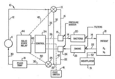
Referring to FIG. 1, the closed circuit CO2 gas cir-
,:- '
~ ~33~
1 culation system 10 embodying the features of the invention
includes a CO2 pump 11 connected with a discharge line 12
extending to a solenoid Elow control valve 13. A CO2
discharge line 14 is connected from the valve 13 through a
pressure transducer 15 and a bacteria filter 20 into a
patient 21 upon whom the surgical procedure is being per-
formed. A recirculating or return line 22 is connected from
the patient through a smoke removal filter 23 and a pressure
transducer 24 into a solenoid flow control valve 25. A
recirculating line 30 connects through a fluid trap 31 into
the suction side of the CO2 pump 11. A bypass or shunt on
line 32 is connected from the valve 13 into the recir-
culating line 22. Another shunt or bypass line 33 is con-
nected from the valve 25 to the supply or pump discharge
line 12. A control module 34 is connected by electrical
lines 35 and 40 to two pressure transducers 15 and 24,
respectively. A foot operated switch 41 is connected by an ;
electrical line 42 with the control module. The control
mcdule is also connected by a electrical line 43 with a
relay 44 which is connected with and operates the valves 13
and 25 by electric lines 45 and 50, respectively. An
insufflator 51 is connected through a flow line 52 into the
patient cavity to maintain the distention of the cavity
while the operation is being carried out and to supply make
:
:~ ~ '` .
.
~2~3~
1 up CO2 gas lost through leakage and tissue absorption. The
flow lines 14, 22, and 52 may connect the recirculating -~
system and the insufflator into the patient maybe as shown
: ~ .
in either FIG. 5 or FIG. 6. In the two puncture technique
of FIG. 5, the input line 14 of the recirculating system and ~-
the flow line 52 from the insufflator are connected into the
patient through a single laparoscope tube 53 which also
directs the laser beam to the operation site and has means
for viewing by the surgeon. The laparoscope tube 53 is a `
standard available laser surgical instrument. In the two
puncture technique, the return or recirculating of the CO
gas and the smoke generated by the laser operation passes
through a second laparoscope tube 54 which is connected to
the recirculating line 22. The laparoscope 54 also is a
standard available laser surgical instrument.
Alternatively; as illustrated in FIG. 6, the close cir-
cuit system and the insufflator may communicate with the
cavlty of the patient through a three puncture arrangement
in which the laser beam is directed through a laparoscope
.
20 ~ tube 55 the input CO2 gas and the CO2 gas from the insu~fla- ~-
tor flows through a second laparoscope 60, and the return of
C2 gas and smoke is through line 22 connected with a third
laparoscope 61. Each of the laparoscope tubes 55, 60, and
:
61 are all standard available laser sur~ical instruments.
-- .
; :
: ~:
; ~ . :. ~ . . : : . -. . ,
~2~3~
8-- .
:.
1 The electrical supply and control cixcuitry i5 shown in
FIGS. 2, 3, and 4~ Referring to FIG. 2, a suitable source
70 of 115 volt alternating current is connected with a
~ransformer 71 which reduces the electrical power to 12
volts direct current. The transformer is connected through
the foot switch 41 and the control circuitry 34 to the solid
state relay 44 which is connected with and operates the pump
11 and the solenoid valves 13 and 25. The pressure trans-
ducers 15 and 24 are connected with the control circuitry
34. As shown in FIG. 3, the control circuitry 34 includes
comparators 70 and 71, a timer 72, and a missing pulse
detector 73, which are connected with the pressure trans-
ducers 15 and 24 as illustrated. More specific details of
the circuitry of the control 34 are shown the wiring diagram
of FIG~ 4. The pressure transducers 15 and 24 convert CO2
gas pressure in the closed circuit of the system 10 to
voltages representing the pressures which are conducted to
the comparators 70 and 71. The voltages from the trans- ~ .
ducers are compared to preset the voltages in the com-
paritorsO If the voltages crosses the preset value set at `
~the comparitors 70 or 71, the output of the comparators
changes:state. The timer 72 normally produces a pulse train
monitored by missing pulse detector 73. When the preset
values of a comparator have been crossed, the timer 72 stops
:: ~
, .
~33~3~S,f2
1 producing pulses and the missing pulse detector begins to
time out. If no pulses are received by the missing pulse
detector within an adjustable time period of approximately
one to five seconds, the pump relay and the solenoids are
shut down. ;~
The laparoscopic tubes schematically illustrated in
FIGS. 5 and 6 are representative of a variety of different -
tubular structures which may be used in the system of the
invention for accessing the laser beam and the flows of CO2
gas and smoke removal from the site of the operation of the
lasex. These various laparoscopic tubes are designed to
allow visualization of the surgical field within the pelvis,
access of the laser beam, and input of CO2 gas to form the
closed circuit flow removal of the laser smoke, and CO2 gas
and to insufflate and thus distend the abdomen by imposing
and maintaining a steady state pressure within the abdomen.
The pressure may range from plus 16-20mm of the gas relative -~
to atmospheric pressure. The insufflation and resulting
abdominal distention are necessary to provide adequate
increased volume to move pelvic organs about and achieve the
necessary visulization and surgical access to the operation
site. One form of the laparoscopic tube, such as the tube ~
53 in FIG. 5, has three channels along its length. One ~-
channel carries a fiber optical cable bundle for
,~,
-10
1 transmission of illuminating light and the image for visuli-
zation of the working field through the eyepiece. A second
channel includes a stopcock or valve for controlling the
input of insufflating CO2 gas. A third channel is provided
for the input of CO2 gas in the closed circuit recirculation
system from the pump 11 through line 14. I'he tube alkso
directs the laser beam to the operating site. Other forms
of laparoscopic tubes may contain one or two channels. For
example, the tube 61 in FIG. 6 includes two channels, one
connected to the line 14 for the input of CO2 gas in the
closed circuit system and another channel connected with the
line 52 for gas from the insufflator. The other tube 60
shown in FIG. 6 utilizes only a single channel for the
return of CO2 gas and laser generated smoke in the recir-
culation system through the line 22.
The procedure for emplacing laparoscopic tubes includes
the following steps. A small incision is made in the skin
of the patient, in the naval or just below the naval, and a
large needle inserted into the abdominal cavity. The abdo-
mlnal cavity is then distended with carbon dioxide gas using --~
an abdominal insufflator raising the pressure to the equiva~
lent to 17 to 20mm of Hg. When the abdomen is properly
distended, a trochar is inserted through the same small
incision used for the large needle. The trochar is then ~ -
- :,
::
. '~ ' . ' . , . . . ' , . , . , ' , . .: , .
1 removed leaving a sleeve for the insertion of a laparoscope.
To this point, this is refered to as a "single puncture"
technique and typically is used for diagnositic laparoscopy.
Where a patient is undergoing laser surgery, usually one or
two additional small incisions are made in the lower abdomen
above the groin on either side. These incisions then are
used for the insertion of the necessary instruments
including the laser beam carrying tube, the CO2 gas return
flow tube, and insufflator input tube as referred to above
I0 in connection with FIGS 5 and 6.
After the laparoscopic tubes have been inserted in the
patient in both the two puncture and three puncture tech-
niques, the required equipment is connected as illustrated
in FIG. 1 to provide closed circuit flow of CO2 gas for the
evacuation of laser generated smoke, for the makeup supply
C2 gas, and to maintain abdominal distention from the
insufflator. The pump ll is controlled by the pu~p impeller
speed to provide a maximum flow rate of 1800 in3/min. The
gas flow from the pump 11 flows through the line 12, the
valve 13, the line 14, the pressure sensor 15, the bacteria
filter 20, and into the patient through the laparoscopic
:
tube 53 in the two puncture technique of FIG. 5 or the
laparoscopic tube 60 in the three puncture t~chnique shown
in FIG. 6. The return flow of the CO2 gas from the patient
.:-. . -
~ 33~-~2
1 with the laser generated smoke passes out of the patient
through the laparoscopic tube 54 in the two puncture tech-
nique, or the laparoscopic tube 61 in the three puncture
technique into the line 22, through the smoke filter 23, the
pressure sensor 24, through the valve 25, the line 30, the
fluid trap 31, and into the suction or return side of the
pump 11 to be recirculated to the patient, the smoke and
vapor having been removed from the closed circuit flow o
the CO2 gas. During this closed circuit flow, flow line ~ -
pressure values at the pressure sensor 15 and the pressure
sensor 24 are maintained at ~25mm Hg and +lOmm Hg,
respectively, relative to the atmospheric pressure. During
this closed circuit circulation of the CO2 gas, CO2 gas is
also injected from the insufflator through the line 52 into
the patient through the laparoscopic tube 53 in the two
puncture technique or the laparoscopic tube 60 in the three
puncture technique. The average abdominal pressur~ main-
tained by the insuffla~or is 16-20mm Hg. Slow leakage
around the laparoscopic tubes and tissue absorption re~uires
only a very small steady state input of CO2 by the insuffla-
tor. The control system is set so that if the pressure at
~the sensor 15 exceeds +25mm Hg or the vacuum at the sensor
24 falls below lOmm Hg, the central control module 34 acti-
vates the solid state relay 44 effecting the closing of the
;
-13-
1 solenoid valves 13 and 25 isolating the patient from the
pump 11. Additional patient safety features include the
bacteria and smoke filters 20 and 23 and the fluid trap 31.
The smoke filter which typically has a 3 micron pore size
removes laser created smoke particles. The bacteria filter
filter 20 is designed to capture any filterable microrga-
nisms greater than 0.2 microns in size. The fluid trap 31
removes condensed water vapor which is produced by tissue
lasing. Control of safety and over-ride of the operator
controlled on-off functions is controlled by the module 34
which is connected in the system as shown in FIG. 4 and `
schemetically represented in FIG. 3. The electrical output
signals from the pressure transducers 15 and 24 are fed
into the control module 34. The signals are compared
electrically to preset values. If a variation occurs for
times exceeding those of typical minor system induced
pneumatic transients, typically CS seconds, an electrical
ouput signal from the control module 34 shuts off the solid
state relay 44 which interrupts the 110 volt power supply to
the solenoid valves 13 and 25, and to the pump 11, effecting
closure of the valves and stopping the pump. The patient
is, thus, isolated. Further, the operator has manual
control of the on-of state of the system by means of the
pedal foot switch 41. The electrical signal from the foot
,
~ ,
~ .
, . . ~ .
~ 3~
-14-
1 switch 41 enters the system through the control module 34
which will prevent the system rom being turned on if a
pressure malfunction produces adverses pressure or vacuum
circumstances. The system of the invention effectively
removes smoke from a pelvis cavity at a rate of up to 1800
cu.in3/min. while maintaining essentially constant volume
and average background pressure in the pelvis.
Each of the valves 13 and 25 are three-way diverting
valves operated by 110 volt current. The solid state relay
44 is controlled by a 12 volt activation current for
switching the 110 volts supply to the pump 11 and to the
solenoid valves 13 and 25. The pump 11 is typically of non-
corrosive plastic polymer vane and chamber construction.
The pressure sensors 24 and 15 are linear voltage differen-
tial transformer type with a moveable piston ssnsing Pg ~
76mm Hg and Pg ~ -76 mm Hg. The foot switch 41 uses a 12~ ~ -
VDC source voltage. Both of the filters use an inert
plastic polymeric medium. The power sources typically 115
VAC, 60 HZ, 1.8 AMPS.
When the valve 13 is closed, flow along the discharge
side of the pump in the line 12 to the valve 13 is shut off
and~the valve shunts the pressurs in the vacuum side of the
patient connecting the lines 14 and 32 thereby equalizing
.
~ the pressure across the patient. Simultaneously, when the ~
~2~3~
15-
1 valve 25 closes t the flow along the line 22 into the valve 25
is shut off and the pressure lines 30 and 33 are com-
municated shunting the in and out ports of the pump iso-
latin~ the patient from the pump.
It will now be seen that a new and improved apparatus
and method for laser smoke evacuation from an operating si~e
in an abdominal cavity has been described ana illustrated.
The apparatus includes a closed circuit circulation system
for CO2 gas wherein CO2 gas is circulate past the operating
site, laser smoke produced during the operation is filtered
from the CO2 gas and the CO2 gas is recirculated past the
operating site. ~dditionally, CO2 gas is introduced from an ~ :
insufflator in a separate system for replacing recirculating
C2 gas lost through leakage and tissue absorption, and to
maintain the necessary cavity distention for visualization
of the operating site and to maintain adequate space for the
operation. The procedure and apparatus eliminates the need
for large replacement volumes of CO2 gas. Normal problems
in maintaining equilibrium physiological blood CO2 gas
balance are avoided. The system is useful with any standard
laser laparoscopic system.
. - . .: . . - . . : , .
, . . . . .. . . . . . -