Note: Descriptions are shown in the official language in which they were submitted.
Thls invention relates to a method of incorporating
a foreign substance into livlng cells.
In the field of anaiysis of gene expression the
behavior of those genes which determine the character of a
living thing can be studied by incorporating selected genes
into living cells and by examining the transformation of
the cells having added genetic material. For example, DNA
can be extracted from cancerous cells, and divided into
minute fragments of different sizes. These fragments are
classified in terms of size, and fragments of different size
are incorporated into living cells. As a result of such
incorporation, some of the cells are found to be cancerous,
presumabl~ as a result of incorporation of the foreign DNA
fragments. By this technique, cancer-associated fragments
can then be identiEied in terms of size.
A prior art implantation method comprises the steps
of: putting livlng cells in a solution containing DNA frag-
ments; making a small hole in each living cell with the aid
of a fine needle under an optical microscope; allowing DNA
fragments to enter the cells through the hole; and confining
the fragments in the cell when the hole in the cell heals
~See Japanese Patent Application No. 56~171347, disclosed
as Public Disclosure Number 58-76091 on May 9, 1983). Another
prior art implantation method comprises the steps of: pre-
cipitating DNA using calcium phosphate in a culture medium;
and making use of the phagocytosis of living cells to in-
corporate the precipitated DNA into the living cells.
~8~3~
The prior art implantation methods, however, are
not en~irely satisfactory. The former implantation method
requires skilled manipulation of the needle. Otherwise, no
holes can be made without injuring the living cells. Also
the work is tedious and laborious although the implantation
succeeds at a relatively high rate. Furthermore~ it is impos-
sible to make holes in certain cells, regardless of the type
of needle used.
The latter implantation method is capable of
handling a great number of cells at one time. The success
rate for incorporation of DNA fragments into host cells by
this method, however, is very low, say one in ten thousand
(1/10,000) at best. Thus, the rate at which the character
of the implanted cells is transformed is very small, and
accordingly a very large number o cells need to be implanted
with DNA fractions. This demand cannot be met. Also, dis-
advantageously the method requires addition of calcium
phosphate of so high a concentration that the additive tends
to injure living cells.
The inven-tors have found that a living cell when
exposed to a laser of appropriate energy is par~ly and
temporarily modified to be permeable to a foreign substance,
thereby allowing the substance to enter the living cell
and confining the same in the living cell upon recovery to
the original condition. This inventio~ is based on that
discovery.
-2-
3~'~
One object of this invention is to provide a method
of implanting a very large number of cells with a foreign
substance such as DNA with ma~imum possible efficiency.
Another object of this invention is to provide a
method for implanting a very large number of cells with a
foreign substance such as DNA in the minimum possible time.
Still another object of this invention is to
provide a method for making living cells temporarily per-
meable to a foreign substance without the necessity of
subjecting living cells to a special treatment.
According to one aspect of the invention, there
is provided a method of implanting living cells with a foreign
substance, comprising the steps of:
ta) exposin~ living cells to a laser beam to modify
at least a part of the living cells to be temporarily per-
meable to the foreign substance; and
(b) placing the living cells and the foreign subs-
tance together under conditions such that the foreign subs-
tance will encounter modified living cells.
A laser beam has good directionality, and can be
focused to a spot of minimal diameter using an optical
microscope. These properties make it possible to use a laser
beam to make a minute "hole" tsubmicron in diameter) in a
host cell. tThe word "hole" is intended to mean that the
area of a living cell which is made permeable to a foreign
substance resembles a hole.) A pulse laser beam can be used,
in which case the tempera-ture rise of the cell due to expo-
sure to the laser beam can be advan-tageously suppressed by
reducing the pulse duration of the laser. This prevents the
cell from being thermally killed.
Lasers produce monochromatic irradiation. In
accordance with the invention, the wavelength of this irra-
diation can be selected as appropriate for the purpose of
making "holes" in a given type host cell, considering the
optical characteristics of the cell ~all and the cell mem-
brane as well as the characteristics of a foreign substance
to be lncorporated.
The strength of a laser also can be adjusted over
a wide range using appropriate electr~ic control. The focusing
dep-th in the host cell can be easily controlled with appro-
priate optical means. These together provide a great advan-
tage to the microsurgery to which this invention is to be
applied. An apparatus for making "holes" in living cells
according to this invention uses a laser microbeam appropriate
for the purpose of conducting a microsurgery on a living cell
without injuring its self-healing capability.
The length of time for which cells are exposed
to a laser microbeam should be controlled so as to be long
enough to make "holes" in the cells, but not enough to
thermally kill the cells. The exposure time can be controlled
by using a train of laser pulses each having a duration con-
trolled appropriately for the purpose, or by scanning with a
laser microbeam in an appropriate length of time a field
in which a large number of living cells are suspended in a
solution. Also, a laser microbeam can be directed to selected
portions of a single cell selected from among those cells
floating in a solution.
3~
Thus, according to another aspect of this invention,
there i5 provided an apparatus for making "holes" in living
cells, comprising a laser source for providing a pulsed or
continuous wave laser microbeam, an optical system for pro-
jecting a laser microbeam to living cells, and means for
monitoring living suspended cells in a solution.
According to a further aspect of the invention,
there is provided a microsurgery apparatus comprising a laser
source, an optical system for pro~ecting a laser microbeam
to living cells, means for monitoring living cells suspended
in a solution, means for determining the position of a cell
selected among those appearing in the field of the monitoring
means, and means responsive to a start signal from the
positions determining means for controlling the -supply of the
laser from the laser system.
According to still another aspect of this invention,
there is provided a microsurgery apparatus comprising a laser
source, an optical system for projecting a laser microbeam
to living cells, means for m~nitoring living cells suspended
in a solution, means for determining the position of a cell
selected among those appearing in the field of the monitoring
means, means responsive to cell position signals from the
position determining means for storing, and means for control-
ling the laser microbeam and directing the same to selected
cells one after another.
~ urther features and advantages of the invention
will become more readily apparent from the following descrip-
tion of preferred embodiments~ with reference to the accom-
panying drawings, in which:
~ . i
Flg. 1 is a copy of microscopic photograph showing
NRK cells planted with gene (Ecogpt) according to this
invention;
Fig. 2 shows a sequence of microscopic photographs
showing the formation of a hole in an NRK cell using a laser
microbeam and the subsequent healing of the holei
Fig. 3 is a copy of microscopic photograph showing
punched cells (human red blood cells);
Fig. 4 is a schematic block diagram of a laser
punching apparatus according to a first embodiment of this
invention;
Fig. S is a perspective view of a laser microbeam
deflector used in the laser punching apparatus of Fig. 4;
Fig. 6 is a schematic block diagram of a laser
punching apparatus according to a second embodiment of this
nventlon;
Fig. 7 is a schematic block diagram of a laser
punching apparatus according to a third embodiment of this
inve~tion;
Fig. 8 is a schematic block diagram of a laser
punching apparatus according to a fourth embodiment of this
invention;
Fig. 9 is a schematic block diagram of a laser
punching apparatus according to a fifth embodiment of this
inventioni
Fig. 10 is a copy of microscopic photograph showing
details of portions of cells swept by a laser microbeam~
As described earlier, the implantation method
according to this invention comprises the steps of exposing
-6-
.
L~
living host cells to a laser microbeam to make the surface
of khe host cells temporarily permeable to a foreign substance,
i.e. to make "holes" in -the host cells, and allowing these
permeable cells to meet with fragments of a foreign subs-
tance, such that the foreign substance enters at least some
of the permeable cells before these cells heal their holes,
and confining the foreign substance in the host cells when
the holes close.
There are a varlety of modes in which fragments of
a foreign substance can meet with permeable host cells and
enter the cell through the holes. For`example, cells and
fragments of the foreign substance are suspended together
in a solution, and cells are exposed to a laser microbeam one
after another, thus causing permeable cells to coexist with
the fragments in the solution. In a second approach, a solu-
tion containing host cells is supplied in drops to another
solution containing fragments of a foreign substance~ and
each drop on the way to the lower solution is exposed and
punched by a laser microbeam. Third, a liquid carrying host
cells and fragments of a foreign substance is made to flow
across the field of a pulse or continuous wave laser. This
final mode is most appropriate for the purpose of handling
a large number of host cells.
Example 1
Referring to Fig. 1, living cells implanted with
fragments of a foreign substance and dead cells which could
not survive owing to there being no foreign substance im-
planted therein are shown under microscope. NRK cells origi-
nating from the kidney of an Osborn Mendel rat were modified
--7--
so as to be unable to survive without Ecogpt (Xanthine-guanine
Phosphoribosyl Transferase) being incorporated therein, and
the NRK cells thus modified were put in an Ecogpt-containing
medium (DMEM added with 10-percent unborn calf's blood serum).
An infrared beam (~ = 1.06 microns) from a laser device
(YAG laser) was converted to an ultraviolet beam ( A = 335 na-
. .
nometers), and the ultraviolet beam was introduced into a
laser microscope. Then, living cells floating in the medium
in the field of the microscope were exposed to pulsed laser
beam irradiation having a pulse duration of 10 nanoseconds.
The laser beam was applied to the cells in one half of the
field at the rate of ten pulses per second, and a large number
of living cells were treated.
The results are shown in Fig. 1 where cells which
were exposed to the laser (left half) are alive, while those
that were not exposed to the laser (right half) are dead.
The cells exposed to the lasex too~ up Ecogpt fragments of
the foreign substance and therefore survived~ but the cells
that were not exposed to the laser could not take up Ecogpt
fragments, and therefore died.
Example 2
Living cells can heal and close their holes essen-
tially immediately after being made. Fig~ 2 is a photograph
of a video sight showing NRK cell immediately after being
punched by a laser microbeam. Specifically, Fig. 2(A) shows
the appearance of cells at the instant they were punched.
Fig. 2(B) shows the appearance of cells immediately after
being punched, and there is already a decrease in the size
of -the hole. Fig. 2(C) shows the appearance of cells after
,.,~.,`
3~,
healing of the hole.
Example 3
Fig. 3 shows human blood cells which have been
dyed and punched with a laser microbeam. These photcgraphs
show the appearance of living cells immediately after being
punched, proving that a single cell can be punched at it
selected portions thank to the good controllability of laser
beams. As is apparent from the above, the method of this
invention permits punching for the sake of implanta-tion of a
foreign substance in the cell. Also, this invention can be
equally applied to microsurgery of cells, as for instance
breaking a particular minute organ in a single cell.
Application of this mYen~ion to the incorporation
of genes into cells permits: production of useful substances
in cells (for instance, synthesis of insulin or any other
useful human substance within living cells); and improvement
of domestic animals and agricultural products (by substituting
different genes for each other in different kinds of plants;
or by lncorporating good genes without recourse to fertiliza-
tion).
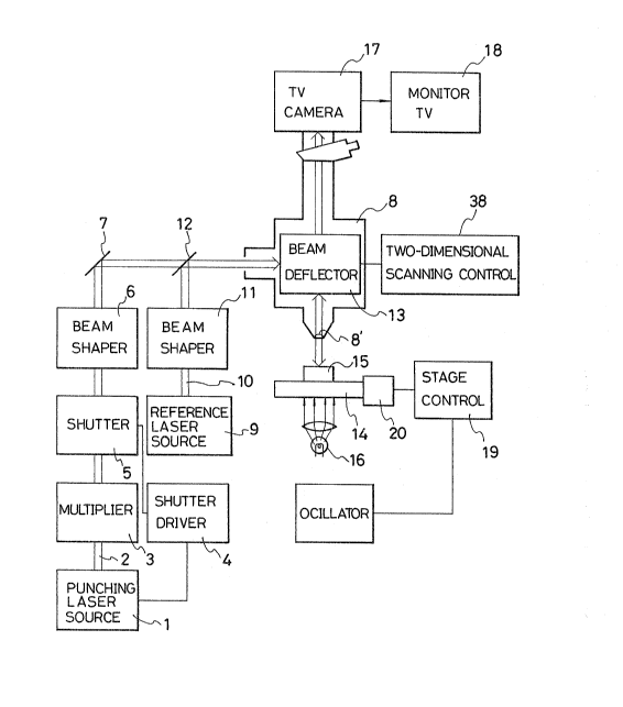
A cell-punching apparatus for performing the method
of~ this inventionis described with reference to Fig. 4.
A laser beam (~ =1060 nm) for punching living
cells is generated by a laser source 1 and passes through a
frequency multiplier 3 essentially composed of KDP or any
other crystal which is appropriate for the purpose of con-
verting the infrared light to ultraviolet (~ =335 nm or
~65 nm). The ultraviole-t laser beam passes through a shutter
_g_
j ~,
~" , .,
~2~3~3~
which is controlled by an associated shutter driver 4. Then,
the laser beam 2 is shaped by a beam shaper 6. The laser beam
thus shaped is directed toward a microscope-and-beam combiner
8 by reflector 7. A reference laser beam 10 functioning as a
pilot or tracing beam (for instance, He-Ne laser ~ = 633 nm)
is generaged by a visible laser source 9. The reference laser
10 is shaped by a beam shaper 11, and then the shaped laser
beam is reflected by a reflector 12 to travel toward the
beam combiner 8 alc,ng with the punching laser beam 2. The
punching and reference laser beams 2 and 10 after passing
through a beam deflector 13 are combined by a condenser lens
8'. The combined laser beam strikes cells floating in a so-
lution in which fragments of a foreign substance such as
DNA are suspended. When cells are exposed to the laser beam,
the cells are punched and become permeable to the fragments.
A sample holder 15 is illuminated by a lamp 16 under the
holder, thereby projecting an image of the cells in the
sample holder to a TV camera 17 through the condenser lens
8', and producing a visible image of cell distribution on a
TV monitor 18. A stage 14 carrying the sample holder 15 is
composed of an ~-Y stage which is driven by a stepping motor
20.
When shutter 5 is closed, the punching laser beam
does not reach the sample holder 15, and the visible laser
beam 10 from the laser source ~ functions as a pilot beam,
thus indicating the place where the punching laser beam will
strike. When the shutter 5 is open, the visible laser beam 10
is combined wlth the punching laser beam 2 and functions as
--10--
~2~
a tracing beam, thus making visible the trace on which the
punching laser beam travels.
In punching cells, the stage 1~ is driven until the
image of a congregation of cells appears in the field of the
monitor 18. Then the shutter 5 is kept open, thereby permit-
ting the continuous irradiation of the sample holder 15 by
the punching laser beam 2. Cells are exposed to the punching
laser beam 2 one after another simply by moving the stage
14. Fragments of a foreign substance floating in the vicinity
of punched cells enter the cells via the "holes" in these
punched cells. The living cells heal their holes in a few
seconds, thus confining the foreign substance in the cells.
As a result the healed cells may now carry a particular gene
present in the fragments of foreign substance.
Moving the sample holder with respect to the
stationary pulse or continuous wave laser beam causes the
laser beam to sweep the cell-floating area in the solution.
This is most efective to treat a lot of cells within a
relatively short time.
Fig. 5 shows a laser deflector 13 as comprising a
combination of two galvanometers 13' and 13" each equipped
with a reflector. The laser deflector 13 is driven by an
associated two-dimensional scanning control 38 so as to
cause the visible laser beam 10 to scan a selected small
area in the field of the sample holder. ~hen, resultant
reflected rays, luminescence rays and scattered rays fall
on the TV camera or a still camera after passing through
the condenser 8', thus producing a clear image showing, in
detail, the inner structures of selected cells. Fig. 10 is a
`: ~
copy of mlcroscopic photograph taken by sweeping wlth the
visible laser beam, showing human red blood cells.
The area encircled with white line is the one swept
by the visible laser beam 10, showing details of the inner
structures of selected human red blood cells/ in contrast
with the rest area of the photograph illuminated by the lamp
16. Although the reason for providing such a clear detailed
image of the inner structure of the cell is not known, it
appears -to the inventors that the laser after passing through
the condenser lens focuses on a point at a determined depth,
thereby causing the appearance of a clear image of the inner
structure of the cell taken along the focal plane at the
depth. Thus the cell punching apparatus equipped with a
laser sweeping which permits the monitoring of modiEication
of the inner structure of a cell punched and implanted with
a foreign substance.
Fig. 6 shows a second cell punching apparatus use-
~ul in the method of this invention. As shown, cells descend
one after another in a fine transparent tube 20 so that they
are exposed to the punching laser beam from a laser source 21.
Specifically, a solution 22 containing living cells and
a protection liquid such as physiological saline 23 are fed
to the fine tube 20. A probe laser beam is emitted by a
probe laser source 24 to a detector 25, and passes through
the, descending flow upstream of the place at which cells
are exposed to the punching laser beam. The detector 25
detects a cell passing by the detector to generate and send
a detection signal to a central processing unit 26, and then
the central processing unit 26 times the start of the punching
laser source 21, thus causing the punching beam to hit the
descending cell to make a hole therein. Fragments of a
foreign substance to be incorporated in cells such as DNA
may be put in the solution 22 or the physiological saline
23. Thus, the cell punching apparatus can punch about 1000
cells per second. If use is made of a detector capable of
determining the angle of diffusion over which the laser beam
spreads when falling on a cell, cells can be classified in
terms of size, and hence kind. Thus, it is possible to select
and punch a particular klnd of cells among different ones
in a solution 22. The casting of the punching laser beam on
cells may be controlled by controlling a shutter (not shown)
provided between the fine tube 20 and the laser source 21
rather than by controlling the punch:Lng laser source 21.
Fig. 7 shows a third cell-punching apparatus useful
in the invention. This apparatus is so designed that a solu-
tion containing living cells is supplied in drops across the
punching laser beam. Specifically, a suspension 22 contain-
ing living cells, and a protection liquid 23 such as physio-
logical saline are fed to a nozzle 28 under pressure by airpumps 27 and 27'. A mixture of suspension and protection
liquid falls in drops 30 under the action of a supersonic
nozzle vibrator 29, which may be composed of, for instance,
a piezoelectric element. In operation, the fall of a drop
30 is detected by a probe laser falling on a detector 25, and
then the detector 25 sends a detection signal to a central
processing unit 26. The central processing unit 26 signals a
-13-
~2~ 3~
punching laser source 21 to emit a punching laser beam at
the instant the drop is about to cross the punching laser
source 21, thereby making holes in cells in the drop.
When drops 30 are exposed to the probe laser, it
is possible to determine which kind (or size) of cells are
contained in each drop with the aid of a conventional laser
analyzing system, and if drops are charged with electxicity
of which the polarity and/or quality varies with the kind of
the cell, and if these drops fall across the electric field
between opposite electrodes 32 and 32', they will be classi-
fied in terms of the polarity and/or ~uantity of the electriccharge, and will be put in different receptacles 33,33',
thus ciassifying punched cells in terms of kind.
As an alternative, if the drops are charged with
electricity of the same sign and quantity, the strength of
the electric field may be varied with the kind of the punched
cells. Fragments of a foreign substance to be incorporated
into cells may be put in the suspension 22, the protection
liquid 23 or in the receptacles 33 and 33'. The casting of
a punching laser beam from the punching laser souxce 21 may
be controlled by controlling a shutter ~not shown) provided
between the path of drops and the punching laser source 21
rather than by controlling the laser source 21. In some
instances the continuous casting of the punching laser beam
may be preferred.
Fig. 8 shows a fourth cell punching apapratus
useful in the invention.
The cell punching apparatus of Fig. 8 is different
from that of Fig~ ~ in that the former is equipped with a
-14-
`~`
L3~ *;~,
light pen 34 for indicating cells appearing in the field
of the TV monitor 18, an associated spot position determining
means 35 for determining -the~coordinates of point of the
monitor field on which the light pen is put and a spot posi-
tion control 36 for controlling the laser deflector 13 so as
to direct the laser beam to the same point as the light pen
indicates. The spot position control 36 is responsive to a
coordinate signal from the spot position determining means
35 for driving the laser deflector 13 to direct the laser
beam to the position indicated by the light pen. In operation,
the distribution of living cells in the sample holder 15 is
watched by the TV monitor 18, and the light pen 34 is put on
a selected part of a desired living cell selected among those
appearing in the field of the TV monitor 13. The coordinate
of the point indicated by the light pen 34 is determined by
the spot position determining means 35. ~ position signal
representing the position indicated by the light pen is
directed from the spot position determining means 35 to the
spot position control 36. Then, the spot position control 36
drives the laser deflector 13 to direct the laser beams 2
and 10 to the point indicated by the light pen 34.
At the same time as the light pen indicates a given
position, the position determining means generates a start
signal, and the shutter driver 4 is responsive to the start
signal for opening the shutter 5 for a predetermined period.
Thus, a corresponding number of laser pulses 2 are thrown
onto the point indicated by the light pen 34. The punched
cell allows fragments of a foreign substance to get therein,
and then the cell heals its hole to confine the fragment
therein as described earlier.
In this particular embodiment the spot position
control 36 is used to drive the laser deflector 13 for throw-
ing the laser beam to a given position. As an alternative
the stage position control 19 is used to drive the stage 14
to attain the same effect.
Fig. 9 shows a fifth cell punching apparatus useful
in the invention.
A light pen 34 is used to indicate selected point
or points Gn each of selected cells or every cell appearing
in the field of a TV monitor 18, and à spot position deter-
mining means 35 determines the coordinates of the points
indicated by the light pen 34. Then, signaIs representing ~
these coordinates are directed to memory 41 through a central
processing unit 40 so that the coordinates of the points
indicated by the light pen are stored in the memory 41.
These coordinates are read out one after another to input in
the spot position control 36 under the control of the central
processing unit 40. The spot position control 36 controls
the laser deflector 13 in the same way as the cell punching
`apparatus of the fourth embodiment. On the other hand, the
central processing unit 40 directs a drive signal to the
shutter driver 4, thereby opening the shutter 5 for a pre-
determined period to throw a punching laser beam 2 to the
points indicated by the light pen one after another. Thanks
to the use of memory, living cells appearing in the field of
the TV monitor are punched in rapid succession. It is pos-
sible to make a decision as to whether a cell is present or
not in terms of the amplitude of video-signal from the TV
-16-
: ~. ,.~..,
3~:
camera 17, and if the spot position detector 35 is designed
to make such a decisionr the cell punching will be completely
automated. Specifically, the so designed spot position
detector 35 may analyze video signals from the TV camera 17,
thereby determining the positions of cells appearing in the
field of the TV camera, and then the position signals repre-
senting the positions of cells are directed to the memory 41
for storing. In this case a pattern identification means may
be used to identify cells in terms of the contour of cell.
As shown in Fig. 9, a spectrometer 42, a photon-
counter 43 and a multichannel analyzer 44 together constitute
an analyzing system, which may be used as a monitor. Specif-
ically, the optical system can make a decision as to whether
a cell is present or not at a given coordinate (and in some
instances a decision as to whether a cell core is present or
absent at a given coordinate), in terms of spectrographic
characteristics.
The embodiments described above use two different
laser sources, that is, a punching laser source and reference
laser source. It, however, should be noted that if a contin-
uous or non-pulse visible laser beam is used as a punching
one, no reference laser beam is necessary because the spot
on which the punching beam focuses is visible in the field
of a T~ monitor. Also, it should be noted that a shutter for
controlling the throwing of the punching laser beam is not
limited to the mechanical one, and that a conventional photo-
switch may be used for the purpose. The expression, "fragments
or fractions of a foreign substance" used herein is intended
to include virus, every kind of protein, and full genome of
DNA.
Finally, in the examples and embodiments described
herein above, selected portion or portions of each living
cell are modified when exposed to a laser beam. This should
not be unders-tood a limitative. Indeed, the whole area of
the living cell may be modified if use is made of a laser
beam larger in diameter than the living cell, indeed.
-18-
;, ~