Note: Descriptions are shown in the official language in which they were submitted.
~ 4~7'7
1 ~ACXGROUND OF THE INVENTION
~ The invention pertains to an apparatus for
3 modifying the refractive properties of the ey~. More
4 particularly, the invention pertains to a method and apparatus
for correcting reractive errors by modifying the cornea of the
6 eye.
7 In general terms, the human eye functions by sensing
8 light rays. Such light rays tend to be focused as they pass
9 through the cornea, the aqueous humor, the lens and the
vitreous humor. Ideally, the focal point of light, after
ll passing throuqh these component~, will be at the retina.
12 Emmetropia, or the lack of refractive error, i~ thus
13 characterized by the focal point of the light entering the eye
14 from an infinite di~tance and falling on the retina.
Billions of human beings suf~er impaired vision due to
16 refractive errors o~ the eye charac~erized by the focal point
17 of light failing to be at the retina, but rather falling short
18 of or behind the retina. Common refractive errors of the eye
19 fall into three main categories: myopia, hyperopia, and
astigmatism.
21 As is well known, th~ cornea provides approximately
22 two-thirds ~2~3) of the refractive power of the eye. This i5
23 primarily due to the optically powerful air/cornea interface
24 created by the large disparity of refractive indices between
the air ~1.00) and the cornea (approximately 1.37). The
26 aqueous~lens interface causes further refraction within the eye.
27 Becaus~ the cornea is such an important facto-r in
28 refrac~ion of the eye, a wide variety of methods and
29 apparatuses have been applied in the pas~ to alter the cornea
in an effort to eliminate refractive errors. For example,
31 contact lenses, which are also commonly used as refractive
4~i7'7
1 entities in themselves, have been intentionally malfitted to
2 temporarily alter the corneal curvature. The later technique
3 is known as ~orthokeratology~ and generally results in only a
4 temporary change in the corneal curvature. Orthokeratology h~s
s a ~urther deficiency in that it is known to induce potential!y
6 serious corneal inflammation and scarring.
7 Several other techniques are known for altering the
8 cornea in various ways to compensate for refractive errors o.
9 the eye. For example, radial keratotomy involves the making o~
radially orientated slit-like incisions in the cornea, in
11 various patterns, to attempt to correct myopia and~or
12 astigmatism. At present, however, the results of radial
13 keratotomy are unpredictable and are often not reproducible in
14 the same patient. Additionally, it is as yet unclear how long
the results of radial keratotomy last. Further, there have
16 been reports of corneal degenerations, infections and
17 distortions after radial keratotomy, such conditions obviously
18 having the potential for serious visual loss.
19 It is also known to use lasers for altering the
condition of the cornea. U.S. Patent 4,461,294 illustrates the
21 use of the thermal effect of a laser to induce corneal-
22 recurving scars by imbedding, under pressure, light absorbing
23 colored bodies in the cornea in a radial pattern. The colored
24 bodies in the cornea are e~posed to a thermal laser through a
matched, slitted diaphram. In the technique disclosed by the
26 4,461,294 patent, corneal tissue is burned for the pur-pose o~
27 creating scar tissue.
28 Another technique for modifying the cornea, known as
29 lamellar keratoplasty involves the taking of a slice of a
patient~s cornea, or a~donor's cornea, freezing the portion and
31 lathing it in a hard-frozen state to a new curvature prior to
77
1 suturing onto the eye of the patient. Particular method~
2 employing this technique include keratomileusis,
3 keratophakia, and epikeratophakia, each of which requires
4 cutting and suturing the patient's cornea.
Yet another cornea modification technique is
6 disclosed in U.S. Patent No. 4,665,913 issued May 19, 1987
7 to Francis A. L'Esperance, which discusses a device for
8 exposing the cornea to an excimer laser in perpendicular
g fashion to reshape the cornea. The European application
discloses a laser which is directed at the eye substantially
11 along the visual axis of the eye. Removal of tissue is
12 effected by exposing the cornea head-on to varying flux
13 densities and exposure times in either rectilinear or
14 spiralling fashion. This head-on exposure to the radiation
of the laser would presumably expose the eyes delicate
16 internal structures, such as the iris, the lens and the
17 retina, to potentially damaging levels of radiatio~.
18 Additionally, in such a device, if the output of the laser
19 were inadvertently increased, deeper levels of tissue
penetration could result in accidental perforation of the
21 cornea or irregular corneal refracting surfaces.
22 To date, there are no known non-~nvas~ve devices or
23 methods which provide effectlve, safe, predictable and
24 reproduc~ble modificat~ons of corneal carvature for effectively
Compen~ating for the refractiv~ error~ Of the eye.
26 It is therefore an ob~ect of the invention to
27 provide an apparatus for accurately shaping the cornea to
28 compensate for refractive errors of the ey~. -
29 It is a further ob~ect of the invention to
provide an apparatus for reshaping the cornea of the eye in
31 vivo w~thout the need for removing and then suturing the
32 corneal tissue,
-- 3 --
4~
1 It i~ a further ob~ect of the invention to
2 provide an apparatus for shapin~ the cornea of the eye in which
3 there is no necessity for ~reezing the corneal tissue prior to
4 shaping but which may be used ln conjunct~on with ~reezing
techniques.
6 It is a still further ob~ect of the invention to
7 provide an apparatus for shap~ng the cornea which reduces
8 or eliminates the risk of accidental damage to the cornea
9 and the other components of the eye, such as the iris, the
lens and the retina.
11 It is a s~ill further object of the invention to
12 provide an apparatus which meets the foregoing ob~ectives
13 and which is safe, predictable, and reproducible.
14 SUMMARY OF THE INVENTION
In one aspect of the invention there is provided
16 an apparatus for modifying the refractive properties of the
17 cornea of an eye including laser means for emitting a laser
18 beam, means for deflecting the laser beam to strike the
19 cornea tangentially about the optical zone thereof and means
for rotating the deflecting means through 360. The center
21 of rotation of the deflecting means is positioned sub-
22 stantially along the visual axis of the cornea such that
23 through rotation of the deflecting means the laser beam
24 strikes the cornea tangentially over an area substantially
centered about the visual axis of the cornea.
26
27 A method for modification of the reactive
28 properties of a cornea may comprise the steps of:
29 (a) activating laser means to emit a laser beam;
(b) directing the laser beam to strike the cornea
31 tangentially about the optical zone thereof; and
~ 4 ~7~
1 (c) rotating the laser beam through 360 while sub-
2 ~tantially maintaining tangential striking of the cornea by
3 the laser beam.
4 In a specific embodiment the invention provides
an apparatus for reshaping the cornea of the eye, including
6 a laser source, computer controlr and a rotating and
7 tran~lating deflective member such as a mirror. The
8 rotating and translating defective member is guided by the
g computer to direct a laser beam emitted from the laser
source to tangentially strike the cornea of the eye,
11 causing controlled corneal ablation and thereby modifying
12 the radius of curvature of the cornea. With the invention,
13 the cornea is lathed tangentially in either its natural or
14 frozen state, in vivo or in vitro, in an area centered
about its optical axis and encompassing its optical zone by
16 either a non-thermal emission such as an infra-red laser,
17 under automated or manual control.
18 The invention is effective to modify the corneal
19 curvature and thereby its refractive properties by shaving or
vaporizing part of the optical zone of the cornea in a
21 precisely calibrated and predictable manner, eliminating the
22 need for cutting blades or mechanical lathing which were
23 heretofore provided. The invention also obviates the need ~or
24 suturing and scarring of the cornea. In addition, since the
.
1~ 77
1 incident laser beam in the invention strikes the cornea only
~ tangentially, changes in the frequency or power output of the
3 laser would not carry the risk of perforation, and delicate
4 intraoccular structures would not be directly exposed to the
laser.
7 BRIEF DESCRIPTION OF THE DRAWINGS
8 The invention will be described in greater detail
9 below by way of reference to the following drawings, in which:
Figs. 1-3 are schematic illustrations of human eyes
11 illustrating the conditions of emmetropia, myopia and
12 hyperopia, respectively;
13 Fig. 4 illustrates the ablation of corneal tissue in
14 the treatment of a myopic patient;
Fig. 5 illustrates the ablation of corneal tissue in
16 the treatment of a hyperopia patient;
17 Fig. 6 is a schematical plan view of an apparatus
18 according to the invention positioned about a human eye;
19 Fig. 7 is a partial elevational view of one embodiment
of the invention positioned about a human eye illustrating a
21 rotating arm for trans~tting a laser beam to a deflector;
22 Fig. 8 illustrates a method of fixing the eye in
23 position prior to and during an operation according to the
24 invention;
Fig. 9 is a control chart illustrating the functions
26 of a command computer in an apparatus according to the
27 invention;
28 Fig. 10 illustrates the alignment of directing
29 elements to correct myopia and hyperopia in an apparatus
according to the invention;
1~4~i7'7
1 Fig. 11 is an operational flow chart describing the
eunctioning o an apparatus according to the invention;
3 Fig. 12 illustrates the "red refle~ motion technique
4 used in monitoring eye fi~ation in one apparatus according to
the invention;
6 Fig. 13 ill~strates the functioning of a light scatter
7 detector used to determine tangentiality in one apparatus
8 according to the invention;
9 Fig. 14 is a partial perspective view of a curved
de1ector element according to one embodiment of the invention;
11 Fig. 15 is a schematical view of a further embodiment
12 of the invention; and
13 Fig. 16 is a partial perspective view of a further
14 embodiment providing a contoured laser beam.
16 DETAILED DESC~IPTION OF THE DRAWINGS
17 Fig. 1 illustrates the condition of emmatropia. As
18 shown in Fig. 1, light enters the eye through the cornea 61 and
19 passe~ through the cornea, the aqueous humor 2, the crystalline
lens 3 and the vitreous humor 6. The light is focused by the
21 refractive power of the cornea 61, the aqueous humor 2, the
22 crystalline lens 3 and the vitreous humor 6 to a focal point Pl
23 which, in the case of e~metropia as shown in Fig. 1, is at the
24 retina 4. The globe of the eye is generally indicated at
numeral 5 in Fig. 1.
~ 67~
1 Myopia (Fig. 2), also known as nearsightedness,
2 results when the focal point P2 of the eye is located anterior
3 to the retina 4. Hyperopia (Fig. 3), also known as
4 farsightedness, results when the focal point of the eye is
located posterior to the retina 4. Astigmatism results when
6 the eye has different refractive errors at different
7 meridians. Thus, astigmatism may be present as a combination
8 of any two of emmetropia, myopia and hyperopia in the same
9 eye. For example, in an astigmatic eye, light entering the eye
in a horizontal meridian may be focused anterior to the retina,
11 while light entering the eye in a vertical meridian may be
12 focused posterior to the retina.
13 Surgical procedure~ for mod~fying corneal refractive
14 properties in treating the condition~ of myopia and hyperopia
with an apparatus according to the invention are described by
16 way of reference to Figs. 4 and 5. Figs. 4 and 5 illustrate
17 schematic cross-section~l views of the eye of a myopic patient
18 (Fig. 4) and a hyperopic patient (Fig. 5). In both Figs. 4 and
19 5, RCI depicts the initial or pre-operative corneal radius of
curvature, RCF depicts the final or post-operative corneal
21 radius of curvature, Tl depicts the,initial or pre-operative
22 corneal thickness and TF depicts the final or post-operative
23 corneal thickness.
24 Fig. 4 i}lustrates the treatment of myopia with an
apparatus according to the invention. For treatment of the
26 myopic patient, corneal tissue 40 may be removed from the apex
4~77
1 41 outward to the periphery 42, altering the cornea from its
~ initial corneal thickness T~ and initial corneal radius of
3 curvature RC, to a lessened final corneal thickness TF and
4 a lessened final corneal radius of curvature RCF . Treating
myopia (Fig. 4) in accordance with the invention should thus
6 result in a flatter cornea.
7 In correcting or treating hyperopia (Fig. 5), the
8 resultant corneal radius of the curvature should be smaller,
9 resulting in a steeper cornea. Thus, in treating the hyperopic
patient, the corneal radius of curvature and corneal thickness
11 are both reduced as corneal tissue 50 is removed primarily from
12 the mid-pheriphery 52 as illustrated in Fig. 5.
13 The treatment methods of Figs. 4-5 have been practiced
14 in the past using, for example, lamellar keratoplasty
techniques.
16 Fig. 6 is a partial planar cross-sectional view and
17 Fig. 7 is a partial elevational view of a laser delivery device
18 according to the invention provided about a human eye 5
19 including cornea 61. Shown in Fig. 6 are a laser source 72, a
rotating arm 70 and three mirrors: an incident mirror ~", and
21 intermediate mirror M2`and a variable mirror M3. The laser
22 source 72 and the incident mirror M, are positioned on the
23 optical axis of the cornea. Fig. 7 also illustrates an
24 autorefraction system including an autorefractor emitter 71 and
an autoreractor detector 75.
26 In the embodiments of Figs. 6 and 7, incident mirror
27 Ml and intermediate mirror M2 are mounted on rotating arm
28 70 at fixed angles such that a laser beam 73 generated by laser
29 source 72 is deflected off mirror M, along rotating arm 70 to
intermediate mirror M2 in a plane perpendicular to the
-- 8 --
1~4~i7'~
l optical axis. Intermediate mirror M2 is positioned to
~ deflect the laser beam 73 to the variable aiming mirror M,,
3 which is shown in three example positions M~A~ M,13 and
4 M3C in Fig. 6.
Aiming mirror Ml may be bearing-mounted within a
6 parallel-bar slide 74 suspended from arm 70, for translation in
7 the direction of the double-headed arrow 76 and may be
8 pivotally mounted within the slide 74 for rotation in the
9 direction of the double-headed arrow 77 of Fig. 6. Both
rotation and translation of variable mirror MJ may be
ll achieved by the action of lightweight stepping or pulse motors
12 (not shown) under the control of the system computer (see Fig.
13 9). Variable mirror M3, (Fig. 6) which rëceives the laser
14 beam from intermediate mirror M2, may thus be adjusted under
the direction of the computer controller 90 to deflect and
16 thereby aim the laser beam to effect tangential lathing of the
17 cornea in accordance with the invention.
18 Referring to Fig. 7, the rotating arm 70, which may be
l9 hollow or comprised of fiber optic or other transmitting
elements, t,ransmits the deflected laser beam from the incident
21 mirror Ml to the inter~diate mirror M2. In accordance
22 with this embodiment of the invention, arm 70 rotates in the
23 direction of the arrow to provide prescribed tangential lathing
24 symmetrically about the entire circumference o~ the cornea 61.
Slide 74 (Fig.6) may be fixed or ot~erwise secured to aim 70 so
26 as to join in the rotating movement of the arm 70 about the
27 optical axis of the cornea 61. Thus, as the arm 70 rotates,
28 carrying with it incident mirror M" intermediate mirror
29 M2, slide 74 and aiming mirror Ml, a properly aimed laser
beam (aimed by rotating and/or translating mirror M3) will
_ g _
4 ~7~
1 lathe the cornea symetrically in accordance with prescribed
surgical procedure.
3 In general, the pattern of tangential lathing will be
4 a microscopically-fine spiral pattern having components from
both the rotation of arm 70 and the translation of mirror M,.
6 As Fig. 4 illustrates, treatment of the myopic patient
7 requires ablation from the apex 41 outward to the pheriphery
8; 42, the tissue to be ablated being designated by numeral 40.
9 Referring to Fig. 6, to effect such ablation for the myopic
case, variable element M, must be capable of rotation along
11 one axis as well as translation in the direction of the arrow
12 76. Fig. 10 thus illustrates, for treatment of myopia, that
13 the variable element M3 should be aligned with the visual
14 axis. The spiralling pattern will thus be centered about the
visual axis.
16 Treatment of hyperopia, on the other hand, requires
17 ablation of primarily the mid-periphery 52 as shown in-Fig. 5,
18 j with the corneal tissue to be ablated being indicated by
19 numeral 50. To achieve such ablation, the variable element
Ml of Fig. 6 must be capable of rotation along two,
21 perpendicular axes as w`ell as translation. Thus, Fig. 10
22 illustrates that for the treatment of hyperopia, element M~'
23 will be directed off of the visual axis and thus should be
24 capable of rotation about an additional axis. In this case,
the spiralling pattern will be off-axis.
26 The invention, in preferred embodiments, may include a
27 variety of safety features in addition to interactive monitors
28 under computer control. As the corneal thickness will be
29 altered in the treatments illustrated in Figs. 4 and 5, it is
important to determine pre-operatively how thick the cornea
-- 10 --
4~7'7
1 will be post-operatively, so as not to perforate the cornea
2 while attempting to change its curvature. For this reason,
3 referring to Fig. 9, pre-operative data of keratometry 91 (i.e.
4 the corneal curvature) and pachymetry 92 (i.e. corneal
thickness, "normal" being approximately 0.5 mm thickness at the
6 corneal apex) are inputted into a system command computer 90.
7 Data of refraction 93 (e.g. expressed in diopters) may also
8 determined pre-operatively and provided to the system computer
9 controller. An autorefractor emitter 71 and detector 73 (Fig.
7) may be provided to interactively obtain refractive error
11 conditions. The computer is programmed to calculate whether
12 the amount to be lathed off of the cornea to achieve the
13 desired refractive correction exceeds the amount that would
14 alter the structural integrity of, or perforate, the cornea.
If the amount to be lathed exceeds a programmed safe amount,
16 the computer 90 will not allow the system to commence laser
17 operation 94. If the calculations prove lathing to be a viable
18 option, laser operation 94 will be activated. During laser
19 operation, the computer 90 will monitor, in real time, the
current refractive error 95, the visual axis 97, and the
21 fixation 96 of the eye.`.
22 As Figs. 6 and 7 illustrate, the laser beam 73 is
23 incident on mirror M " perpendicular to the corneal apex and
24 exactly on the visual axis. In preferred embodiments of the
invention, the visual axis of the patient will be determined
26 automatically using a visual fixation device testing for either
27 centration of corneal light reflexes or for the "red reflex"
28 response to a collimated light beam entering the pupil and
29 reflecting off the retina and exiting the pupil. In preferred
embodiments, fixation will be closely monitored during the
-- 11 --
677
1 lathing procedure tsee Fig. 9, item 96), such that if fixation
~ along the visual axis is lost, the laser will automatically
3 cease operation.
4 As discussed above, aiming or variable mirror M3 is
capable of both rotation and translation, these movements
6 preferably being controlled by calculations of the system
7 control computer 90 based on both pre-operative and real time
8 data. More particularly, the translation and rotation of
9 element M, enable adjustment to the path of the laser beam
such that the laser beam can be directed to vaporize, ablate,
11 burn or slice microscopic, tangential sections of the cornea 61
12 from the apex outward to the periphery on-a~is in the myopic
13 case (See Fig. 4) and primarily from the mid-periphery off-axis
14 in the hyperopia case (see Fig. 5). In preferred embodiments,
these procedures are done under constant guidance of the
16 computer controller 90 and an autorefractor including an
17 autorefractor emitter 71 and an autorefractor dectector 75
18 (Fig. 7) to insure that the refractive end-point (presumably
19 emmetropia, Fig. 1) is not passed.
The system autorefractor may be provided along the
21 visual axis to intra-op~ratively monitor the refractive
22 properties of the eye and to provide real-time data to the
23 computer as to the changing refractive power of the cornea.
24 The computer may be programmed to interpret this data and,
2S based on the data, to make appropriate adjustments to mirror
26 M, or to terminate the procedure.
27 In one embodiment, the system autorefractor may
28 comprise a rotating "chopper" disc to break-up a continuous
29 light beam into moving pulse trains. Alternatively, a phased
array of light emitting sources may be provided to accomplish
- 12 -
4~)7'7
1 the same effect. The moving pulse train, projected through an
appropriate lens though the pupil, would generate apparent
3, "against motion", ~with motion" or "no motion~ with respect to
4 the directionality of the pulse train to signify to the system
5I computer 90 that more myopic, more hyperopia or no additional
6 corneal curvature correction would be indicated to achieve
7 emmetropia. Fig. 12 illustrates apparent red reflex motion in
8 a pupil 120, indicating "against motion~ and "with motion~ with
9 respect to the incident motion of the pulse train. Such
autorefraction mechanisms are, of course, well-known.
11 Throughout the procedure according to the invention,
12 the globe 5 (see Fig. 7) of the eye should be precisely
13 fixated. This may be accomplished by use of a known vacuum
14 fixation ring 30 (Fig. 8) which may be attached to an X-Y axis
movement device to automatically adjust the axis of fixation of
16 the eye. An electronic output of such an X-Y axis movement
17 device will preferably signal the control computer 90 (Fig. 9)
18 to deactivate the laser 72 upon detection of loss of fixation
19; and should signal the computer when fixation has been
re-established so that the lathing procedure can recommence.
21 Although the e~bodiment of Figs. 6 and 7 discloses
22 fixed angle mirrors Ml and M2, mirrors Ml and M2 can be
23 easily replaced by a wide variety of non-mirror elements such
24 as prisms, lenses, fiber-optic elements or holographic
elements. Likewise, variable elemept M" disclosed in Figs.
26 6 and 7 as a mirror, can be replaced with a prism, a lens, a
27 fiber optic element or a holographic element. Additionally, it
28 is contemplated that the invention may be embodied in
29 apparatuses where the angles o~ the incident and intermediate
elements M" Mz are not fixed as shown in Figs. 6 and 7 but
- 13 -
~4~i7'~
1 rather may be variable and provided in a wide variety ofgeometries. Similarly, the arm 70 and slide 76 may be replaced
3 by a variety of components provided the same enable a laser
4 beam to be directed tangentially at the cornea about the entire
5; circumference thereof.
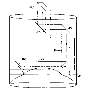
6 Fig. 14 illustrates a further embodiment of the
7 invention wherein a curved optical element 140, such as a
8 planar mirror with a curved inferior border, replaces the
9 deflector M~ of the embodiment of Figs. 6-7. The
cross-sectional projection 142 of the curve of the mirror 140
11 may correspond to the desired post-operative shape of the
12 altered cornea. In the embodiment of Fig. 14, element 140,
13 which may be one of a series of specially configured mirrors,
14 deflects laser beam 141 into a fat, finitely-thick
curved-profile beam 143 to effect the above-described
16 tangential corneal ablation resulting in a particular curve
17 shaped cornea. In some embodiments, a wide variety of-such
18' curved elements 140 may be provided in a ~kit~ such that a
19 particular element may be chosen to fit a particular corneal
profile. The curved profile of element 140 avoids the need to
21 provide rotation of the`~aiming element in the direction of
22 arrow 77 in Fig. 6 while maintaining the curved pattern of the
23 striking laser beam. Translation of curved element 140 in the
24 direction of arrow 146 (Fig. 14) may be provided using a
mechanism similar to that described for providing translation
26 of element M, in Figs. 6-7.
27 Figure 16 similarly illustrates an embodiment o~ the
28 invention wherein a curved profile laser beam is provided to
29 effect corneal ablation. In the embodiment of Fig. 16, the
laser beam 163 deflected by a rectangular mirror 160 passes
- 14 -
4~i7'7
1 through a mask 161 having a curved opening 164 provided
~ therein. The resultant contoured laser beam 163 has a
3 cross-sectional profile 162 which matches the desired
4 post-operative corneal contour. A variety of masks may be
provided in "kit~ form to match a variety of desired corneal
6 , proiles.
7 Figure 15 illustrates a further emodiment of the
8 invention using only two rotating deflective members 150, 151
9 to direct the laser beam to strike the cornea 61 tangentially.
In this embodiment, light from the laser source strikes
11 incident element 150 which deflects the beam to element 151.
12 The geometric relationship between the first element 150 and
13 the second element 151 eliminates the need for the intermediate
14 element M2 of the embodiment of Figs. 6-7. Alternatively,
the curved optical element 140 of Fig. 14, or the masked
16 element 160, 161 of Fig. 15, may be provided as the second
17 deflector in this two-element embodiment.
18 " Since the laser beam in an apparatus according to the
19, invention is preferably maintained consistently tangential to
the corneal surface, only the portion of the cornea that
21 actually touches the be~m is subject to ablation. Studies have
22 indicated that precise control of corneal cutting by an
23 ultraviolet laser can yield negligible effects on corneal
24 tissue immediately adjacent to the beam, yielding extremely
precise effects. Particularly preferred are ultraviolet lasers
26 such as the excimer type of far-ultraviolet laser. One example
27 is a Model 201E excimer laser from Lambda Physik, Gottingen,
28 west Germany. Such lasers, charged with argon-fluoride gas,
29 have been shown to be precise at wavelengths of 193 nm. The
laser output of such a laser may be pulsed with typical pulse
- 15 -
4~77
energies of more than 300mJ at a repetition rate of as much as
~ 400 pulses per second. Alternatively, radiation from a
3 frequency doubled or quadrupled Nd:YAG laser (Quanta-Ray DCR)
4 may be employed giving frequencies in the ultraviolet range.
As the light emitted from a laser is coherent,
6 tangentiality can be monitored utilizing perpendicular light
7 scattering techniques for detecting when a laser beam
8 intersects the surface of the cornea tangentially. Fig. 13
9 illustrates the function of such a light scatter detector 130
which may be provided in an apparatus according to the
11 invention so as to be in close proximity to the cornea 61
12 during operation of the invention. A first output signal may
13 be provided by the light scatter detector 130 when a laser beam
14 132 is in a non-tangential pattern relative to the cornea 61
lS and a second output signal may be provided to the computer when
16 the laser beam 132 intercepts the cornea tangentially.
17 Preferably, the laser source 72 (Fig. 7) of the
18 invention will be a non-thermal laser such as an ultraviolet or
19 excimer laser as described above. However, thermal or
infra-red lasers can also be used. The ultraviolet lasers are
21 currently peeferred as ~hey provide precise beams of energy
22 which break apart protein bonds, ablating or vaporizing the
23 cornea as opposed to burning the cornea as caused by thermal
24 lasers. Of course, a wide variety of laser or other radiation
sources may be provided within the spirit and scope of the
26 invention.
27 Fig. 9 illustrates the high degree of computer
28 coordination which can be provided in an apparatus according to
29 the invention. For example, pre-operatively, data can be ~ed
into the system computer 90 to determine whether an operation
- 16 -
1 according to the invention is viable. Such data can include
~ pre-operative refraction data 93 to determine how much
3 refractive error needs to be corrected, keratometric data 91 in
4 terms of the diopters of corneal refractive power, and
pachymetric data 92 or the measurements of the pre-operative
6 corneal thickness. Using known calculation techniques, the
7 computer can determine whether the procedure will be viable and
8 accordingly signal the operator.
9 Intra-operatively, the computer 90 may also be used to
monitor intraoperative data to insure that the system is
11 operating safely and effectively. For example, the computer 90
12 can monitor the visual a~i5 determination 97, fixation 96, as
13 well as intra-operative autorefraction 95; Individual
14 apparatuses for determining these individual conditions are
generally known.
16 Additionally, the computer 90 operates the laser
17 including signalling of the laser 72 (Fig. 6) to emit the laser
18, beam 73, controlling the frequency and speed of rotation of the
19,l arm 70 and controlling the rotation and translation of variable
mirror M, or the translation of element 140 (Fig. 14). It
21 should be understood th~t the degree of lathing and the length
22 of the lathing procedure will depend on various factors,
23 including the strength and the diameter of the laser beam, the
24 speed of arm rotation and the number of arm rotations
required. Thus, accurate computer assistance will preferably
26 be provided in an apparatus according to the invention to
27 coordinate the many variables and provide the various
28 calculations.
29 Fig. 11 illustrates the operation of an apparatus
according to the invention. First, pre-operative refraction
- 17 -
1~4~77
1 data is provided to the system computer to establish whether
~ hyperopia or myopia is present and to determine how much
3 refractive error to correct. Reratometry and pachymetry data
4 are then provided to the computer to determine whether the
patient has enough corneal tissue to safely carry out the
6 lathing procedure. If not enough corneal tissue is available
7 to safely carry out the procedure, then the operator will be so
8 noti~ied and the laser procedure will be disabled. Depending
9 upon the pre-operative data, beginning positions of the
variable mirror will be established. If sufficient corneal
11 tissue is present, the computer may then be set to check that
12 fixation has been maintained. If fixation has not been
13 maintained, the laser will be disabled. If fixation is
14 present, the laser will be activated. Note that at any time
fixation is lost, the computer will automatically disable the
16 laser. While the laser is activated, the tangent monitor will
17 be constantly operating to determine whether the laser is
18 tangential to the cornea. If the laser is not tangential to
19 the cornea, the variable element will be adjusted until
tangentiality is achieved. Tangentiality may be constantly
21 monitored through eithe`r system interrupts or polling
22 procedures. Assuming tangentiaiity is achieved, and the laser
23 lathing process is under way, the system autorefractor will
24 constantly monitor the intra-operative refractive state of the
patient until either emmetropia or a pre-set condition has been
26 reached. At this point, the laser will be deactivated.
27 The invention thus provides a method and apparatus ~or
28 reshaping the cornea of the patent in vivo without the need ~or
29 removing, reshaping and then suturing the modified corneal
tissue back in place. In addition, with the invention, there
- 18 -
4~77
-
1 is no need for freezing the corneal tissue prior to reshaping
~ it, although the technique could easily be used with corneal
3 tissue in the frozen state in conjunction with known lamellar
4 keratoplasty techniques.
In the invention, radiant energy, from either a
6 non-thermal laser such as an ultraviolet or "excimer" laser or
7 a thermal laser such an an infra-red laser can be used to
8 modify the corneal curvature, and thereby its refractive
9 properties, in vivo or in vitro to shave away or vaporize part
of the optical zone of the cornea in a precisely calibrated and
11 predictable manner. The invention obviates the need for blades
12 for cutting or mechanical lathing and also obviates the need
13 for suturing or unpredicably scarring the cornea of the patient
14 as was required in various prior known methods. Additionally,
since the incident laser beam touches the cornea only
16 tagentially, rather than perpendicularly, changes in the lasers
17 power output would not carry the risk of perforation of the
18 ~ cornea, as there would be no additional tissue in the path o~
19 the beam to be further exposed to damaging irradiation. Thus,
the delicate intraoccular structures would not be directly
21 exposed to the effects bf the laser. The invention thus
22 provides a safe, effective and reproducible method and
23 apparatus for modifying corneal refractive properties.
24 Although the invention has been described in great
detail above by way of reference to the accompanying drawings,
26 it should be understood that a wide variety of embodiments may
27 be provided within the spirit and scope of the invention and
28 that the invention should not be limited to the specific
29 embodiments herein disclosed, but should be interpreted only in
accordance with the claims which follow.
-- 19 --