Note: Descriptions are shown in the official language in which they were submitted.
~.2~3~i33~3 ~
ANALYTE DETECTION BY MEANS OY ENERGY TRA~SFER
BACRGROUND OF TEIE INYEN'rIOR
The present invention relates to a method for
determining the presence of an analyte by means of an
energy transfer that results in the generation of
bathochromic and/or delayed fluorescence emission.
luorescence radiation, emitted from a first energy
emitter (El), is absorbed by a second energy emitter
(E2). This second energy emitter emits fluorescence
radiation of a longer wavelength than the first energy
emitter. The second energy emitter may in addition emit
fluorescence for a substantially longer period than the
first energy emitter ~in a delayed manner). The
detection of either the bathochromic fluorescence or of
any ~luorescence ater a time period during which
fluorescence radiation from background sources has
decayed verifles the presence of the analyte.
Methods for the _-vitro detection of analytes are well
known in the art. The methods include the formation of
antibody-antigen complexes (immunodetection), and the
formation of nucleic acid complexes (polynucleotide
hybridization). The analyte can be an intact cell or a
component of the cell. Examples of analytes are
bacteria, viruses, antigens, antibodies, and
polynucleotides.
The immunoassay for detecting antigen (or antibody)
analytes is well established in the art. The assay
involves the formation of antigen-antibody complexes.
In radioimmunoassay (RIA), a radioactive isotope is used
to report the presence of the analyte. In enzyme
immunoassay, chromogen or fluorescence generated by
--1--
~.2~
means of an enzyme is used to report the presence of the
analyte. Several enzyme immunoassays are currently is
use. They include the enzyme multiplied immunoassay
technique tEMIT) and the enzyme-linked immunosorbent
assay (ELISA). The ELISA method comprises the
"sandwich" technique for antigen, the antibody assay,
and the competitive assay for antigen.
A typical ELISA assay using the sandwich technique is
carried out by adsorbing an antibody to the surface of a
support. The test specimen is added to the support and
the antigen allowed to complex to the antibody. Unbound
antigen is washed away. An enzyme-conjugated antibody
is added and allowed to react with a different set of
determinants on the bound antigen which are not blocked
by the support-absorbed antibody. After the reaction,
the excess of unbound enzyme-linked antibody is washed
away and a substrate of the enzyme is added to the
support. The generation of a colored product indicates
the presence of the antigen in the test speci~en. See
Enzyme Immunoassays by S. 8akerman in Laboratory
Management, August 1980, p. 21.
A drawback of these methods is that they cannot be
carried out in one-step, to achieve detection, i.e., by
adding the antibody to the antigen or the antigen to the
antibody. One or more washing steps are required to
remove antibody unbound to antigen tor vice versa).
Also, a number of these methods involves compe~ition
kinetics which in some instances can provide ambiguous
results.
Polynucleotide hybridization assays using a
polynucleotide probe for verifying the presence o~ a
target polynucleotide analyte is a well known method.
l~ybridization is based on complementary base-pairing~
--2--
3~ ~
When single-stranded polynucleotide probes are incubated
in solution with single-stranded target polynucleo~ides,
that are immobilized on a support complementary base
sequences pair to form double-stranded hybrid molecules.
The double-stranded hybrid molecules remain immobilized
on the support while unbound polynucleotide probe
molecules are washed off. See M. Grunstein and J.
Wallis, ~ETHODS IN ENZYMOLOGY, volume 68, R.W.U (Ed)
(1979) pp. 379-469; A.R., Dunn, and J. Sambrook, MET~ODS
IN ENZYMOLOGY, volume 65; part 1, (1980) pp. 468-478;
Modified Nucleotides And Methods Of Preparing And Using
The Same by D.C. Ward, ~.A. Waldrop, and P.R. Langer,
European Patent Publication Number 0,063,879 published
November 3, 1982; DNA Probes for Infectious Disease by
- A.J. Berry and J.B. Peter, Diagnostic Medicine (March,
1984~ pp. 1-~3; and ~eco~binant DNA Technology;Some
Applications In Clinical Microbiology by Wie-Shing ~ee
and James L. Bennington, Laboratory Manage~ent (April,
19~5) pp. 21-26.
The polynucleotide probes generally comprise a
polynucleotide segment and a signalling segment which is
attached to the polynucleotide. The polynucleotide
segment of the probe has the ability to base-pair, i.e.
hybridize to a sequence of interest, namely the analyte
~5 or target polynucleotide. The signalling segment of the
probe has or produces the means by which the presence of
the analyte moiety can be verified. The m~ans can be,
for example, fluorescence, phosphDrescence,
radioactivity, chromogen, or electron density.
The method of detecting the presence of a target
polynucleotide generally involves several steps or~e of
which is the separation of hybridized polynucleotide
probe from unhybridized probe. The separation can be
facilitated by immobilizing either the probe or the
--3--
ii3~ ~
targ~t onto a solid support. Typically, dou~le-stranded
polynucleotides are isolated from a sample suspected of
containing a target polynucleotide. The double-stranded
polynucleotides are cut into smaller segments by means
of restriction endonuclease enzyme digestion, the
segments are separated by gel electrophoresis, and the
segments are transferred from the gel onto a support,
for example, nitrocellulose paper. Alternatively, the
double-stranded polynucleotide are fixed directly onto
the support without any prior enzyme digestion. The
fixed polynucleotides are contacted with a solution
containing the polynucleotide probe, and the support is
heated to about 80-90C to denature the polynucleotide
double-strands. ~The double-strands can alternatively
be denatured by means of alkali). The system, which now
contains the denatured target polynucleotide and the
polynucleotide probe, is allowed to cool to an
appropriate temperature to allow hybridization to take
place. After sufficient time has elapsed for
hybridization to be complete, which can be for ten
minutes to several hours, the fixed target
polynucleotide is washed to remove all unbound
polynucleotide probes. The signalling moiety of the
polynucleotide probe is now detected, either directly,
for example, by means of radioactivIty or fluorescence,
or indirectly, for example, by means of a chromogen
formed through an enæymatic reaction.
A drawback of this method is that it requires several
steps before the presence of the ta~get polyi~ucleotide
can be verified. Namely, it requires the fixation of
the target polynucleotide to a support, the contacti.ng
of the target polynucleotide with a polynuc~eotide
probe, and the removal of all unhybridized
polynucleotide probes from the support. Besides being
time consuming, the method is not readily a~enable to
8533~
automation and requires some expertise for obtaining
reproducible results. In addition, hybridiza~ion and
detection of the target polynucleotide in a one phase
system is not possible.
One method seeking to overcome the above drawbacks by
detecting the presence of a target polynucleotide with a
homogenous (one-step or one phase) nucleic acid
hybridization assay has been reported. The method
comprises hybridizing first and second single-stranded
polynucleotides, both of which contain light-sensitive
labels, with a complementary single-stranded
polynucleotide target from a sample such that
non-radiative energy transfer occurs between the
light-sensitive labels of the f;rst and second
polynucleotides. At least one of the light-sensitive
labels is of the absorber/emitter type such tha~ energy
absorbed by this label from the emission of the other
light-sensitive label is reemitted at a different
wavelength. These secondary emissions can only occur if
hybridization of both the first and second
single-stranded polynucleotides to the target
polynucleotide has taken place. The quantity of the
target polynucleotides in the sample is related to the
amount of secondary light emitted. See European Patent
2S Publication No. 0,070,685 by Michael Ja~es Heller,
published January 26, 1983.
A drawback of this method is that it reguires two
se~arate polynucleotide strands to detect the presence
of a target polynucleotide. In addition, the method
requires the presence of a chemiluminescent catalyst, ar,
absorber/emitter moiety, and chemiluminescent reagents
effective for causing light emission in the presence of
the chemilumi~escent catalyst. Furthermore, only one
label can be attached per polynucleotide probe because
;3~
the light-sensi~ive label is attached to the sugar
moiety of a terminal nucleoside. Also, the bulky labels
may prevent hybridization of the bases adjacent to the
labels.
S Another method for detecting the presence of a target
polynucleotide by means of a homogeneous assay has been
recently reported. The method involves forming a hybrid
between the target polynucleotide and the polynucleotide
probe, wherein the hybrid has binding sites for two
specific binding reagents, one of which comprises a
first label and the other a second label. The
interaction of the first and second labels provide a
detectable response which is measurably different when
the two labeled reagents are both bound ~o the same
hybrid, as compared to when the two labeled reagents are
not so bound. The formation of the hybrid assay product
brings the two labels within approximate interaction
distance of one another, e.q., as in the cases of
sequential catalyst ~enzyme~ interaction and energy
transfer. Since the labels provide a response which is
distinguishable when the labels are associated with a
hybridized probe, no separation step is required. See
European Patent Application No. 0,144,914 by James P.
Albarella et al., published November 29, 1984.
The method has two main embodiments. The first
embodiment involves the generation of a component which
subsequently produces a color. This embodiment has a
drawback in that it requires the use of two ~istinct
chemical reactions, namely, the reaction of ithe first
label to produce a diffusible mediator produ~t, and the
reaction of the mediator product with the se~ond label
to yield a detectable product. In addition, detection
depends on the formation and maintenance of a higher
localized concentration of the mediator product in t:he
;330 ~
vicinity of the first label as compared to elsewhere in
the solution. Furthermore, both reactions eequire the
use of bulky enzyme molecules attached to the
polynucleotide probe. These bulky molecules may
sterically "clash" with each other.
S '
A second embodiment involves that of energy transfer,
namely the emission of photons from a first label, for
example, fluorescence, followed by absorption of the
photons by a second label, to either quench the
emission, or to provide a second emission. This has a -
drawback in that when an intercalator is the first
label, it is attached to the polynucleotide probe
covalently. In addition, the method requires the
formation of two complexes, namely the formation of a
polynucleotide/polynucleotide complex, and the formation
of an antigen/antibody complex. Furthermore, one aspect
involves the quenching of emitted photons, and since
hybridization of probe to target is usually no more than
a few percent, such minute quenching would produce
ambiguous results.
Fluorescence detection is wide~ly used in hybr~idization
assays. In fluorescence spectroscopy the substance to
be determined which is present in a liquid or a solid
phase is subjected to a radiatior~ vith a known spe~ctral
distribution, for instance~ light with a limited band
width. The fluorescent radiation thereby emitted has a
longer wavelength than the exciting radiation and this
radiation is specific for the substance to be
determined. The mea~urement of the ir~tensity of the
fluorescent radiation constitutes a quantification o
the substance to be determined. Fluorescent moieties
attached to polynucleotide probes are most eficient
when they have a high intensity, a relatively long
emission wavelength~(more than 500 nm), a high Stoke's
3S33~ ~,
shift, and the ability to be bound covalently to a
polynucleotide probe without negatively affecting its
hybridization capabilities. Aromatic agen~s used in
biological systems that give a rather strong
fluorescence and are relatively stable include, for
example, fluorescenisothiocyanate (FITC), rhodamines
(RBITC, TRITC, RB-200-SC), dansil chloride (DNS-Cl), and
fluorescamine (FL~.
Fluorescence is generally measured with a spectro-
fluorimeter. A disadvantage of current methods for
detecting signalling moieties with spectrofluorimeters
is that the detection sensitivity is limited because of
interfering fluorescence or noise in the exciting and
detecting systems that increases the background.
Interfering fluorescence is generated from substances
such as substrate molecules, non-specifically bound
compounds sample holders, air particles, and the
intrinsic fluorescence of the biological sys~em.
The background is also affected by a heavy scattering
which gives rise to an interference, especiaIly when
aromatic organic agents with a small Stoke's shift ~less
than 50 nm) are used.
Several approaches have been described that attempt to
overcome the background problem with fluorescence
detection One approach, described in U.S. Patent No.
4,058,732, measures delayed fluorescence using a
signalling moiety comprising a substance with a
fluorescence emission having a duration that
considerably exceeds the duration of the fluorescence of
the noise sources. A laser pulse is used to excite a
sample, and the detection of the fluorescence from the
signalling moiety takes place only when a sufficiently
long time has passed for the fluores~cence fr~om the noise
sources to have decayed. This method has drawbacks in
--8--
~.z~533a~ ~
that i~ is not readily adaptable to co~mercial use, and
is not amenable for a homogenous assay.
A second approach, described in U.S. Patent No.
4,374,12~, by E. Soini and I. Hemmilia, discloses a
method for determining the presence of an antigen by
attaching a first ligand to an antibody, complexing a
lanthanide metal to the first ligand, and complexing a
second ligand to the lanthanide metal. The
antigen-containing sample is fixed to a support,
antibodies are then contacted with the sample, and
unbound antibodies are washed away. A radiation pulse
of short durati~n is used to excite the second ligand.
Energy is transfered from the triplet state of this
ligand to the chelated metal which emits radiation at a
longer wavelength and for a longer time period than the
noise sources. Detection of this delayed fluorescence
verifies the presence of the antigen. This method has a
drawback in that it cannot be carried out in one step;
all unbound antibodies must be washed away from the
support-
BRIEF SUM~ARY OF T~E INVENTION
.
It is an object of this invention to provide a method
for detecting an analyte by complexing it to a binding
entity comprising a first partner of an energy transfer
system, wherein the formation of the complex induces or
allows for the localization of a reporting entity
comprising a second partner of the energy transfer
system within a proximate distance of the first pa~tner
so that energy emitted by one partner, the energy ~onor
or El, can be absorbed by the other partner, the elnergy
acceptor or E2, and wherein, the f~uoresce!nt energy
emitted by the second partner i5 of longer wavelength
than that emitted by the first partner and in addition
~ 35330 ~
may have fluorescence (~ ~,ub~-tanti~]l~ gr~ater durati.on
than the first partner or o the 'aac~:ground
fluorescence.
It is another object of this invention to provide a
-' method for detecting an analyte by complexing it to a
binding entity comprising a first energy emitter (El),
wherein the formation of the complex induces or allows
for the locali~ation of a reporting entity comprising a
second energy emitter (E2) within a proximate distance
of El so that energy emitted by El can be absorbed by
E2, and wherein, the fluorescent energy emitted by E2 is
of longer wavelength than that emitter by the El and in
addition may have fluorescence of substantially greater
duration than El or backgro~nd fluorescence.
It is an additional object of this invention to provide
a method for detecting an analyte by comple~ing it to a
binding entity eomprising a second energy emitter (E2),
wherein the formation of the complex induees or allows
2~ for the localization of a reporting entity eomprising a
first energy emitter (El) within a proxi~ate distanee of
E2 so that energy emitted by El can be absorbed by E2,
and wherein, the fluorescent energy emitted by E2 is of
longer wavelength than that emitted by the El and in
addition may have fluoreseenee of substantially greater
duration than El or baekground fluoreseenee.
It is another object of this invention to provide a
method for deteeting the presence of an antigen in
solution by complexinq it to a specifle antibody
eomprising an E2 (or El), eontaeting the formed complex
with Clq ~of eomplement) comprising an El (or E2) or a
seeond antibody comprising an El (or E2) to form a unit,
3r irradiating the El with appropriate energy, and
~ measuring the fluorescellce emission.
--10--
~.285330
It is a further object of t~lis invel)ti.on to provide a
method for detecti~g the pres(-llcl- of a~ ntigen by
fixing the antigen to a support, contacting the antigen
with a solution containing a speci~ic antibody
' comprising an E2 (or El) to form an antigen/antibody
complex, contacting said complex with Clq comprising an
El (or E2) or a second antibody comprising an El (or E2)
to form an entity, irradiating the El with appropriate
energy, and measuring the fluorescence emission.
' O
It is an additional object of this invention to pro~ide
a method for detecting the presence of an antigen in
solution by fixing a specific antibody comprising an ~2
~or El) to a support, contacting the antibody with a
solution containing the antigen to form an
antigen/antibody complex, contacting said complex with
Clq comprising an El (or E2) or a second antibody
comprising an El (or E2) to ~orm an entity, and
measuring the fluorescence emission.
It is also an object of this invention to provide a
method for detecting the presence of an antigen by
fixing the antigen to a support which has attached to it
the El (or E2), contacting the support with a solution
containing an antibody comprising an E2 (or El),
allowing the antibody to complex with the antigen,
irradiating the El with appropriate energy, and
measuring the fluorescence emission.
It is a further object of this invention to provide a
method for detecting the presence of a target
polynucleotide in solution by hybridizing it ~o a
polynucIeotide probe comprising an E2, permitting an E
to intercalate into the formed hybrid, irradiating the
El with appropriate energy, and measuring the
1~'8~
fluorescence emission.
It is also an object of this invention to provide a
method for detecting the presence of a target
polynucleotide by fixing the target polynucleotide to a
support, contacting the target polynucleotide with a
solution containing a polynucleotide probe comprising an
E2 to form a hybrid, permitting an E1 to intercalate
into the formed hybrid, irradiating the ~1 with
appropriate energy, and measuring the fluorescence
emission.
It is another o~ject of this invention to provide a
method ~or detecting the presence of a target
polynucleotide by fixing a polynucleotide probe
comprising an E2 to a support, contacting the
polynucleotide probe with a solution containing the
target pol~nucleotide to form a hybrid, permitting an
E1 to intercalate into the formed hybrid, irradiating
the E1 with appropriate energy, and measurin~ the
fluorescence emission.
It is yet another ob~ect of this inventlon to provide a
method for detecting the presence of a target
polynucleotide by fixing the target polynucleotide ta a
support which has attached to it the E1 tor E2),
contacting the support with a solution containing a
polynucleotide probe comprising an E2 ~or E1),
allowing the target polynucleotide to hybridize to the
polynucleotide probe, irradiating the E1 with
appropriate energy, and measuring the fluorescence
emission.
It is an additional object of this invention to provide
a method for detecting the presence of a target
polynucleotide in solution by hybridizing it to a
-12-
polynucleotide probe comprising a hapten, binding an
antibody specific for the hapten or for a specific
double-stranded polynucleotide comprising an E1 (or
E2) to said hybrid to form a complex, contacting said
complex with Cl~ comprising an E2 (or E1) to form an
entity, irradiating the E1 with appropriate energy,
and measuring the fluorescence emission.
A method is disclosed herein for detecting the presence
of an analyte in a homogeneous or one-step assay. The
assay can ~e carried out either in one phase (liquid) or
in two phases (li~uid and solid). The method comprises
first complexing an analyte with a binding entity. The
binding entity and the analyte can both be dissolved in
the li~uid phase or one o~ them can be dissolved in the
liquid phase and one of them can be ~ixed to a solid
support. A reporting entity which is dissolved in the
li~uid phase or comprises the solid support, is then
brought into contact with the complex to ~orm a unit.
The analyte comprises an antigen, antibody, or
polynucleotide. The binding entity comprises a
recognition segment and a signalling segment. The
recognition segment comprises an antibody, antigen, or
polynucleotide. The signalling segment comprises either
an E1 (an energy donor) or an E2 (an energy
acceptor). The reporting entity comprises an E1 or an
E2 depending on what the signalling entity does not
comprise. The actual composition of the binding entity
and the reporting entity depend on the composition of
the analyte and the embodiment used for carrying out the
detection.
The EI and E2 constitute the two partners in the
energy transfer system. The E1 or E2 can be either
a fluorescent aromatic agent or a lanthanide metal.
~13-
~.~8533 [)
When the E1 is a fluorescent aromatic agent, then the
E2 can be a fluorescent aromatic agent or a lanthanide
metal. When the E1 is a lanthanide metal, then the
E2 must be a fluorescent aromatic agent.
The E1 always absorbs the initial energy and then
emits some of this energy at a wavelength which is
absorbed by the E2. The E2 then emits some of this
energy as ~luorescence of a longer wavelength than the
E1 and in addition may emit fluorescence whose
duration considerably exceeds the duration of the E1
and of the bacl~ground fluorescence. The presence of
this bathochrornic and/or delayed fluorescence emission
indicates the presence of the analyte.
BRIEF DESCRIPTION OF THE FIGURES
Figure la depicts the detection of an analyte antigen in
solution with a ~inding entity comprising an antibody
and the E2 and a reporting entity comprising Clq and
the E1.
Figure 1~ depicts the detection of an analyte antigen
fixed to a solid support with a binding entity
comprising an antibody and E1 and a reporting entity
comprising Clq and the E2.
Figure lc depicts the detection of an analyte antigen
fixed to a solid support with a binding entity
comprising an antibody and the E2 and a reporting
entity comprising the solid support and the E1.
Fj.gure ld depicts the detection of an analyte target
polynucleotide in solution with a binding entity
compri`sing a complementary polynucleotide and the E2
and a reporting entity comprising an intercalating agent
-~ -14-
:,
;33(3
as the E1.
Figure le depicts the detection of an analyte target
polynucleotide fixed to a solid support with a binding
entity comprising a complementary polynucleotide and the
E2 and a reporting entity comprising an intercalating
agent and the E1.
Figure lf depicts the detection of an analyte target
polynucleotide fixed to a solid support with a binding
entity comprising a complementary polynucleotide and the
E2 and a reporting entity comprising the solid support
and the E1.
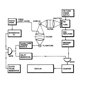
Figure 2 shows a schematic diagram of a fluorimeter
which can be used to carry out the detection an analyte
with a probe comprising a lanthanide metal.
DETAILED DESCRIPTION OE THE INVENTION
1. GENERAL DESCRIPTION OF THE INVENTION
This invention discloses an homogeneous assay for
determining the presence of an analyte. An homogeneous
assay, also known as a one-step assay, permits the
detection of an analyte upon the contacting of the
analyte with a binding entity and a reporting entity
(and other components) in an assay medium. There is no
need to remove unbound binding entities from the assay
medium before detection can be achieved.
The method comprises the use of a first energy emitter,
the E1 (energy donor), and a second energy emitter,
the E2 (energy acceptor). The E2 is capable of
absorbing some o~ the energy emitted by the E1. The
complexing of the binding entity to the analyte causes
or allows the reporting entity to contact the formed
.~
-15-
complex to form a unit. The formation of this unit
places the E1 sufficiently proximate to the E2 such
that energy emitted by the E1 can be absorbed by the
E2. The E2 emits its absorbed energy as
fluorescence of a longer wavelength ~bathochromic) than
the fluorescence of the E1, and in addition, ma~ emit
fluorescence of substantially greater duration (delayed)
than the E1 (or other background fluorescence). The
presence of this bathochromic and/or delayed
fluorescence indicates the presence of the analyte.
The method is applicable to the detection of analytes
which include, for example, antigens haptens,
antibodies, hormones, enzymes, or polynucleotides, and
can be carried out in a one phase system i.e. in a
s,olution, or in a two phase system, i.e. in a solution
o~er a solid support. The detection is carried out by
forming a complex between the analyte a~d a binding
entity.
The binding entity contains a recognition segment and a
signalling segment. The recognition segment is the part
of the binding entity that complexes to a part of the
analyte. The signalling segment is the part that is
involved in the ~ormation of an energy-transfer system
to produce a signal indicating that recognition of the
analyte by the binding entity has occurred. If the
analyte is an antigen, then the binding entity comprises
an antibody. If the analyte is an antibody, then the
binding entity comprises an antigen. If the analyte is
a target polynucleotide, then the binding entity
comprises a complementary polynucleotide. The
signalling segment comprises either the E1 or the
E2. The E1 can be a fluorescent aromatic agent; the
E2 can be a fluorescent aromatic agent or a lanthanide
metal.
-16-
3533~
The reportin~ entity comprises either the E1 or the
E2. When the signalling segment comprises the E1,
then the reporting entity comprises the E2. When the
signalling segment comprises the E2, then the
reporting enti-ty comprises the E1.`
In some embodiments of the assay, all of the components
are dissolved in a solution (liquid phase). In other
embodiments, one or more of the components are fixed to
a solid support while the remaining components are
dissolved in a solution. A number of various
embodiments are described below. These embodiments are
not meant for limitation.
1. The analyte is an antibody and the binding
entity comprises an antigen and the E1.
The E2 is attached to Clq (o~ complement)
or to an antibody. All the components are
dissolved in the liquid phase.
2. The analyte is an antibody and the binding
entity comprises an antigen and the E2.
The E1 is attached to the Clq or to an
antibody. All the components are dissolved
in the liquid phase.
3. The analyte is an antigen and the binding
entity comprises an antibody and the E1.
The E2 is attached to Clq or to an antibody.
All the components are dissolved in the liquid
phase.
4. The analyte is an antigen and the binding
entity comprises an antibody and the E2.
The E1 is attached to the Clq or to an
antibody. All the components are dissolved
in the liquid phase.
~T
-17-
~ ~85330
5. The analyte is an antibody and is fixed
onto a solid support. The binding entity
comprises an antigen and the E1. The E2
is attached to Clq or to an antibody. Both
the binding entity and the Clq or antibody
are dissolved in the liquid phase.
6. The binding entity comprising an antigen and
the E1 is fixed onto a solid support. The
analyte is an antibody. The E2 is attached
to Clq. Both the analyte and the Clq or
antibody are dissolved in the liquid phase.
7. The analyte is an antibody and is fixed onto
a solid support. The binding entity comprises
an antigen and the E2. The E1 is attached
to Clq or to an antibody. Both the binding
entity and the Clq are dissolved in the liquid
phase.
8. The binding entity comprising an antigen and
the E2 iS fixed onto a solid support. The
analyte is an antibody. The E1 is attached
to Clq or to an antibody. Both the analyte
and the Clq or antibody are dissolved in the
liquid phase.
9. The analyte is an antigen and is ~ixed onto
a solid support. The binding entity comprises
an antibody and the E1. The E2 is attached to
Clq or to an antibody. Both the--binding entity
and the Clq or antibody are dissolved in the
liquid phase.
10. The binding entity comprising an antibody and the
-18-
~r
~.~8533~
E1 is fixed onto a solid support. The analyte
is an antigen. The E2 is attached to Clq or to
an antibody. Both the analyte and the Clq or
antibody are dissolved in the liquid phase.
11. The analyte is an antigen and is fixed onto
a solid support. The binding entity comprises
an antibody and the E~. The E1 is attached to
Clq or to an antibody. Both the binding entity
and the Clq or antibody are dissolved in the
li~uid phase.
12. The binding entity comprising an antibody and the
E2 is fixed onto a solid support. The analyte
is an antigen. The 1 is attached to Clq or to an
antibody. Both the analyte and the Clq or antibody
are dissolved in the liquid phase.
13, The analyte is an antibody and is ~ixed onto
a solid supporc. The binding entity comprises
an antigen and ~he E1. The E2 is attached
onto the solid support. The binding entity is
dissolved in the liquid phase.
14. The analyte is an antibody and~is~ixed onto
a solid s~pport. The binding entity comprises
an antigen and the E2.__~he El is attached
onto the solid support. The binding entity is
dlssolved in the liquid phase.
15. The analyte is an antigen and is fixed onto
a solid support. The binding entity comprises
an antibody and the E1. The E2 is attached
onto the solid support. The binding entity is
dissolved in the liquid phas~. ~
-19-
~'
~3533(~
16. The analyte ls an antigen and is fixed onto
a solid support. The binding entity comprises
an antibody and the E2. The E1 is attached
onto the solid support. The binding entity is
dissolved in the liquid phase.
17. The analyte is a target polynucleotide and the
binding entity comprises a complementary
polynucleotide and the E2. The E1 is either
an intercalating agent or attached to an
intercalating agent. All the components are
dissolved in the liquid phase.
18. The analyte is a target polynucleotide and
the binding entity comprises a complementarY
polynucleotide, a hapten attached to the
polynucleotide, and an E2 which is attached
to an antibody bound to the hapten. The El
is an intercalating agent or attached to an
intercalating agent. All the components are
dissolved in the liquid phase.
19. The analyte is a target polynucleotide and is
fixed onto a solid support. The binding entity
comprises a complementary polynucleotide and the
E2. The ~1 is an intercalating agent or
attached to an intercalating agent. Both the
binding entity and the E1 are dissolved in the
liquid phase.
20. The binding entity comprislng a complementary
polynucleotide and the E2 is fixed onto a solid
support. The analyte is a target polynucleotide.
The ~1 is an intercalating agent or attached to
an intercalating agent. Both the analYte and the
E1 are dissolved in the li~uid phase.
-20-
$
.
33~3
21. The analyte is a target polynucleotide and is fixed
onto a solid support. The binding entity comprises
a complementary polynucleotide, a hapten attached
to the polynucleotide, and an E2 which is
attached to an antibody bound to the hapten. The
E1 is an intercalating agent or attached to an
intercalating agent. The binding entity, the
antibody, and the E1 are dissolved in the liquid
phase.
22. The binding entity comprising a polynucleotide,
a hapten attached to the polynucleotide, and a
E2 which is attached to an antibody bound to
the hapten is fixed onto a solid support. The
analyte is a target polynucleotide. The E1 is
an intercalating agent or attached to an
intercalating agent. The analyte, the antibody,
and the E1 are dissolved in the liquid phase.
23, The analyte is a target polynucleotide and is fixed
onto a solid support. The binding entity comprises
; a complementary pol~nucleotide and the E1. The
E2 is fixed onto the solid support. The binding
entity is dissolved in the liquid phase.
24. The analyte is a target polynucleotide and is fixed
onto a solid support. The binding entity comprises
a polynucleotide and the E2. The E1 is fixed
onto a solid support. The binding entity is
dissolved in the liquid phas~e.
The method of the assay involves irradiating a
fluorescene-emitting agent (E1), generally an aromatic
agent, causing some of its electrons to "jump" to an
excited state. This agent emits fluorescent energy when
-21-
~' '
~ 28~;33~
its electrons return to the ground state. Some of this
energy can be absorbed by a proximate lanthanide metal
or another fluorescent aromatic agent (E2), which then
emits some of this energy also as fluorescent energy.
However, the fluorescent energy of the E2 is emitted
at a longer wavelength (batho~romic) than the
fluorescence of the E1 and in addition, the
fluorescence energy of the E2 may last longer than the
fluorescence of the E1 or is "delayed" as compared to
that of the E1. Thus, the detection of bathochromic
and/or delayed fluorescence indicates the presence of
the analyte.
Important limitations are that the radiation energy used
to excite the E1 must be absorbed only by the E1 and
not by the E2, and that the ~1 is brought within the
required proximate distance of E2 only if the binding
entity is complexed to the analyte. Therefore, the
concentrations of the E1 and the E2 should not be of
a value that they are placed within the required
distance of each other even without the analyte first
complexing to the binding entity. The required distance
between the E1 and the E2 should not be greater than
about the Furster's radius, preferably not more than
about 3OA.
By way of illustration, an example of a one phase assay
where the analyte is an antigen is the addition of a
binding entity comprising an antibody as the recognition
segment and a chelator-lanthanide metal complex (E2)
as the signalling segment, and a reporting entity
comprising Cl~ and a fluorescent aromatic agent (E1),
to a solution comprising the test antigen. The
concentration of the E1 and E2 is such that random
diffusion of the E2 does not place it sufficiently
proximate to the E1 that the E2 can absorb energy
emitted by E1, The complexing of the antibody to
antigen, however, allows the Clq to blnd to the formed
-22-
,
. . ,, ~ ' .
... .
~.~8533~)
complex. This brings the E1 (which is attached to the
Cl~) within a distance of E2, that energy emitted by
the E1 is absorbed by the E2. Irradiation of the
E1 with energy of the appropriate wavelength induces
the E1 to emit fluorescent energy. Some of this
energy is absorbed by the E2 which then emits some of
this energy as fluorescent energy of a long waveIength
as compared to the wavelength of the fluorescent energy
emitted by the E1 and also in some instances as
delayed fluorescent energy. This emitted fluorescence
can then be measured. If test antigen was not present
in the sample, then no complex comprising antigen and
antibody would be formed to which Cl~ could bind. No
E1 would thus become localized proximate to the E2,
no energy would be transferred from the E1 to the
E2, and accordingly, no fluorescent energy shift or
delayed fluorescence would be observed.
An example of a ~wo phase assay where the analyte is an
antigen is the addition of a solution containing the
binding entity comprising the E2 to a solid support
onto which the antigen has been fixed. The E1 is
provided in one of two ways. The first way is the
addition to the solution of a reporting entity
comprising Clq and the E1. The reporting entity binds
to the complex to form a unit. The second way is the
attachment of the E1 onto the solid support by means
of a linker arm. Upon the formation of the
antigen/antibody complex on the support to form a unit,
the linker arm permits the E1 to be sufficiently
proximate to the E2 that an energy transfer can occur.
If antigen was not present in the sample, then no
complex would be formed, and accordingly, the E1 -
attached to the support would not be sufficiently
proximate to the E2 that an energy transfer from E
to E2 could occur.
-23-
~ ?af~85330
,~n example of a one phase assay where the analyte is a
target polynucleotide is the addition of a binding
entity (binding entities for polynucleotides are
generally known as polynucleotide probes) comprising a
polynucleotide as the recognition segment and a
chelator-lanthanide metal complex (E2j as the signalling
segment, and a reporting entity comprising a fluorescent
aromatic intercalatiny agent (El) to a solution
comprising the test target polynucleotide. The
concentration of the E1 and E2 is such that random
diffusion of the E2 doesn't place it sufficiently
proximate to the El that the E2 can absorb energy
emitted by the El. The hybridization of the
polynucleotide probe to the target polynucleotide to
produce a target polynucleotide/ polynucleotide probe
hybrid, however, allows the El to intercalate into this
hybrid. This intercalation brings the E~ withi~ a
distance of E2, that energy emitted by the El is
absorbed by the E2. Irradiation of the El with energy
of the appropriate wavelength induces the El to emit
2~ fluorescent energy. Some of this energy is absorbed by
the E2 which then emits some of this energy as
fluorescent energy of a longer wavelength and also in
some instances as delayed fluorescent energy. If target
polynucleotide was not present in the sample, then no
hybrid comprising target polynu~leotide and
polynucleotide probe would be formed into which E1 could
intercalate. No El would thus become localized
proximate to the E2, no energy wo~ld be transferred fron~
the El to the E2, and accordingly, no fluorescent energ~
shift or delayed fluorescence would be observed.
An example of a two phase assay where the analyte is a
target polynucleotide is the addition o~ a solution
containinq the polynucleotide probe comprising ~he E2 t~a
3~ a solid support onto which the target p~lynucleotide has
-24-
d ~S33~
been fixed. The E1 is provided in one of two ways.
The first way is the addition to the solution of a
reporting entity comprising a fluorescent aromatic
intercalating agent. The second way is the attachment
of a fluorescent aromatic agent onto the solid support
by means of a linker arm. The agent need not be an
intercalating agent. Upon the formation of the target
polynucleotide/polynucleotide probe hybrid on the
support, the linker arm permits the E1 to be
sufficiently proximate to the E2 that an energy
transfer can occur. If target polynucleotide was not
present in the sample, then no hybrid would be formed,
and accordingly, the E1 attached to the support would
not be sufficiently proximate to the E2 that an energy
transfer from E1 to E2 could occur.
2. DESCRIPTION OF THE BINDING ENTIT~
A. THE BINDING ENTITY COMPRISES
AN ANTIGEN OR A~TIBODY.
1. THE RECOGNITION SEGMENT
This is the portion of the binding entity which
recognizes a structure or shape of a segment of the
analyte and thus enables the binding entity to form a
complex with the analyte. When the ana~yte is an
antigen then the recognition segment comprises an
antibody. When the analyte is an antibody, then the
recognition segment comprises an antigen.
The reaction of antibodies (Ab) with antigens (Ag) is a
well known and described reaction in the field of
immunology. An antigen has two properties: (a)
immunogenicity, i.e., the capacity to stimulate the
formation of the corresponding antibodies, and (b) the
-25-
~.~8~330
ability to react specifically with these antibodies.
Haptens are substances that are not immunogenic but they
react selectively with antibodies of the appropriate
specificity. They provide antigenic determinants to an
antigen molecule. Antibodies are proteins that are
formed in response to an antigen and which react
specifically with the antigen. All antibodies belong to
a special group of serum proteins called
immunoglobulins.
The antibody should be specific for at least one
antigenic determinant site or epitope on the antigen.
The antibody is prepared by exposing immunoglobulins to
the antigen. Methods of purifying antibodies are based
on the dissociability of antibody/ligand complexes. At
least two steps are usually involved: (1) Antibodies are
precipi-tated from the serum with soluble anti~ens or
absorbed by insoluble antigenic materials; (the latter
are often prepared by coupling smaIl haptenic groups or
soluble proteins to an insoluble matrix, such as
agarose); and (2) After the extraneous serum is washed
away, the antibodies are eluted from the insoluble
complexes by means of specific or nonspecific
procedures.
A number of antibodies can be purified by specific
procedures. With aggregates whose stability depends
largely on specific ionic interactions, such as those
involviny types 3 and 8 pneumococcal polysaccharides,
strong salt solutions (e.g., 1.8 M MaCl) elute purified
antibodies effectively. When the specific antigenic
determinants are simple haptenic groups, such as
2,4-dinitrophenol, small univalent haptens that
encompass the crucial part of the determinant (e.g.
2,4-dinitrophenol) are useful for competitive
displace~ent from the precipitating antigen or
-26-
.
.
~533~
absorbent, yielding soluble antibody hapten complexes.
Depending upon the properties of the antigen, the
absorbent, and the hapten, diverse procedures are then
used to isolate the soluble antibody-hapten complexes
and finally to separate the hapten from the antibody
(e.g., ion-exchange resins, dialysis, gel filtration).
When small univalent haptens are employed for specific
elution of antigens it is desirable to use those haptens
that are both 1) weakly bound by the antibody and 2)
highly soluble. Highly concentrated solutions of hapten
can then be used to elute the antibody in high yield,
and the weakly bound hapten is easily separated from the
soluble hapten-antibody complex, e.~., by dialysis or
gel fLltration.
Nonspecific procedures are used for the isolation of
other antibodies to protein antigens. It is usually
necessary to expose specific antigen/antibody aggregates
to conditions that cause reversible denaturation of the
antibody, allowing it to dissociate from the antigen.
Organic acids at pH 2 to 3 are often effective; various
procedures are then used to separate the denatured
antibody and antigen, depending u~on the properties of
the antigens. Since antibodies usualIy regain their
native structure on being restored to physiological
conditions, neutralization of the antigen-free material
yields active antibody, usually without excessive losses -
due to persistent denaturation.
Though antibodies can be isolated from serum in high
yield (50 to 90%) and with high purity (90% of the
recovered antibodies usually react specifically with
anti~en) the purified molecules are usually
heterogeneous with respect to affinity and with respect
to many other physical and chemical properties.
-27-
~3?
.
'
~.~8~rj33~1
Antigens generally comprise proteins, polysaccharides,
or polynucleotides. A variety of methods are available
for purifying antigens. They include chromatography,
electrophoreses, centrifugation, and immunodiffusion.
These methods are well known to one skilled in the art.
2. THE SIGNALLING SEGMENT
This is a moiety of the binding entity which is involved
in the generation of a signal by means of energy
transfer. The signal consists either in the emission of
bathochromic fluorescence with respect to the El or in
the emission of delayed fluorescence. The presence of
the signal indicates the presence of the analyte.
The signalling segment is attached to the recognition
segment. The attachment can be by covalent or
non-covalent means. The attachment can also be through
a linker arm. The signalling segment comprises either
an El or an E2. The El is generally a fluorescent
aromatic agent; the E2 is either a fluorescing
aromatic agent or a lanthanide metal. Details of
attachment of the signalling segment to the recognition
segment are described hereinbelow.
B. THE BINDING ENTITY COMPRISES
A POLYN~CLEOTIDE G
1. THE RECOGNITION SEGMENT
This is a moiety of the binding entity that recognizes
the structure of a target polynucleotide, and comprises
a polynucleotide. This type of binding entity is known
in the art as a polynucleotide probe. The target
-28-
~.~8533~
polynucleotide complexes with the polynucleotide probe
to form a target polynucleotide/polynucleotide probe
hybrid.
The polynucleotide portion of the polynucleotide probe
comprises at least one single-stranded base sequence
substantially complementary to the base sequence to be
detected (target polynucleotide). The sequence should
comprises at least about twelve bases to impart
specificity to the probe. However, such a base sequence
need not be a single continuous complementary
polynucleotide sequence, but can be comprised of two or
more individual complementary sequences interrupted by
non-complementary sequences. In addition, the
complementary region of the probe can be flanked at the
3'- and 5' termini by non-complementary sequences, such
as those comprising the DNA or ~NA of a vector into
which the homologous sequence had been inserted for
propagation. In ei.ther instance, the probe as prese~ted
as an analytical reagent will exhibit detectable
hybridization at one or more points with sample nucleic
acids o~ interest.
.
Methods for preparing a polynucleotide that is
`substantially complementary to a tar~et polynucleotide
are~well known and routine in the art.~ The most
commonly used methods are that of recombinant DNA and
cloning. One widely used vector is the M13 phage.
Brie~ly, the~method entaiIs (1)~ c~eaving the M13 RF
- ~ ~(replicative~form) DNA with the~one~o~ the restriction
enzymes having a unique recognition sequence in the
cloning region (2) ligating the desired polynucleotide
into the cleaved insertion site (3) transformation E.
coli host cells (4) growing these host cells on
nutrient-containing plates and selected the colorless
-29-
:
: : . ' - '
~.~8533(~
plaques (5) amplifying the phages from single plaques in
small cultures (6) harvesting the phages from culture
supernatant and removing the protein coat by treatment
with phenol, and (7) precipitating the purified DNA with
ethanol. Greater detail can be found in M13 CLONING AND
SEQUENCING HANDBOOK Published by Amersham Corporation
(1983) and in MOLECULAR CLONING by T. Maniatis, E.~.
Fritsch, and J. Sambrock, published by Cold Spring
Harbor Laboratory (1982).
Specific polynucleotides can also be prepared with a DNA
Synthesizer Instrument such as on manufactured by
Applied Biosystems, 850 Lincoln Centre Drive, Foster
City, California 94404, using the appropriate nucleotide
precursors. According to the manufacturer, one can
prepare polynucleotides of about 120-200 bases with
great specificity. The synthetic schemes involve the
use of phosphoramidites to link together predetermined
bases. Other manufacturers of polynucleotide
synthesizers include Biosearch Inc., 2980 Kerner
Boulevard, San Rafael, CA 94901, and Beckman
Instruments, 1050 Page Mill Road, Palo Alto, CA 943904.
The polynucleotide can also be prepared by the method of
nick translation. This~method involves removing
selected bases from double-stranded polynucleotides and
replacing some of them with other predetermined bases.
This method however produces a double-stranded probe.
Since this invention requires the use of single-stranded
probes, as discussed hereinbelow, where only one of the
two complementary strands are present during the assay,
the two strands of the probe must be separated from each
other. This can be achieved by column chromotography
using, for example, metylated albumin columns. The
separation however depends on the two strands having
different ratios of G-C/A-T. Thus, where the G-C
-30-
~r
~l.?~85~3~
content of the probe is about 50%, nick translation
cannot be used to prepare the probe.
2. THE SIGNALLING SEGMENT
This is a moiety of the binding entity that is involved
in the generation of a signal by means of energy
transfer. The signal is the production of bathochromic
and/or delayed fluorescence. The signalling segment is
attached to the recognition segment o~ the binding
entity. The signalling segment can be attached to the
recognition segment directly or through a linker arm.
The signalling segment can also be attached to the
recognition segment covalently or non-covalently. An
example of covalent attachment is where a chelator/metal
complex is attached by means of an allylamine to a
complementary polynucleotide. The allylamine is the
linker arm. An example of non-covalent attachment is
where a chelator/metal complex is covalently attached to
an antibody, and the antibody is non-covalently bound to
a hapten which is covalently attached to the
complementary polynucleotide. In this instance, the
hapten and antibody comprise the linker arm.
;
The signalling segment is either an E1 or an E2.
The E1 is generally an aromatic fluorescence-emitting
agent while the E2 is an aromatic
fluorescence-emitting agent or a lanthanide metal.
Details of attachment of the signalling segment to the
recognition segment are described hereinbelow.
3. POL~NUCLEOTI~E PROBE FORM
A bathochromic and/or delayed fluorescence emission
should only occur when the polynucleotide segment of the
~polynucleotide probe is hybridized with the target
-31-
~8S~3~
polynucleotide. The shift or delay in fluorescence
should not occur in the presence of hybrids if not one
of the hybrid strands is that of the target
polynucleotide. The target polynucleotide to which the
polynucleotide portion of the polynucleotide probe
hybridizes must be one originating from the sample.
Thus, the polynucleotide probe must be provided to the
sample single-stranded and none of the prov~ded
single-strands should be complementary to each other.
If the polynucleotide probe is provided to the sample
double-stranded and then denatured in the sample, the
signalling segment of the probe will assist in the
generation of a shift in fluorescence emission or of
delayed fluorescence when one polynucleotide probe
strand hybridizes with the complementary probe strand to
which it was originally hybridized. This will produce a
false positive result.
The formation of hairpin loops can also result in the
production of a false positive result when the reporting
entity comprises on interelating agent. This can be
minimized by using polynucleotide probes not longer than
about 30 base se~uences, or by carrying out the assay at
elevated temperatures or under stringent conditions.
It is preferable that the polynucleotide probe comprise
an integral strand. That is, bathochromic and/or
delayed fluorescence emission should be generated with
the assistance of the signalling segment upon the
hybridization of only two strands. This permits the
detection of a target polynucleotide with only one
polynucleotide probe molecule. However, there may be
instances where the polynucleotide probe will comprise
two different polynucleotide strands. This can be, for
example, where each polynucleotide strand contains
different signalling segments and the two polynucleotide
-32-
; .
3~
strands hybridize to adjacent non-overlapping sequences
on the target polynucleotide. The signalling segment of
each strand itself does not produce a detectable
bathochromic and/or delayed fluorescence, but the
interaction of the two signalling segments together,
produces a detectable bathochromic and/or delayed
fluorescence. Such a situation is contemplated as being
covered by this invention.
3. THE REPORTING ENTITY
This is an entity other than the binding entity which
comprises one partner of the energy transfer system.
The partner can be either the E1 or the E2. The
reporting entity and the binding entity together
comprise a unit. The unit contains the means for
generating an energy transfer system, because one part
of the unit com~rises the E1 and the other part of the
unit comprises the E2. The reporting entity also
comprises a component which can be either Clq, an
antibody, an aromatic intercalating agent, or a support,
depending on the assay. The energy transfer partner of
the reporting entity is attached to this component which
can be either to Clq or to an antibody, or to an
aromatic intercalating agent when the analyte is in
solution, and can also be attached to a support when the
analyte or binding entity is fixed to a support. The
E1 must be brought within the required proximate
distance of the E2 only when the binding entity is
complexed to the analyte. Thus, the concentrations of
the E1 and the E2 should preferably be such that
random diffusion does not place significant amounts of
the two within a distance that the E2 can absorb
energy emitted by the E1. Amounts are considered
significant if they are greatly increase the background
and make accurate measurements difficult.
-33-
. : .
3~
Clq is one of the complement proteins. Complement (C)
is now known to consist of 11 proteins. The proteins
make up about 10% of the globulins in normal serum of
man and other vertebrates. These proteins are not
immunoglobulins (IGA), and they are not increased in
concentration by immunization. They react with a wide
variety of antibody-antigen (Ab-Ag) complexes, and exert
their effects primarily on cell membranes, causing lysis
of some cells and functional aberrations in others,
e.g., degranu~ation of mast cells with release of
histamine ! increased permeability of small blood
vessels, directed migration of polymorphonuclear --
leukocytes, increased phagocytic activity by leukocytes
and macrophages, and bacteriolysis. The 11 proteins of
~ complement are Clq, Clr, Cls, and C2-C9. Cl~ is the
; recognition unit of the complex. It consists of five
subunits, each with one binding site for the heavy
chains o~ those Ig classes (e.g., IgG-1, IgG-2, IgG-3,
IgM) that can trlgger the entir.e C sequence. Unlike
~- other C proteins, Clq has stable combining sites and
requires no activation. The clq has a striking chemical
similarity to collagen, i.e., it has~a high content of
glycine, hydroxyproline, and hydroxy~lysine, with a
galactose-glucose disaccharide attached to the hydroxyl
`or hydroxylysine, and it can be inactivated by
collagenase.
The E1 or E2 can be attached to Clq when the analyte
is an antigen or antibody. The Clq does not bind either
to antigens or to antibodies individually. Only
following the complexing of the antigen to the antibody
does the Clq bind to the formed complex. Thus, for
example, when the assay is carried out in solution, the
Clq will bring the E1 (or E2) within the required
distance of E2 ~or E1) only when the antigen analyte
-34-
: :
.
~.~8533~
is bound to the antibody binding entity comprising the
E2 (or E1). In the presence of analyte, bindin~
entity, and Clq, irradiation of the E1 with
appropriate energy will result in a transfer of energy
from the E1 to the E2. Methods for attaching an
E1 or E2 to Clq are similar to those used for
attaching linker arms which are described hereinbelow.
The E1 or E2 can be attached to an antibody when the
analyte is an antigen, antibody or polynucleotide. The
analyte polynucleotide includes RMA/DNA, RNA/RNA and
DNA/DNA hybrids. The antibody would be one that would
not bind to either the analyte or binding entity
individually. The antibody would only bind to a complex
comprising the analyte and t~e binding entity. The
reporting entity would thus comprise an antibody to this
complex and an E1 or E2
The isolation of an antibody that is only specific for a
complex is readily achieved by one who is skilled in the
art. It involves the isolation of antibodies from
~animal of an inbred strain, and creating a tolerance in
one of these animals for the particular antigen. An
antibody can be isolated that is specific only for the
analyte/binding entity complex. An E1 or E2 can be
attached to the particular antibody by the method
described hereinbelow.
The E1 or E2 can be attached to a support when the
analyte is an antigen, antibody or polynucleotide. The
support can be glass, plastic, cellulose, or a gel
matrix (such as sepharose). The E1 or E2 can be
attached to the support by means of a linker arm. Some
support may need to be siliconized prior to the
attachment of a lin~er arm.
-35-
_~
~.~85330
The E1 can be a fluorescent aromatic interelating
agent that is unattached to any other moiety when the
analyte is target polynucleotide. This is when the
intercalating agent emi-ts fluorescence at a wavelength
which can be absorbed by the E2. ~owever, if the
intercalating agent does not emit at a wavelength at
which the E2 can absorb, then the E1 can be
attached to an aromatic intercalating agent. The E
can be attached to the intercalating agent. The E1
can be attached to the intercalating agent by means of a
linker arm. The intercalating agent becomes inserted
into the hybrid formed from the target polynucleotide
(analyte) and the polynucleotide probe (binding entity).
This all.ows the E1 to lie at the periphery of the
double helix adjacent to on E2 which is part of the
polynucleotide probe. An energy transfer ~rom the E
to the E2 can then occur. Without prior
~ybridization, no energy trans~er occurs.
4. DESCRIPTION OF_LINKER ARM
A. GENERAL DESCRIPTIOM
The signalling segment is generally attached to the
recognition segment of the binding entity by means of a
linker arm so that there is minimal steric interference
between the signalling and recognition segments of the
binding entity, and so that the signalling segment
allows the E1 to be within the required distance of
E2. The linker arm refers to the fragment in the
binding entity attaching the signalling segment to the
recognition segment.
B. THE LINKER ARM WHEN THE RECOGNITION
SEGMENT COMPRISES AN ANTIBODY OR ANTIGEN.
In this embodiment, the linker arm attaches a
-36-
~353~
fluorescent aromatic agent or a chelator-metal complex
to either an antibody or an antigen. The linker arm
should not be one that interferes, however, with the
formation of an antigen/antibody complex.
Antibodies and/or antigens comprises a number of primary
and secondary amino and hydroxy functional groups. Some
antigens also comprise one or more sulfhydryl groups.
The covalent attachments of a linker arm by means of
electrophilic addition to most of these functionalities
would not significantly interefere with the formation of
an antibody/antigen complex since the active site of the
antibody comprises, relatively speaking, only a few of
these atoms. Functional groups by which a linker arm
can be attached to an antibody or antigen and other
characteristics of the linker arm are described
hereinbelow in section C.
C. THE LINKER ARM WHEN THE RECOGNITION
SEGMENT COMPRISES A POLYNUCLEOTIDE
In this embodiment, the linker arm attaches a
fluorescent aromatic agent or a chelator-metal complex
to a polynucleotide. The linker arm should be one that
does not substantially interefere with the hybridization
of the polynucleotide probe to the target
polynucleotide. Therefore, the linker arm and/or
chelator: (A) should not prevent the base to which it
is attached from pairing with its complementary base;
(b) should not prevent the complexing of the
complementary bases, so as to prevent the hybridization
of the polynucleotide probe to the target
polynucleotide; (c) should not prevent the
incorporation of nucleotides to which the linker arm is
attached by the polymerase enzymes (unless it is at a
terminal position of the polynucleotide sequence); and
-37-
~ r~
35330
(d) preferably, should not change the conformation of
the sugar moieties in the polynucleotide.
The lin~er arm is generally attached covalently to the
polynucleotide, but can also comprise non-covalently
attached moieties. The attachment is preferably to the
base moiety, although it can be to the sugar moiety, or
the phosphate moiety. The base moiety can be either a
purine or a pyrimidine. As mentioned hereinabove, the
attachment of the linker arm to the base moiety should
preferably be to a position at which the linker arm does
not interfere with Watson-Crick pa_ring of the bases.
Suitable positions are, for example, positions 5 and 6
of uracil, positions 5, 6, and the exocylic 4-amino of
cytosine, Positions 7 and 8 of deazapurine, position 8
of guanine, and positions 8 and the exocyclic 6 amino of
adenine. A preferred linker arm for attachment to the
base moiety is allylamine. See European Patent
Publication Mo. 0,063,879 by David Ward et al.,
published November 3, 1982.
Preferred positions on bases are the 5 and 6 positions
of pyrimidines and the 7 position on deazapurines, since
8-purine nucleotides are poor substrates for the
polymerase enzymes, and the exocyclic amino group of
either adenine or cytosine is involved in base-pairing
to thymine and uracil, or the guanine respectively,
Although a substituent at an exocyclic amino group of a
base does not prevent that base from pairing to its
complementary base in some instances, the substituent
may alter the optimum orientation between the two bases.
Preferred pyrimidines are uracil and cytosine, with 5
being the preferred position. Preferred purines are
deazaadenine and deazaguanine.
. .~
~ ?.d~533~
D. METHODS FOR ATTACHING A LINKER ARM
In the instance when the recognition segment is an
antigen, any condition which does not result in the
modification or blocking of required epitopes is
satisfactory. In the instance when the recognition
segment is an antibody, any condition which does not
denature the antibody or result in the modification of
the active site is satisfactory. In the instance when
~he recognition segment is a polynucleotide, any
condition which does not result in the modification or
blocking of the functional groups of the bases required
for hybridization or the cleavage of the base from the
sugar is satisfactory. The optimum conditions including
those of pH, temperature, solvent, or reaction time can
readily be determind by one skilled in the art.
The linker arm comprises the group of atoms joining the
recognition segment to the chelator-metal complex or to
the fluorescent aromatic agent. The linker arm can be
joined to the recognition segment by any number of
methods.~ The linker arm must have a first functional
group by means of which it can be attached to the
recognition segment, and a second functional group by
means of which it can be attached to the chelator-metal
complex or fluorescent aromatic agent. The linker arm
can be attached by means of a carbon-carbon single bond,
carbon~carbon double bond, carbon-nitrogen single bond,
carbon-nitrogen double bond, carbon-oxygen single bond,
carbon-sulfur single bond, or carbon-silicon single
bond. Suitable functional groups include but are not
limited to amino groups, thio groups, aklyl sulfates,
and halides.
It is not necessary that the linker arm be attached to
the recognition segment as one fragment. The linker
-39-
330
arm can be constructed by attaching a first fragment to
the recognition segment, followed by the attachment of a
second fragment to the first fragment. Examples of
suitable first fragments are:
-CH=CH-CH2-NH-; -CH=CH-CH2-CH2-SH; and
-cH=cH-cH2-o-cH2-cH2-NH
Examples of suitable second fragments are:
N-O-~-R; R-~-OR; R-C-O-~-R
N-hydroxysuccinimide imidates anhydrides
esters
R-N=C~S; and R-C-SR
iso~hiocyanates thioesters
General methods for attaching a linker arm onto a base
of a polynucleotide are discussed in J. L. Ruth and D.
E. Bergstrom, J. Org. Chem., 43, 2870 (1978); D. E.
Bergstrom and M. K. Ogawa, J. Amer. Chem. Soc. 100,
8106, (1978); and C. P. Bigge, P. Kalaritis, J. R. Deck,
and M. P. Mertes, J. Amer. Chem. Soc. 102, 2033 (1980).
One preferred method is the one disclosed in detail in
European Patent Application Number 0,063,879, by David
C. Ward, et al., published in November 3, 1982, which is
hereby incorporated by reference. The method involves
reacting a linker arm or a linlcer arm fragment
containing an alpha vinyl group with a mercurated base
in the presence of K2PdCl4, wherein the mercury is
-40-
~ .
D
~85330
bound as Hg+ to the position of the base which is to
react the linker arm. The scheme is shown below.
H ~l(~ PJC/~ PJ I~ h~ H~ ~C
R r. 1~ 'Y h~ H~ ~ og~
A ccT~c~e b u~ficv-
~/1 Y- S '~ s ~/1 B (- ~
There are no particular size or content limitations :Eor
the linker arm, The linker arm can contain from about
two carbons to about any number of carbons, as long as
the chelator is within the required distance from the
recognition segment. The linker arm can contain
heteroatoms and unsaturations. The linker arm can
comprise aliphatic, alicyclic or aromatic moieties. The
actual size or content of the linker arm will depend on
the recognition segment to which it is attached and on
the chelator-metal complex or fluorescent-aromatic agent
chosen.
; ~
Attachment of the linker arm to the sugar moiety of a
polynucleotide can be my means of a Schiff base to the 1
aldehyde following depurination or depyrimidation of
preselected bases, or it can be to the 2 hydroxy in the
case when the sugar is ribose. The linker arm when
attached to the 1~ aldehyde can comprise, for example,
an amine, hydrazine, or hydrazide functionally.
~1-
~.7~3S330
Attachment of a linker arm to the phosphate moiety can
be by alkylation of the phosphate moiety. See U.S.
Patent No. 4,469,863 by P.O.P.Ts'O and P.S. Miller.
When the linker arm is attached to the base moiety, it
is preferable to attach it to the base at the nucleoside
or nucleotide level. This is because the reaction
conditions that may be required to attach the linker arm
to the base may cause undesirable side reactions to a
polynucleotide. ~urthermore, attachment at the
polynucleotide level may give inconsistent and
irreproducible yields. Attachment at the nucleoside or
nucleotide level permits the modified nucleoside or
nucleotide to first be purified, and then to be
incorporated into a polynucleotide. The incorporation
can be either by cloning, for example, in an M13 vector,
or by synthesis with a polynucleotide synthesizer
instrument as disclosed hereinabove.
For incorporation by an M13 vector, the modified
nucleotide must be a relatively efficient substrate for
the commonly studied nucleic acid polymerases. Thus,
the linker arm should not sterically interfere either
with the active site on the enzyme or with the
complementary base-pairing of the modified nucleotide.
Substitution at positions that alter normal "anti"
nucleoside conformation should also be avoided since
such conformational changes usually render the modified
nucleotide a poor substrate for the polymerase enzymes.
When the linker arm is attached to the 1~ aldehyde of
the sugar, the linker arm must be attached following the
formation of the polynucleotide portion of the
polynucleotide probe. This is because attachment of the
sugar requires a free aldehyde at the 1-position of the
-42-
,
330
sugar. The free aldehyde is formed by depurination or
depyrimidation. A moiety comprising a sugar and
phosphate without a base is not a substrate for the
polymerase enzymes. Thus, the linker arm must be
attached b~ first selectively depurinating or
depyrimidating the desired polynucleotide sequence, and
then attaching the linker arm, to the sugar by means of
the aldehyde. When the linker arm is attached to the 2
hydroxy of a ribose sugar, the linker arm can be
attached at the nucleoside, nucleotide or polynucleotide
level. This is because nucleotides modified by a linker
arm can be incorporated into a polynucleotide by means
of a gene synthesizer instrument. When the linker arm
is attached to the phosphate, the linker arm must be
attached at the nucleoside or nucleotide level so that
the attachment is not at positions other than at the
phosphate.
5. AT~ACHMENT OF THE CHE~ATOR
A chelator is a moiety which can sequester and bind a
metallic cation. The chelator has two or more
functional groups which interact non-covalently with the
metal. The chelator can be a-ttached to an antigen,
antibody, polynucleotide or support. The attachment of
chelator-metaI groups to antibodies is known in the art.
See U.S. Patent No. 4,374,120 by E. Soini and I.
~emmilia. The attachment of me-tal-chelating groups to
polynucleotides is also known in the art. See European
Patent publications: No. 97,373 by D. Engelhardt et.
al., published on January 4, 1984; No. 150,844 by J.
Stavrianoupoulos, published on August 7, 1985, and No.
157,788 by J. Stavrianoupoulos, published on September
18, 1985, which are coassigned to the same assignee of
this patent application.
-43
'~-
~ 8~3~
Examples of chelators, not meant ~or limitation, are
ethylenediaminetetraacetic acid (EDT~) which can be
derived from l-(p-benzenediazonium) EDTA (I);
~1ooc c~ ,coott
~Iooc ~1~ ~ \c~tlc11
~ t
diethylenetriaminepentaacetic acid (DTPA) II;
~ c~o
OC C~la a~ ~ -c~
>~ J / \ Ch~ coc, ~ (~ )
o oc~ \ Ch co~ ~
and trans-diaminocylohexanetetraacetic acid (DCTA) III.
~ cf~,C~o
20 ~' ~ c~,C6~0- ¢~ )
C~t~,c~7o
C~CC)o
Other chelators are listed in "DiEfusion-Enhanced
Fluorescence Energy" by L. Stryer, D.~. Thomas, and C.F.
Meares, Ann. ~er. Biophys. Bioeng. (1982~ 203-32
:
The chelator can be attached ~o the linker arm by a
number of groups. Examples o~ such groups, not intended
for limitation, are:
-0-, -NII-CO, -NH-CNH-, -N=N-, -Nl~-SO2-, -S-,
-O-PO2-O-~ -52-~ -NI~-N=N_, -Nll-CI~ -
-CH2-NII-, -N-, -O-C112-, O-CO-, -Nll-CO-C1~2-S-,
-N~l-CO-cll2-Nll-, --Cl~2-C~2-o- ~ -0-co-cl5
-S-CH2-, and -O-CO-NII.
--44--
.,
~ :
:
` : .
:
:: .
. .
33~)
Varying conditions can be used for attaching a chelator
to a linker arm. Generally, any pH range from about 4
to about 10, preferably from about 5 to about 8, any
temperature from about 20C to about 100C, preferably
from about 40C to about 65C, any solvent, and any
buffer or catalyst can be used as long as the pH,
temperature, solvent, or buffer does not modify any of
the groups or moieties of the antigen, antibody or
polynucleotide. Thus, for example, reagents or
conditions that can depurinate or deaminate the
polynucleotide should be avoided. There are also
relatively few limitations as to reaction times. The
optim~tm pH, temperature, solvent, or reaction time for
attaching a chelator to a linker arm will depend on the
linker arm, the chelator, and the functionalities to be
reacted. The conditions can readily be determined by
one skilled in the art.
The stoichiometry of the reactants required for these
reactions can vary widely. Generally, an excess of the
component that is more easily prepared will be used for
the attachment of the chelator to the antigen, antibody
or polynucleotide. In practice, the amounts will vary
depending upon the required reaction conditions, the
chelator, the linker arm, and their reacting functional
groups.
The chelator can be attached to the linker arm after
incorporation of the linker arm-containin~ nucleotide
into the polynucleotide or before incorporation of the
linker arm-containing nucleotide into the
polynucleotide. The only limitation is that the
chelator cannot be attached before incorporation if it
interferes with polynucleotide synthesis.
-45~
~.'
- .
. ~ ,, :, .
8S33~ ~
The bind;ng entity can comprise one chelator or ~ore
than one chelator. When the recognition segment is a
polynucleotide, the chelator can be attached at terminal
positions or at non-terminal positions of the
polynucleotide probe. The greater the number of
chelators, the more sensitive the binding entity will
be. However, the chelators should not be present in
such numbers that effective complexing of ~he analyte to
the binding entity is substantially prevented. The
number of chelators that can be attached will depend on
the composition, the size and length of the recognition
segment.
6. DE:SCRIPTION OF T~ LANTEIAWIDR 2iET~L
Certain lanthanide metal chelates~fluoresce for a time
period considerably longer than aromatic compounds. Two
such metal chelates are europium and terbium which
fluoresce for about several milliseconds. Tebium (Tb~3)
emits in the 480 to 630 nm range and europium (Eu*3)
emits in the 580 to 700 nm range. Both have long
excited-state ~ifetimes because of the forbidden nature
of the transition between their ground state and lowest
excited state. The absorbance coefficients of these
lanthanides are of the order of 0.1 M-l cm 1 compared
with 103 to 105 M-l cm 1 for most fluorescent organic
chromophores. This longer fluorescence permits the
detection of these metals after background ~luorescence
due to aromatic compounds has decayed. These metal
chelates have additional advantages in that their
absorbance is very strong (about 104), their excitakion
maximum is within the short uv range (terbium chelates
are excited at 270-320 and about 488 nm, while europium
chelates are excited at 320-360 and about 580 nm), their
excitation maximum are independent of the complexed
ligands which makes it possible to excite them with
-46-
,
~8S~ 0 j~
commercially available lamps or lasers, their emissions
can be monitores with a narrow band width, and they have
the ability to laser in different solutions and at
~, different temperatures.
The fluorescence emission of the lanthanide metal
chelates can arise from the absorption of energy by an
energy absorbing species which can be a proximate ligand
or aromatic compound of excitation radiation, conversion
of this energy from the singlet state to the triplet
state, and transfer of this energy from the
energy-absorbing species to that of the metal. The
energy is then emitted as by the metal fluorescence for
]5 a relatively long interval at a narrow band width and
long wavelength characteristic of metals. The
fluorescence of terbium green while that of europiu~ is
purple.
7~ D}~SCRIPTION OF T1~13 FLUORESCENT AROMAT~ C AGE~T
The fluorescent aromatic agent can he either the El or
the ~2. When it is the El, it must emit fluorescence of
a wavelenth that can be absorbed by the E2. when it is
the E2, it must emit so~e fluorescence at a wavelength
that is longer than that emitted by the El. Detection
of the E2 can be carried ou~ by measuring the
fluorescence with a filter that cuts off all
fluoresecence emission of the~El but allows the longer
wavelength fluorescence of the E2 to pass through.
When both the El and E2 are~ fluorescent aro~atic agents,
any combination of agents which satisfy the criteria
listed in the preceeding paragraph are satisfactory.
3'j When the El is a fluorescent aromatic agent and the E2
is a lanthanide metal, then when the analyte is a target
polynucleotide, the binding entity can comprise, for
-4~-
.
;33~
example, tryptophan as the ~1 It has becn demonstrated
that tryptophan can efectively transer energy to an
E2. Even a single tryptophan residue boond to a
polynucleotide is an excellent ~l because its emission,
centered about ~30 nm, overlaps the absorption of many
, potential energy acceptors, including the lanthanide
metals. See the article by W. D. ~Iorrocks, Jr., B.
~lolm~uist, and B. L. Vallee n Proc. Natl. ~cad. Sci.
USA ~1975), 72: 4763-68. When the analyte is an antigen
or an antibody, the fluorescent aromatic agent can be,
for example, lumichrome, 9-aminoacridine, and auromine 0.
The fluorescent aromatic agent is attached to the linker
arm by means o a suitable functional group. Such
methods are described heeeinabove.
8. D~SCRIPTIO~ OF T~E ~NTERCh~ATING ~GEN~
~ n~mber of fluorescent aromatic agents or dyes are able
to intercalate into double-stranded polynucleotide
helices. The double-strand polynucleotide can be
DNA/DNA, RNA/RNA, or DNA/RNA. These agents show a shift
in fluorescence emission after intercalation into a
double-stranded helix. This shift is caused by a change
in the hydrophobic environment o these agents.
Generally, the intercalating agents are aromatic dyes.
These intercalating aromatic ~yes have a planar ring
structure and have distinct fluorescence emission
spectra. The fluorescence is indicative of the electron
delocalization of the intercalating agent, and is
aected by the inductive effect of substi~uent groups
attached to the dye and by quenching agents.
The result of intercalation is t~le spceading o adjacent
base pairs to about twice theic normal separation
distance, leading to an increase in molecular len~Jth o
-48-
533~
the duplex. Further, unwinding of the double helix of
~bout 12 to 36 degrees must occur in order to accomodate
the intercalator. General reviews and further
information can be obtained from Ler~an, J., Mol. Biol.
3:18 (1961); nloomield et al, "Physical Chemistry of
Nucleic Acids", C~apter 7, pp. 429-476, ~arper and ~owe,
NY (1974~; Waring, Nature 219:1320 (196~3); Hartmann et
al, Angew. Chem., Engl. Ed. 7:693 (196B); Lippard,
Accts. Chem. Res. 11:211 (1978); Wilson, Intercalation
Chemistry (1982), 445; and ~erman et al, Ann. Rev.
Iq Biophys. ~ioeng. 10:~7 (19~31). Exemplary of inter-
calators are acridine dyes, e.g., acridille orange, the
phenanthridines, e.g., ethidium, anthracyclines, e.g.
adriamycin, the phenazines, furocoumarins, phenothiazines,
and quinolines.
9. ~NALYT~
A. ANTIGENS RND ANTIBODIES
This method can be used to detect most antibodies and
antigens. The antibody can be monoclonal or
polynclonal. The epitope of the antigen can comprise a
protein, a carbohydrate, or both. The antigen can
comprise one unit or a number oE subunits. The antigen
can be from a micr~organism, a plant cell, or a
mammalian cell~ The miceoocganism can be a bacteria,
fungus, virus, or yeast. The antigen can be an epitope
of the microorganism or cell, or can be p~oduct secreted
3~ by the microorganism or cell. The antigen can be, for
example, a membrane receptor, a blood cell, or a ~uscle
protein.
B. TARGET POLYNUCLEOTIDE
This method can be used to detect a target
polynucleotide, for example, from a microorgani~m, a
-49-
~.~85330
plant cell, or a mammalian cellO The microorganism can
be a bacteria, fungus, virus, or yeast. The target
polynucleotide can be one that is unique for a
particulac pathogenic virus, one that is present in a
mutated mammalian gene that results in the production of
~j a non-functioning protein, or one that imparts
antibiotic resistance to a bacteria. For example, it
can be one that imparts penicillin resistance in
Streptococcus ~y~ or Neisseria meninqitidis;
. _ __ _ ___ __ _ _
tetracycline resistance in Staphylococcus aureu_,
1~ Candida albicans, Pseudomonas aeru~osa, Streptococcus
Pvoq~es, or eisseria qonorrhoeae; and aminoglycoside
resistance in MYcobacterium tuberculosis.
C. ANALYTE SOURCE
The test sample to be assayed can be any medium oE
interest, and will usually be a liquid sample of
medical, veterinary, environmental, nutritional, or
industrial significance. Human and animal specimens and
body fluids particularly can ~e assayed by the present
method, including urine, blood ~serum or plasma),
amniotic fluid, milk, cerebrospinal fluid, sputum, fecal
matter, lung aspirates, throat swabs, genetal swabs and
exudates, rectal swabs, and nasopharnygal aspirates.
Where the test sample obtained from the patient or other
source to be tested contains principally double stranded
nucleic acids, such as contained in cells, the sample
will be treated to denature the nucleic acids, and i~
necessary first to release nucleic acids from cells.
Denaturation of nucleic acids is preEerably accomplished
by heating in boiling water or alkali treatment (e.g.,
0.1 N sodium hydroxide), which i~ desired, can
simultaneously be used to lyse cells. ~Iso, rele3se of
nucleic acids can, ~oc example, be obtained by
mechanical disruption ~freeze/thaw, abrasion,
sonication), physical/chemical disruption (deter~;ents
such as Triton*, Tween* sodium dodecylsul~ate! alkali
--50--
* Trademark
~3533C~
treatment, osmotic shock, or heat), or enzymatic lysis
(lysozyme, proteinase K, pepsin). The resulting test
medium will contain nucleic acids in single stranded
form which can then be assayed according to the present
hybridization method.
This approach can be extended to the diagnosis of
genetic disorders, such as thalassemia and sickle cell
anemia. The polynucleotide gene whose presence of
absence (in the case of thalassemiaj is associated with
the disorder can be detected following hybridization
with a polynucleotide probe according to this invention.
The mapping of genes or their transcriPtS to specific
loci on chromosomes has been a tedious and
time-consuming occupation, involving mainly techniques
of cell-fusion and somatic cell genetics. Although
_-si.tu hybridization has been employed successfully for
mapping single-copy gene sequences in species that
undergo chromosome polytenization, such as that of
Drosophila, detection of unique sequence genes in most
higher eukaryotic chromosomes has been extremely
difficult, if not impossible, using standard
hybridization methods. The necessity for polynucleotide
probes of very high specific radioactivity to facilitate
autoradiographic localization of the hyridization site
also results in rapid radiodecomposition of the
polynucleotide probe and a concomitant increase in the
background noise of silver grain deposition. The use of
hybridization probes with low to moderate specific
radioactivities requires exposure times of many days or
weeks, even to detect multicopy sequences, such as
ribosomal RNA genes or satellite D~A. ~ince recombinant
DNA technology has made feasible the molecular cloning
of virtually every single-copy sequence found in
eukaryotic cells, it would be extremely beneficial to
have a rapid and sensitive method for mapping the
chromosomal origin of such cloned genomic fragments.
-51-
3533(1
Finally tumor cells can be diagnosed by preparing a
polynucleotide probe according to this invention which
is complementary to the messenger ribonucleic acid
transcribed from a deoxyribonucleic acid gene sequence
associated with the production of polypeptides, such as
fetal protein antigen or carcinoembryonic an~igen, the
presence of which is diagnostic for specific tumor
cells. Hybridization and detection of the probe/target
polynucleotide hybrid would provide a method for
detecting the tumor cells.
10. DETECTION OF DE~AYED FLUORESCENCE
:
The detection of delayed fluorescence can be measured by
means of an instrument shown in Figure 2. This
instrument was developed by Erkki Soini and Hannu
Kojola. See Clin. Chem. 29/1,65-68 (1983). The sample
compartment is covered by a light-tight lid and the
sample is changed manually. The samples are held in
small disposable tubes or cuvettes made of polystyrene,
which has a reasonably low long-decay background
~fluorescencé. Because the intensity of the single
flashes from the xenon flashtube was not very
reproducible, we had to ensure stabilization of the
excitation system. An integrator (P1) for a
semiconductor photodiode serves as the stabilizer of the
flash lamp. The flask lamps is activated about 103
times at a frequency of 1 kHz. The exact number of
flashes (N) is controlled by the integrator P1 so that
the integrated intensity of the photon emission is thus
fixed. For the stabilization detector we used a
photodiode (Model UV-215B; EG and G Inc., Electro-optics
Div., 35 Concress St., Salem, MA 01970), operated in
the photovoltaic mode and connected to the optical
system by a fiber light guide. The integrator is made
of an operational amplifier, which provides a control
signal for the flash tube circuit. The inteyrated
-52-
~3533V
photon emission from the flashtube is stabilized by this
method with a precision of +(1/N).100% assuming that the
deviation of the intensity of single flashes is not
greater than ~50%.
This stabilization method has many advantages. First of
all, the system is simple, the flashtube and its power
supply can be made without any stabiIization circuit and
less expensive flashtubes with lower stability can be
used. The temperature dependence of the system can be
minimized by a single compensator element. The
flashtube is operated only during a measurement, thus
ensuring a long practical life. The eventual fatigue of
the flashtube will be automatically compensated by the
integrator.
.
The pulsed-light source used in this fluorometer was an
FX-198 bulb-type xenon flashtube with a 1.5-mm arc cap
(EG and G Inc.), An EG and G Lite Pac Trigger Module
produced the high-voltage trigger pulses required to
operate the flashtube. We operated the flashtube system
at +6D~ V and a flash duration of 0.5 ~s.
To provide optimal excitation and emission bands, we
used intereference band-pass filters (Ferroperm AS,
Copenhagen, Denmark) mounted inside the sample
compartment for easy and quick replacement.
The detector is a side-window photomultiplier tube
(Model R928; Hamamatsu TV co. ~td., 1126 Ichono-cho,
Hamatsu, Japan) operated with negative-bias voltage,
thus obtaining a direct analog signal between the anode
and ground. We found this to be a practical arrangement
for monitoring the total amount of fluorescence and
obtaining an indication of counter saturation.
The photomultiplier tube, operated in the single-photon
mode, is connected to a fast preamplifier and
. ~
-53-
: ~ . ' . ,' ' ~ , ",
" `
~8~33~
discriminator and to a fast scaler having a digital
display of seven decades. The counting speed of random
events is limited to 40 MHz by the preamplifier and
single-photon discriminator.
ll. MET~OD FOR DET~CTI~G T~E ANALYT~
A. THE ANALYTE IS AN ANTIBODY OR ANTIGEN
The antigen or the antibody is generally purified from
circulating body fluids. In addition, the detection of
the antigen may involve lysing a cell. Antigens and
antibodies are generally purified using, for example,
affinity columns, ammonium sulfate fractionation, in
exchange chromatography, gel eletrophoresis, and
immunodiffusion. The purified antigen or antibody
analyte is added to a solution comprising the binding
entity and the reporting entity. Alternatively, either
the analyte or the binding entity is immobilized t~ a
support and the other components are dissolved in
solution. Furthermore, the reporting enti~y may also
comprise the support. The appearance of bathochromic
and/or delayed fluorescence following irradiation of the
El indicates the presence of the analyte.
B~ THE ANALYTE IS A POLYNUCLEOTIDE
The target polynucleotide is generally isolated from
microorganisms or cells. One method using a
polynucleotide probe, wherein a lanthanide metal is the
signalling segment, is carried out, for example, by
lysing the cells in a sample comprising the target
polynucleotide in a solution to release t~e target
polynucleotide in a solution to release the target
polynucleotide from the surrounding membrane. Ly~is can
be, for example, by exposing the sample to sonication,
or to a detergent. The polynucleotides can be separated
from cell debris by centrifugatlon, and purified further
-54-
~,.
- ~ ' -
. ,'' ' ~ ',
, .
~.~?~ 3~)
by alcohol precipitation, or by dialysis. The
polynucleotide probe is then added to a solution,
containing the target polynucleotide. The appearance of
delayed fluorescence indicates the presence of the
target polynucleotide in the sample.
The target polynucleotide must be rendered in
single-stranded form during the hybridization step
before it can hybridize with the polynucleotide moiety
portion of the polynucleotide probe. This can be
achieved either by heat or by alkali. Typically,
hybridization will proceed at slightly elevated
temperatures, e.g., between about 35 and 75C and
usually around 65C, in a solution comprising buffer at
pH between about 6 and 8 and with appropriate ionic
strength (e.g., 5XSSC where lXSSC = 0.15M sodium
chloride and 0.015M sodium citrate, pH 7.0). In cases
where lower hybridization temperatures are desirable,
hydrogen bonding reagents such as dimethyl sulfoxide and
formamide can be included. The degree o~ complementary
between the sample and probe strands required for
hybridization to occur depends on the stringency of the
conditions. Factors which determine stringency are
known in the art.
Following hybridization, the solution is placed in an
instrument in which the aromatic agent is excited with
photons of the proper wavelength. The fluorescence
emission is then measured after a predetermined time
interval which can vary between 4-6 milliseconds.
The polynucleotide probe has attached to it at least one
chelator. A lanthanide metal, for example, terbium, is
complexed to the chelator. The metal has the ability to
absorb energy emitted at particular wavelengths by
selective fluorescent aromatic agents and furthermore
has the ability to emit fluorescence for time periods
substantially longer than that of the aromatic agents
-55-
~.?~3S330
themselves. The presence of the target polynucleotide
is thus determined by contacting the sample suspected of
containing the target polynucleotide with (1) the
polynucleotide probe to which the chelator is covalently
bound and which also comprises a terbium complexed to
the chelator and (2) a fluorescent aromatic
intercalating agent which emits fluorescent energy
capable of being absorbed by the terbium. The emission
of fluorescence at a longer wavelength than that emitted
by the intercalating agent or of fluorescence after a
given interval during which the fluorescence of the
intercalating agent has decayed indicates the presence
of the target polynucleotide.
This assay, by detecting a target polynucleotide in one
step, avoids many limitations of other detection assays.
For exam~le, since there is no need to remove unbound
probe molec~les, there is no requirement that the
hybridization must withstand various conditions or
manipulations, such as elevated temperatures, phenol and
organic solvent extractions, electrophoresis, column
chromotography or low or high pH.
12. REAGENT KITS
The present invention additionally provide~ a reagent
kit, i.e., reagent combination or means, comprising all
of the essential elements required to conduct a desired
assay method. The reagent system is presented in a
commercially packaged form, as a composition or
admixture where the compatability of the reagents will
allow, in a test kit, i.e., a packaged combination of
one or more containers, devices, or the like holding the
necessary reagents, and usually including written
instructions for the performance of assays. Reagent
systems of the present invention include all
configurations and compositions for performing the
various complexing or hybridization formats described
-56-
~.~8S~30
herein.
The reagent system will generally comprise (1) a binding
entity and a reporting entity. A test kit form of the
system for target polynucleotides, for example, can
additionally include ancillary chemicals such as the
components of the hybridization solution and
denaturation agents capable of converting double
stranded nucleic acids in a test sample into single
stranded form. Preferably, there is included a chemical
lysing and denaturing agent, e.g., alkali, for treating
the sample to release single stranded nucleic acid
therefrom.
Although assays for the detection of several analytes
have been described hereinabove, the assays can be used
~or the detection of other analytes using appropriate
binding entities. Examples of various analyte~binding
entity combinations include, but are not limited to,
lectin/sugar; sugar/lectin; hormone/receptor;
recep-tor/hormone; inhibitor/enzyme; enzyme/inhibitor;
cofactor/enzyme; enzyme/cofactor; ligand/substrate; and
substrate/ligand. It is intended that these
combinations be included within the scope of the
inventions.
-57-
,~
.