Note: Descriptions are shown in the official language in which they were submitted.
~;:86178
The present invention relates to an apparatus and
method of maintaining a sterilized operating field and a
sterilized cassette therefor.
The need to maintain an operating field free from
infectious organisms manifests itself in many ways. Surgical
incisions may become infected from airborne organisms or from
wound exudates. Trauma sites, e.g. injuries, burns etc., may
become infected in the same way. This is particularly true in
the case of long term treatment of a patient by means of
introducing nutrients, medication or the like via a catheter or
cannula.
The present invention provides a sterile cassette
having a open top and open bottom. The open bottom of the
cassette is secured to the dermis to enclose an operating
field, after which the operating field is sterilized by W
light and the open top of the cassette is closed by a sterile
cover. Thereafter, a sterilizing gas is continuously flowed
into the cassette, over the operating field and out of the page
~ ` ~?361'7f3
cassette. The operating field is thus at all times kept free
of infectious organisms as long as the cassette is in place.
In addition, there are many instances where a
catheter is inserted in a blood vessel during diagnosis or
treatment of a patient. Catheters are used to supply fluids
intravenously, such as nutrients, pharmaceuticals, dyes etc.
Catheters are also used in arteries, such as in angioplasty.
In all cases where a needle enters a blood vessel, there is the
- potential for great harm due to infection. This risk of
infection also exists for intramuscular and intralymphatic
intubations.
In medical practice today, it is all but impossible
to avoid accidental introduction of infectious organisms into a
dermal puncture site. When this occurs, the results are
serious and sometimes fatal. This can occur in several ways.
First, the patient may harbour infectious organisms
on the skin, which can be introduced into the body when the
catheter is inserted. Second, airborne organisms can land on
the skin after the catether or cannula is in place and can be
transported into the blood vessel by movement of the catether
or cannula caused by movement of the patient. Third, organisms
can be brought to the puncture site by the patient or medical
personnel. Introduction of infectious organisms can also occur
during removal of the catether or cannula.
The present invention also provides a package
comprising a sterile cassette containing a needle assembly that
lX8~;17~
is used to puncture the skin and a tubing for connection to the
needle assembly after it is inserted. The cassette has an open
top and bottom for enclosing the operating field on the
patient. After the cassette is installed on the patient, the
operating field is irradiated and the open top is closed by a
sterile cover. Thereafter, the needle assembly is inserted and
connected to the tubing without contaminating the irradiated
operating field. To enable the user to manipulate the needle
assembly and tubing after the cassette is closed, a flexible
pouch or hag is provided and the cover is made transparent.
Moreover, a sterilizing gas may be flowed into the cassette as
discussed above.
The present invention is illustrated in terms of its
preferred embodiments in the accompanying drawings, in which:
Fig. 1 is a view in perspective of the cassette of
the present invention with parts broken away for clarity;
Fig. 2 is a plan view of the cassette of Fig. 1 with
the top and bottom covers removed;
Fig. 3 is a plan view of the cassette of Fig. 1 with
the cannula inserted into the skin and attached to the tubing,
the cover shown being folded back only for clarity;
Figs. 4 and 5 are detail views of the tubing and
needle assembly;
Fig. 6 is a detail view in section of the needle
assembly support;
~2861~f~
., ,
Fig. 7 is a detail view of an alternative embodiment
of the invention;
Fig. 8 is a detail view of another alternative
embodiment of the invention;
Fig. 9 is a view in perspective of another cassette
of the present invention with parts broken away for clarity;
Fig. lOA is a detail view in section taken along
lines lOA-lOA in Fig. 9;
Fig. lOB is a view similar to Fig. lOA, but with the
protective sheets of Fig. 9 removed and the cover in place;
Fig. ll is a plan view of the cassette of Fig. 9 with
the cover being shown folded hack only for clarity; and
Fig. 12 is a schematic diagram showing the system for
flowing sterilizlng gas into the cassette.
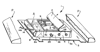
Fig. l shows the cassette l of the invention within
sterile packaging P. Also within packaging P is cover 20 (Fig.
3), which is enclosed within its own sterile packaging (not
shown).
After packaging P is opened and discarded, the user
removes and discards paper or plastic covers 2 and 3 as
described below, which are removably adhesively secured to the
open top 4a and open bottom 4b of wall 4. Fig. 2 shows the
cassette with covers 2 and 3 removed.
Wall 4 is preferably of rigid plastic and is mounted
on and projects from the top surface of the elongated, flexible
8617f3
--5--
support 5, which has an outer edge 5a and an inner edge 5b
defining an opening 6. The interior surface 4c of wall 4 is
located at the inner edge 5b of the support 5 and thus
surrounds the opening 6. Support 5 is suitably made of
flexible plastic or fabric so as to be easily secured to the
skin of a patient. Suitable means is provided, such as
adhesive 5c (Fig. 2) on the bottom surface of support 5, for
this purpose.
As best seen in Fig. 1, wall 4 has an entrance 7 at
one end to permit one to gain access to the interior of wall 4.
Secured to wall 4 is a flexible, transparent bag 8, which is
made of thin but strong plastic, with the open end 8a
hermetically sealed to the cassette 1. Suitably, the open end
8a is sealed to the wall 4 around entrance 7 and to support 5
in front of entrance 7 by means of a suitable adhesive. If the
open end 8a is secured at the entrance 7 as shown, the bag 8
can move freely up or down or from side-to-side. If a portion
of end 8a is secured to the support 5 in front of entrance 7,
the bag 8 can be made taller than shown to provide the desired
degree of vertical movement.
Wall 4 includes a needle assembly support 9
projecting into the interior of the wall 4 adjacent entrance 7.
Removably housed within needle assembly support 9 is assembly A
comprising a hollow needle 10 ~Fig. 5) having a hub lOa and a
pointed end lOb. Detachably connected to hub lOa is syringe 11
1~86~7~
--6--
through a conventional friction fit (Fig. 2). Plastic cannula
12 is telescoped over the needle 10 as is conventional.
As best seen in Fig. 6, needle 10 is removably held
in support 9 with flange lOc between ring 9a and
circumferentially spaced members 9b. Fig. 6 shows the needle
10 held by support 9 with the cannula 12 removed. As can be
seen, members 9a and 9b cooperate with flange lOc to detachably
hold needle 10 in the support 9 whether or not the cannula 12
is carried by the needle 10.
Completing the assembly is tubing 14, which is sealed
to bag 8 and which has an end 14a outside the bag 8 and an end
14b inside bag 8. In particular, end 14b is provided with a
conventional female fitting 14c, which is designed to be
friction fitted within hub 12a (Fig. 3) of cannula 12, as is
conventional.
The cassette 1 is used as follows. First, the
cassette 1 and cover 20 are removed from packaging P, cover 20
being reserved for later use. Bottom cover 3 is removed and
support 1 attached to the skin of the patient. Cover 2 is then
removed and the area exposed within the open top 4a is cleansed
and then irradiated with ultraviolet light in a manner known
` per se. This sterilizes the exposed skin within the operating
field.
Cover 20 is removed from its sterile packaging (not
shown) and is secured to top 4a by suitable means, such as the
bead 21 and groove 22 (Fig. 3). It is noted that Fig. 3 shows
617f~
--7--
cover 20 partially removed. This is for clarity only. Once
cover 20 is installed, it is intended to remain secured to wall
4 until after cannula 12 is removed from the body in order to
maintain the sterility of the operating field at all times.
Needle assembly A is then withdrawn from support 9 by
gripping syringe 11 through flexible bag 8. If desired, bag 8
may have inwardly projecting thumb portion 23 and finger
portion 24 (Fig. 7). The user will hold the syringe 11 with
one hand and will enter a desired blood vessel by means of
pointed end 10b of needle 10, which projects beyond plastic
cannula 12, as is known. When cannula 12 is inserted, syringe
; 11 is used to withdraw blood from the vessel to ensure that a
blood vessel was indeed entered, as is known.
If the cannula 12 was properly inserted into a blood
vessel, syringe 11 and needle 10 are withdrawn, leaving cannula
12 in place. One hand withdraws the syringe 11 and needle 10,
while the other holds the cannula 12 in place, if necessary,
through bag 8. The used needle 10 is then replaced in support
9, as shown in Fig. 6. To minimize growth of microorganisms on
the used needle 10, wall 4 is provided with conduit 25 having
exit ports 26. Inlet 27, made of a self-healing membrane,
closes conduit 25. A suitable anti-microbial agent can be
admitted into the interior of support 9 and into contact with
needle 10 by injecting it into conduit 25 through membrane 27.
Additional self-healing membranes 28 and gas inlet
ports 29 are provided in wall 4 for admitting liquid or gaseous
1.2a6~7~3
media into the interior of the cassette For example,
sterilized gas under atmospheric pressure can be admitted into
the cassette 1 through one port 29 and exhausted from the
cassette 1 by the other port 29, thereby contacting the
operating field and providing the desired effect of sterilizing
the operating field and/or promoting healing. For example,
hish levels of oxygen are known to promote healing and to
prevent growth of anaerobic bacteria. Ozone is known as a
sterilizing gas. Hence, oxygen or ozone, are suitable gases
for use in this aspect of the invention. This is explained in
detail below in connection with Figs. 9-12.
With cannula 12 in place, tubing 14 is connected to
hub 12a by means of fitting 14c. Thereafter, the end 14a of
tubing 14 is connected to a reservoir (not shown) of the
lS desired fluid. End 14a is provided with a conventional cap
(not shown) that is detachably sealed to end 14a to maintain
the sterility of the interior of tubing 14.
While the wall 4 has been shown as rectangular, other
shapes are possible, such as oval or circular.
Where the cassette 1 is used for intramuscular
administration, the syringe 11 may be used simply as a device
for assisting in handling needle 10 and cannula 12 or it may be
omitted from the needle assembly A, in which case needle 10
need not be hollow.
Cassette 1 may be of any convenient size to provide
opening 6 with dimensions suitable for use on the human body,
-` lZ861~1~
such as from about 40 to about 60 mm wide to about 40 to about
90 mm long, depending upon the length of needle 10 and the size
of the area available for the dermal puncture.
Fig. 8 shows a portion of a flexible bag 8 having an
opening or access port 8b closed by lid or cover 8c. Cover 8c
is secured to bag 8 by any rapidly detachable means (not
shown), such as a bead and groove interlock as in elements
21,22 or by a peelable adhesive, in order to gain rapid access
to the operating field via access port 8b in an emergency. Tab
8d facilitates rapid removal of cover 8c.
As seen in Fig. 9, cassette 100 of the invention is
stored within sterile packaging P and comprises a rigid tubular
wall 102 having an open top end and open bottom end, a flexible
bellows-like skirt 103 depending from the bottom end of tubular
wall 102 and a flexible flange portion 104 extending away from
the skirt 103. Protective sheets 105,106 suitably made of
paper or plastic, are removably adhesively secured to the top
of wall 102 and across the entire underside of flange portion
104 (Fig. lOA), respectively. In particular, sheet 105 carries
a pressure-sensitive adhesive (not shown) for removably
adhering to wall 102, whereas sheet 106 removably adheres to
the pressure-sensitive adhesive 104a (Fig. 9) on the underside
of flange portion 104.
Wall 102 is preferably made of rigid plastic so as to
withstand handling during use. Skirt 103 is preferably of
flexible rubber or plastic and may be pleated as shown so as to
12~3~1'7f3
-10-
conform to the area of the dermis of the patient where the
operating field is to be established. Skirt 103 may be secured
to wall 102 by adhesive or by heat welding, as desired. Flange
104 is preferably integral with skirt 103, and is also
sufficiently flexible to conform to the dermis and create a
gas-tiyht seal around the operating field. Adhesive 104a may
be any suitable pressure-sensitive adhesive, such as used in
connection with wound dressings.
Ports 107 communicate with the interior of cassette
100 and are provided with suitable means (not shown) for
attachment to conduits 108 (Fig. 12), which are used to flow a
sterilizing gas into and out of the cassette 100 as will be
described below. Such attachment means may be suitable
fittings, such as used in connecting IV assemblies together as
illustrated in Fig. 11.
Separately stored in its own sterile packaging (not
shown) is transparent cover 120 (Fig. 11), which will be
described hereinafter. Any suitable transparent plastic may be
used for cover 120.
After packaging P is opened, the user removes and
discards protective covers 105,106 from cassette 100 and
secures the cassette 100 to the dermis by means of adhesive
104a on the underside of flange 104. The operating field
within wall 102 is cleansed before or after the cassette 100 is
affixed to the dermis and is irradiated through the open top of
cassette 100 with ultraviolet light in a manner known per se.
~3S1~3
This sterilizes the exposed skin within wall 102.
Alternatively, the operating field may be irradiated with W
light before the cassette 100 is affixed to the patient.
Cover 120 is removed from its sterile packaging ~not
shown) and is secured to wall 102 by suitable means, such as
bead 121 and groove 122 (Fig. 10B). It is noted that Fig. 11
shows cover 121 partially pulled back for clarity only. Once
cover 121 is installed, it is intended to remain secured to
wall 102 in order to maintain the sterility of the operating
field at all times.
With the cassette 100 in place, each conduit 108
(Fig. 12) is attached at one end to a port 107 on either side
of cassette 100 and at the other end to pump 130 and chamber
131, respectively. Pump 130 may be any type of medically
acceptable pump and is used to pump a sterilizing gas, such as
oxygen or ozone, in a closed loop through chamber 131 to
cassette 100 and back to chamber 131. Chamber 131 is provided
with an ultraviolet lamp 132, such as used with drinking water
dispensers, that irradiates the recycled sterilizing gas
exhausted from cassette 100 with an effective dose of
ultraviolet radiation to sterilize the gas.
Sterilizing gas is thus admitted into the cassette
100 through one port 107 and exhausted from the cassette 100 by
the other port 107, thereby contacting the operating field and
providing the desired effect of sterilizing the operating field
and/or promoting healing. For example, high levels of oxygen
lX8G~
-12-
are known to promote healing and to prevent growth of anaerobic
bacteria. Ozone is known as a sterilizing gas. Hence, oxygen
or ozone are suitable gases for use in the present invention.
Sufficient sterilizing gas to start the cycle and to
replenish losses is obtained from reservoir 132. Appropriate
pressure control equipment (not shown) is preferably used to
maintain a pressure slightly above atmospheric, such as about
0.5 to about 5 psig, n order to prevent inflow of airborne
organisms into the cassette 100, although it will usually
suffice to use a fan or the like as pump 130, whereby the
sterilizing gas will be at atmospheric pressure.
Fig. 11 shows a surgical or wound dressing 123 within
wall 102 of cassette 100. Suitably, the dressing 123 can be
applied to the skin before or after the cassette 100 is secured
to the skin, and preferably after the operating field has been
irradiated.
When dressing 123 is to be changed, the flow of
sterilizing gas is discontinued, after which cover 120 is
removed to permit dressing 123 to be removed and replaced, if
necessary. The operating field is then irradiated with W
light as described above and a new sterile cover 120 is removed
from its sterile packaging (not shown) and secured to cassette
100, whereafter the flow of sterilizing gas is again commenced.
Cassette 100 may be of an convenient size to provide
opening 106 with dimensions suitable for use on the human body,
such as from about 40 to about 60 mm wide and about 40 to about
617~
-13-
90 mm long, depending on the size of the area to be enclosed.
The height of wall 102 and bellows 103 may be from about 10 mm
to about 30 mm each, or any other convenient size. Preferably,
flange 104 extends from about 20 to about 50 mm away from
bellows 103. If desired, surgical tape or the like can be
placed over flange 104 and onto the patient to provide more
secure placement of cassette 100.
Cover 120 is transparent in order to allow viewing of
the operating field, and any suitable transparent flexible
plastic material may be used as cover 120.
It is contemplated that cassette 100 will be provided
in a series of sizes, together with templates that define the
area enclosed by wall 102. The templates are used to identify
the location and size of the pre-operative area of the skin to
be prepped so that it will fit within a selected cassette 100.
Each template will be provided within its own sterile
packaging. Preferably, the templates 140 are congruent to the
opening in skirt 103, but they can be a smaller size, if
desired.
Of course, cassette 1 of Figs. 1-8 may be used in the
above-described sterilization method instead of cassette 100,
in w~ich case the sterilizing gas may be flowed into contact
with the operating field therein by connecting conduits 108 of
the system of Fig. 12 hereof to the ports 29 of the cassette 1
after the cassette 1 and cover 2 are installed on the patient.