Note: Descriptions are shown in the official language in which they were submitted.
~ X86~
The present invention relates to a method of determining the
content of a component in a sample of blood, particularly the
antigen or antibody content of a sample of whole blood.
Previously proposed methods of determining the antigen
content of a sample of blood involve forming a complex
between the antigen to be determined and marker antibodies to
form agglomerates. These agglomerates will scatter incident
radiation and by measuring the intensity of the scattered
radiation, the concentration of complex agglomerates, and
hence the concentration of antigen can be determined.
T~ically, the incident radiation has had a wavelength in the
ultra-violet part of the spectrum, usually at 290-340 nm
where the scattering signal is strong.
However, this method suffers from the problems that there is
also a strong scattering at these wavelengths by haemoglobin,
and the red blood cells themselves absorb and scatter
radiation both of which interfere with the signal from the
antigentantibody agglomerates. Consequently, it has been
considered necessary to remove the red blood cells by
centrifuging in which case the time taken from obtaining the
blood sample to obtaining the results of the analysis is
relatively long. Also the use of the radiation in the ultra-
violet part of the spectrum requires a stable power source to
be used which is bulky and requires mains electricity.
2S It is an object of the present invention to obviate or
mitigate the problems outlined above and provide a method of
determining the antibody content of whole blood which can be
carried out quickly, and in the absence of a mains power
supply.
It has been previously proposed to carry out immunological
and other blood analyses using a nephelometer. It has also
been proposed to utilize a flash light source such as a xenon
~8~
flash (DE-A-3020677) in such a nephelometer. However, the
previously proposed designs of nephelometer suffer from the
problems that they are intended for used with existing
methods of blood analysis and consequently utilize
wavelengths of radiation which either require stabilized
power supplies, e.g. ultra violet wavelengths, or wavelengths
which would be absorbed by haemoglobin and so require
separation of red blood cells or haemoglobin from a blood
sample if they are to be useful.
It is a further object of the present invention to provide a
nephelometer which can be used to analyse samples of whole
blood without the need to remove red blood cells or
haemoglobin.
In accordance with a first aspect of the present invention
there is provided a method of quantifying, in a whole blood
sample in which the red cells are lysed, a component which
will react with a reagent to form an antigen-antibody
complex, comprising mixing said sample with said reagent to
obtain said complex, exposing said sample to a source of
radiation and measuring the intensity of radiation scattered
through a given angle by said complex.
In a particular embodiment the intensity of said scattered
radiation is measured at intervals, to determine the rate of
formation of said antigen-antibody complex.
Preferably, the wavelength of the radiation is selected such
that it is a wavelength at which the intensity of the
radiation scattered through said angle by the
antigen/antibody complex is high and the absorption of said
radiation by haemoglobin and other proteins is low. It is
particularly preferred that the intensity of the scattered
radiation is at a local maximum and the absorption of the
radiation is at a local minimum.
~r
22
Typical wavelengths of suitable radiation are 450-530 nm,
more preferably 460-510 nm. The red cells are lysed such
that they fragment into particles of a size which does not
scatter light of these wavelengths and so reduces
interference.
Because the methods described in the previous aspects of the
present invention do not require centrifuging or an
ultraviolet light source and its attendant power supply, it
is possible to construct the apparatus for carrying out these
methods such that it is portable and relies on an internal
power supply.
According to a further aspect of the present invention, there
is provided an apparatus for quantifying an antigen/antibody
complex in a whole blood sample in which the red cells have
been lysed, including means for receiving said sample which
has been treated with a reagent which forms an
antigen/antibody complex with a component of the sample, said
means being transparent to radiation having a wavelength
falling within a given band width, typically 460-530 nm, a
source of radiation having a radiation within said band width
and means for detecting the intensity of said radiation which
is scattered through a given angle by the sample.
Preferably, the apparatus is portable and it is also
preferred that the radiation source is a xenon flash tube
powered by a dry cell battery. The duration of the flash may
be controlled automatically by means of a sensor which
monitors the amount of light reflected or transmitted by the
sample and which is connected to the source to terminate the
flash when sufficient light has been reflected. More
particularly, the amount of haemoglobin in the sample is
measured by measuring the light transmitted by the sample at
a wavelength corresponding to a haemoglobin absorption peak
and this measurem~ent is used to control the duration of the
-- 3
~a
2~
flash and so compensate for the red blood cell content of the
sample.
The present invention will now be described, by way of
example, with reference to the accompanying drawings in
~hich:
Fig. 1 is a chart showing the spectrum of haemoglobin;
Fig. 2 is a spectrophotometer scan of a cuvette containing
saline, polyethylene glycol, zaponin/KCN and whole blood
(line A); the same cuvette two minutes after the addition of
anti IgG antiserum (line B); and the interference filter
employed in the device described below (line C);
Fig. 3 is a scan showing the relative light scattering in
arbitrary units, of the solutions scanned in lines A and B of
Fig. ~;
Fig. 4 is a block diagram of an apparatus according to an
aspect of the present invention;
Fig. 5 is a block diagram of an apparatus according to the
present invention for carrying out simple analyses,
Fig. 6 is a block diagram of a further apparatus according to
the present invention for carrying out more accurate
analysis; and
Fig. 7 is a block diagram of an apparatus according to the
present invention interfaced with a micro-computer for
control and result analysis purposes.
In an example of the present invention, the apparatus
comprises a high intensity light source comprising a xenon
flash discharge tube 10, such as is typically used in
''1~
photography powered by a small dry cell. This tube, in
conjunction with interference filters and suitable
attenuation means 11, 12, gives a narrow band width source of
radiation with a maximu~. at 479 nm. The filters are desired
to give a maximum at 473 but production defects may cause a
small shift of a few nm from this from filter to filter.
Alternatively, a high intensity output light emitting diode
can be used. This has the advantage that the wavelength of
the emitted light (e.g. 480 nm) can be controlled quite
accurately and so reduces the need for extensive filtering.
A cuvette 13 is charged with 3.0 ml of physiological saline/4
percent polyethylene glycol, 20 ~1 of Zaponin/KCN (available
~rom Ortho Pharmaceutical) and 5/1 of whole blood. 40 ~1 of
anti IgG antiserum is added to this mixture. It is possible
to increase the sensitivity of the method according to the
present invention by using particle bound antibodies e.g.
latex bound antibodies which cause agglomerates to form which
scatter radiation more effectively.
The cuvette 13 is made from a suitable transparent material
and is placed in the path of the light 16 from the flash tube
10. Because it is not necessary to effect any pre-treatment
to the blood sample before it is introduced into the cuvette,
it is possible to reduce the amount of handling of the sample
to a minimum. This reduces any contact the operator may have
with the blood sample to a minimum and so increases the
safety of the present method and apparatus.
A photo diode detector 14 is arranged to receive any light 17
which is scattered through a given angle from the sample, in
the present case the angle is 90O. The interference filters
11 are placed between the flash tube 10 and the detector 14
such that only light of a specified wavelength is transmitted
to the detector 14.
`` ~286~
A further detector 15 is positioned to detect light 18
transmitted by the sample at a given wavelength (selected by
means of further filters 20) corresponding to haemoglobin
absorption. This detector 15 is linked via control circuitry
19 to the flash tube 10 and terminates the flash when
sufficient light has been received. This automatically
compensates for the red blood cell content of the sample and
is not necessary in certain applications of the present
invention.
For instance, when it is desired to find the concentration of
a particular protein in a sample of whole blood compensation
may be needed, whereas if it is desired to find only the
concentration of a protein in the serum of a sample the
reading no correction for haemoglobin content is required.
Referring now to Figs 1-3, the wavelength of light used is
chosen such that the signal from the complex is relatively
high and the signal from haemoglobin is at a relative minimum
(x) and the total signal relative to background signal is as
strong as possible. This is done to ensure that the strength
of the signal obtained is mainly effected ky the
concentration of the complex rather than other incidental
factors. In the present case the wavelength chosen is 473 nm
(~ a few nm due to variations between filters). The detector
14 is linked to a calibrated display 21 such that the signal
may be directly displayed in terms of antigen concentration.
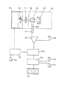
Referring now to Fig. 5 those parts which are the same as
shown in Fig. 4 are given the same reference numerals in the
100 series. Located within an optical chamber 125 are flash
charge and trigger circuitry located within a screened
compartment 126 controlling acti~ation of the xenon discharge
tube llO, said tube 110, interference filter and illumination
aperture 111 and 112, the sample 113, a light sink 127 and
the detector 114 in a scatter aperture pipe 128. The light
-- 6 --
.,~
6~
sink 127 serves to absorb any light which is transmitted by
the sample and so prevent any reflections within the chamber
125 which may affect the readings. The detector 114 and
aperture pipe 12S are arranged in such a way that only light
which has been scattered through substantially 90 falls upon
the detector 114. The detector 114 emits a signal to a
detector amplifier 129 which has means 130 for adjusting the
gain on the amplifier 129 and hence allows adjustment of the
sensitivity of the apparatus.
A signal from the amplifier 129 is taken to a peak detector
131 which is connected to a timing circuit 132 provided with
adjustment means 133. The timing circuit is also connected
to the trigger circuitry in compartment 126. The timing
circuit 132 controls the activation of the tube 110 and the
peak detector 131. Activation of the timing circuits 132
causes the tube to discharge at a given time after the
immunochemical reaction has been initiated in the cuvette
113. The peak detector 131 is activated a short time after
the tube 110 is discharged in order to eliminate any e.m.f.
peak effects caused by the discharge. The peak detector 131
continues to function until a peak is reached when no more
readings are taken.
The signal from the peak detector 131 is fed into a digital
multimeter 134 which is provided with controls for zeroing
135 and calibration 136. The readings from the multimeter
are shown on a digital display 121.
This apparatus does not cater for compensation for
differences between test blanks or in differences in
haemoglobin level between samples.
However, such an apparatus can be used satisfactorily in
application for detecting the presence or absence of a factor
in the sample.
-- 7
~L~86222
Activation of the timing circuits 132 may be achieved
manually by operating a switch when the cuvette 113 has been
inserted, or insertion of the cuvette 113 can cause automatic
operation by use of a micro-switch.
Referring now to Fig. 6 those parts which correspond to those
shown in previous drawings are givlen the same reference
numerals in the 200 series. In this embodiment, the
interference filter 211 is located between the cuvette 213
and the detector 214. A further interference filter 220 is
located between the cuvette 213 and the light sink 227. A
transmission aperture pipe 240 having a further detector 241
therein is provided in the light sink 227 and is arranged to
receive light which has been transmitted through the sample.
The signal from the further detector 241 is passed to an
associated amplifier 242 which in turn sends a signal to the
flash change and trigger circuitry. This arrangement is used
to control the duration of the flash in order to compensate
for the amount of light absorbed by the haemoglobin in the
sample.
The signal from the detector 214 is fed to a peak detector
231 via an amplifier 229 as before, the peak detector being
controlled by timing circuitry 232. However, the output from
the peak detector is fed through a two-way switch 243 to
either a first sample and hold circuit 244 or a second sample
and hold circuit 245. Both sample and hold circuits 244, 245
and the switch 243 are controlled by the timing circuits 232.
The output of the sample and hold circuits 244, 245 is fed to
a digital multimeter 234 with calibration control 236 and
displayed on a digital display 221.
In use, a first reading is taken when the immunological
reaction is initiated and the switch 243 is operated by the
circuit 232 such that the output from the peaX detector 231
is fed to the first sample and hold circuits 244. At a pre-
'~i
J
~.~86~
set time after this first reading, a second reading is taken
and is fed to the second sample and hold circuits 245. The
output from the first sample and hold-circuits is fed to a
zero reference pin on the multimeter 234 and so the output
from the second sample and hold circuits 245 can be displayed
to give the change in scatter intensity after said pre-set
time interval, the zero reading being used to compensate for
any scatter from the sample which is not due to the
immunological reaction.
In Fig. 7 parts corresponding to these parts shown in
previous drawings are given the same reference numerals in
the 300 series. The apparatus shown in Fig. 7 is controlled
by a micro computer which handles timing control and data
analysis operations.
In addition to the detectors 314 and 341, an incident light
detector 353 is included to measure the incident light from
the tube 310. This can be used to improve the accuracy of
the apparatus. The output from each detector 314, 341, 353
is fed to an associated amplifier 329, 342, 350 and then to a
respective peak detector 331a, 331b, 331c. The output from
each peak detector 331a, 33ab, 331c is fed to a multiplexer
354 and then to an analogue to digital converter 355 which is
connected to a micro computer 356. The micro computer 356
replaces the timing circuits shown in previous embodiments
and also controls sample processor 357 which can process the
sample accurately before the readings are taken and so
improve overall accuracy.
~hen there is high interference to the signal from the
complex due to other proteins, it is preferable to use a rate
determining method. In this case, measurements are taken at
specific time intervals after the blood sample and the
antigen are mixed. Typically, a number of readings over a
g
~L2~Z~
few seconds and the detected signals are fed to data analysis
means which allo~s
-- 10 --
~Z8~22;~o
determination of the rate of formation of the
antjgen/antibody complex and hence the concentration of
the antibody.
Although the present invention has been described with
relation to determination of antigen content, it will
be clear that this method may also be employed to
determine the content of a first antigen protein in a
sample of blood by utilizing one or more other
antibodies which is specific to the first antibody.