Language selection

Search

Patent 1288475 Summary

Third-party information liability

Some of the information on this Web page has been provided by external sources. The Government of Canada is not responsible for the accuracy, reliability or currency of the information supplied by external sources. Users wishing to rely upon this information should consult directly with the source of the information. Content provided by external sources is not subject to official languages, privacy and accessibility requirements.

Claims and Abstract availability

Any discrepancies in the text and image of the Claims and Abstract are due to differing posting times. Text of the Claims and Abstract are posted:

  • At the time the application is open to public inspection;
  • At the time of issue of the patent (grant).
(12) Patent: (11) CA 1288475
(21) Application Number: 548210
(54) English Title: INSTRUMENT AND METHOD FOR TESTING FOR FLUID CONSTITUENTS
(54) French Title: INSTRUMENT POUR LE DOSAGE NON INVASIF DU GLUCOSE ET D'AUTRES SUBSTANCESCORPORELLES
Status: Deemed expired
Bibliographic Data
(52) Canadian Patent Classification (CPC):
  • 324/1
(51) International Patent Classification (IPC):
  • A61B 5/05 (2006.01)
  • G01R 33/46 (2006.01)
(72) Inventors :
  • PANOSH, RICHARD L. (United States of America)
  • RAPOPORT, URI (United States of America)
(73) Owners :
  • FOXBORO NMR LTD. (Israel)
(71) Applicants :
(74) Agent: RIDOUT & MAYBEE LLP
(74) Associate agent:
(45) Issued: 1991-09-03
(22) Filed Date: 1987-09-30
Availability of licence: N/A
(25) Language of filing: English

Patent Cooperation Treaty (PCT): No

(30) Application Priority Data: None

Abstracts

English Abstract


ABSTRACT OF THE DISCLOSURE

There is disclosed herein a nuclear magnetic
resonance apparatus for testing body fluids for a
constituent, for example, blood for glucose. The apparatus
includes a principal magnet, a magnetizable coil, and a
circuit for energizing the coil for energizing and
realigning molecules and detecting changes resulting from
relaxation of said field and analyzing said changes. The
apparatus is compact and adapted to receive and test an
extremity or vessel carrying a body fluid. The coil is
constructed to be positioned adjacent the extremity or
vessel to be tested. Circuit means are provided for
energizing the coil to energize and realign molecules
adjacent said coil, so as to permit molecules adjacent said
coil to assume an aligned position and for sensing changes
in position when the coil is deenergized, which is indicated
by spectra having peaks corresponding to various molecular
bonds. The circuit also includes means for comparing the
actual value of a peak for a first constituent to A
predetermined value for the peak of said first constituent
and determining the actual value of a second constituent
from a predetermined relationship between the values of the
peaks for the first and second constituents. Specifically,
predetermined water and glucose peaks are compared with the
measured water and glucose peaks for determining the
measured glucose concentration.


Claims

Note: Claims are shown in the official language in which they were submitted.


WHAT IS CLAIMED IS:

1. A nuclear magnetic resonance apparatus for in
vitro spectroscopic testing of fluid samples for the
presence of a constituent, said apparatus comprising:
principal magnet means partially defining a test
region and having a pair of opposed magnet poles
establishing a substantially uniform field within the test
region;
coil means;
circuit means coupled to said coil means for
producing an energization and realignment field within the
test region and for detecting changes resulting from
relaxation of said field, and for analyzing said changes;
said principal magnet means being positioned to
receive and test said fluid samples;
said coil means constructed to be positioned
adjacent the fluid sample to be tested; and
said circuit means including means for energizing
said coil means to resonate 1H protons adjacent said coil
means, and means for sensing the change as indicated by
spectra having peaks corresponding to various molecular
bonds,
said circuit means also including means for
comparing the actual amplitude of a peak for a first
constituent in said sample being tested to a predetermined
amplitude for a peak for a predetermined quantity of said
first constituent contained in a separate standard sample
and to determine the actual value of a second constituent in


- 23 -


said test sample from a predetermined relationship between
predetermined values of the peaks for the first and second
constituents.

2. In a nuclear magnetic resonance spectroscopy
apparatus for testing fluids for the presence of
constituents, said apparatus being of the type in which a
first magnetic field aligns 1H protons to an initial
position, and in which a second magnetic field is cyclicly
energized and deenergized to cause alignment of the 1H
protons to a second position and realignment to said initial
position, and in which the magnetic changes resulting during
realignment are detected and analyzed, the improvement
comprising:
first magnet means comprising a pair of permanent
bar magnets each including a north pole and a south pole,
second magnet means for creating said second
magnetic field;
mounting means for mounting said first magnet
means,
said mounting means comprising a pair of spaced
apart members for positioning said pair of bar magnets
spaced apart with the north pole of each said bar magnet
secured to one of said members and the south pole of each
said bar magnet secured to the other of said members, said
first magnet means, said second magnet means and said
mounting means providing a cavity defining a test region
therein for receiving a sample of the fluid to be tested,


- 24 -


said first magnetic field being substantially
uniform in field strength and direction throughout said test
region;
said second magnet means being operatively disposed
with respect to said test region for being magnetically
coupled to the fluid sample to be tested;
first and second members connecting said pair of
spaced apart members, said first connecting member including
an aperture therethrough for access of said fluid sample
into said test region;
circuit means for detecting and analyzing said
magnetic changes;
said second connecting member having switch means
operatively associated therewith for initiating operation of
said circuit means;
a sample holder containing a standard sample of
known concentrations of the constituents to be investigated;
said standard sample holder being initially
disposed in said test region; and
means for biasing said sample holder to urge said
sample holder toward said second connecting member and to
initially locate said standard sample in said test region
and capable of being operatively coupled to said second
magnet means when said second magnet means is energized;
said switch means being activated in response to
the movement of said standard sample holder out of said test
region to contact and operate said switch means when said
fluid sample to be tested is moved into said test region.


- 25 -


3. A self-contained portable apparatus for
performing in vitro nuclear magnetic resonance spectroscopy
testing of fluids for the presence of certain constituents
therein, said apparatus comprising:
wall means defining a housing;
a permanent magnet assembly disposed within said
housing,
said permanent magnet assembly at least partially
defining a test region within said housing;
means for defining a relatively narrow access
opening in said housing enabling access to said test region
for the placement therein of a test sample of the fluid to
be tested,
said test region being slightly larger than said
test sample,
said permanent magnet assembly creating a permanent
magnetic field sufficiently effective to align to a first
position 1H protons of said test sample located in said test
region;
said permanent magnetic field being substantially
uniform in field strength and direction throughout said test
region;
a generator and first means for producing gated
radio frequency pulses;
coil means positioned within said housing and in
close proximity to said test region, said coil means being
connected to said generator, and being cyclicly energized by
the gated radio frequency pulses for cyclicly flipping the
1H protons from said first position to a second aligned

- 26 -


position, and for sensing the magnetic changes as analog
data signals during realignment of said 1H protons from said
second positon to said first position;
second gate means connected to said coil means for
receiving the analog data signals during realignment of said
1H protons from said second position to said first position;
an analog/digital converter connected to said
second gate means for converting the analog data signals
into digital data signals;
control means connected to said generator and said
first gate means for receiving data from said analog/digital
converter, said control means comprising means for storing
and analyzing said digital data signals; and
display means for displaying the results of said
analysis to a user.

4. The improved nuclear magnetic resonance
apparatus of claim 1, wherein:
said coil means comprises three coils phased at 120°
for both transmitting energization and sensing.

5. The improved nuclear magnetic resonance
apparatus of claim 2, wherein:
each of said spaced apart members comprises a pole
piece directed into said test region to concentrate the
magnetic flux.

6. The improved nuclear magnetic resonance
apparatus of claim 2, wherein:

- 27 -


said second magnet means comprises a plurality of additively
connected surface coils for both transmitting energization
and sensing.

7. The apparatus of claim 3, wherein said control
means comprises:
a program memory storing an operating program;
a random access memory in said analysis means for
storing and emitting the digital data signals; and
a microprocessor means connected to said generator
and first gate means, to said program memory, to said random
access memory, and to said analog/digital converter for
controlling said apparatus in accordance with the stored
program.

8. The apparatus of claim 7, and further
comprising:
a housing surrounding and enclosing said program
memory and said sample holding means, said housing including
a housing cover;
a power supply for said program memory;
said housing having means to disable said apparatus
responsive to the removal of said housing cover, said
disabling means including switch means connecting said power
supply to said program memory and operated by the removal of
said housing cover to cause erasure of the stored program.

9. An apparatus as in claim 1, wherein the first
constituent is water and the second constituent is glucose,

- 28 -


and including means for calculating and displaying the
actual concentration of glucose as determined from the
following relationship:

Glucose concentration = K (mg/dl)
(mg/dl)

x (Glucose patient peak height)
(Glucose standard peak height)

x (Water standard peak height)
(Water patient peak height)
wherein
K = concentration of glucose in said standard
sample expressed in mg/dl.

10. An apparatus as in claim 1, wherein the first
constituent is water and the second constituent is alcohol,
and including means for calculating and displaying the
actual concentration of alcohol as determined from the
following relationship:

Alcohol concentration = K (mg/dl)
(mg/dl)

x (Alcohol patient peak height)
(Alcohol standard peak height)

x (Water standard peak height)
(Water patient peak height)
wherein
K = concentration of alcohol in said standard

sample expressed in mg/dl.

11. An apparatus as in claim 1, wherein the coil
means comprises at least one surface coil positioned
adjacent the sample to be tested.

- 29 -


12. An apparatus as in claim 1, wherein said coil
means comprises three coil pairs arranged to be in a plane,
whose axes are equally spaced from each other and which
intersect at a center so that their fields are at 120
spacing, said test sample adapted to be positioned at the
center of said pairs.

13. An apparatus as in claim 12, wherein said
circuit means includes RF generator and gate means for
applying an exciting current to said coil means, a receiver
and gate means for receiving signals sensed by said coil
means, display means for displaying test results, memory
means for storing an operational program, and microprocessor
means coupled to said generator and gate means, to said
receiver and gate means, to said display means, and to said
memory means, for selectively activating said coil means,
for processing signals received by said coil means, for
comparing actual test values of said test sample with
predetermined test values and for displaying test results in
accordance with the stored program.

14. An apparatus as in claim 13, including
disabling switch means for disabling said memory means in
the event an unauthorized access to said apparatus is
attempted.

15. A method for determining in vitro the value of
a constituent in a fluid sample using nuclear magnetic

- 30 -

resonance spectroscopy comprising the steps of:
analyzing the sample comprising the substeps of:
(a) exposing said fluid sample to a biasing
magnetic field, providing at least one magnetic field pulse
to decrease a water peak reading in said sample, resonating
a magnetic field across said sample, sampling the field and
recording data from the field and storing spectrum of
chemical shifts versus peak height as test sample data;
(b) analyzing a standard sample having a known
concentration of the constituent being tested for with the
same fields as in substep (a), obtaining a sample pulse and
recording data in memory and storing spectrum of chemical
shifts versus peak height as standard sample data;
(c) checking operation of the system, including the
steps of comparing the standard sample data for the
constituent being tested with an acceptable standard sample
spectrum previously recorded for error within a normal
tolerance;
(d) normalizing the test sample data and standard
sample data for equal water peaks, including the steps of
recalling the test sample data and standard sample data;
adjusting the test sample data peak height of water to match
that of the standard data peak height; and
(e) calculating the test sample constituent level
comprising the substeps of obtaining a ratio of test sample
constituent height to standard sample data constituent
height and multiplying it by the concentration of said
constituent in said standard sample to obtain a test sample
reading and displaying that reading.

- 31 -


16. A method for determining in vitro the value of
a constituent in a fluid sample using nuclear magnetic
resonance spectroscopy, comprising the steps of:
applying a biasing magnetic field to said sample to
align at least the 1H protons in the sample to an initial
orientation;
applying a resonating field to flip the 1H protons
between a further position and the initial position;
sensing magnetic changes as the bonds flip from the
further position to the initial position as analog signals;
converting the analog signals into digital signals;
storing the digital signals as test sample data in
a memory;
multiplying the accumulated responses in the memory
by an exponential decay to improve line broadening;
transforming the multiplied data with a fast
Fourier transform;
repeating the above steps for a standard sample
which includes water and a predetermined amount of the
constituent being tested for;
comparing the spectrum of chemical shifts versus
peak height of the standard sample to stored data of a
previous predetermined spectrum of the standard sample for
allowable error;
scaling the test sample data peak height of water
to match the peak height of water of the standard sample
data;
forming a ratio of the test sample constituent peak
height to the standard sample data peak height;

- 32 -


multiplying the ratio by the known standard sample
constituent/water ratio to obtain a test sample constituent
reading in designated units; and
displaying the test sample constituent level in the
designated units.

17. The method of claim 15 wherein the fluid
sample comprises blood.

18. The method of claim 16 wherein the fluid
sample comprises blood.

19. In a nuclear magnetic resonance spectroscopy
apparatus for testing body fluids for the presence of
constituents, said apparatus being of the type in which a
first magnetic field aligns 1H protons to an intial
position, and in which a second magnetic field is cyclicly
energized and deenergized to cause alignment of the 1H
protons to a second position and realignment to said intial
position, and in which the magnetic changes resulting during
realignment are detected and analyzed, the improvement
comprising:
first magnet means comprising a pair of permanent bar
magnets each including a north pole and a south
pole,
second magnet means for creating said second magnetic

field;
mounting means for mounting said first magnet means,

- 33 -


said mounting means comprising a pair of spaced apart
members for positioning said pair of bar magnets
spaced apart with the north pole of each said bar
magnet secured to one of said members and the south
pole of each said bar magnet secured to the other
of said members, said first magnet means, said
second magnet means and said mounting means
providing a cavity defining a test region therein
for receiving a sample of the body fluid to be
tested,
said first magnetic field being substantially uniform
in field strength and direction throughout said
test region;
said second magnet means being operatively disposed
with respect to said test region for being
magnetically coupled to the body fluid sample to be
tested;
first and second members connecting said pair of spaced
apart members, said first connecting member
including an aperture therethrough for access of
said body fluid sample into said test region,
circuit means for detecting and analyzing said magnetic
changes;
said second connecting member having switch means
operatively associated therewith for initiating
operation of said circuit means;
a sample holder containing a standard sample of known
concentrations of the constituents to be investi-
gated;

- 34 -


said standard sample holder being initially disposed in
said test region; and
means for biasing said sample holder to urge said
sample holder toward said second connecting member
and to initially locate said standard sample in
said test region and capable of being operatively
coupled to said second magnet means when said
second magnet means is energized, said switch means
being activated in response to the movement of said
standard sample holder out of said test region to
contact and operate said switch means when said
body fluid sample to be tested is moved into said
test region.

20. The improved nuclear magnetic resonance
apparatus of claim 19, wherein;
each of said spaced apart members comprises a pole
piece directed into said test region to concentrate
the magnetic flux.

21. A portable nuclear magnetic resonance
apparatus for spectroscopic testing of samples of body
fluids for the presence of a constituent, said apparatus
comprising:
principal magnet means partially defining a test region
and having a pair of opposed magnetic poles
establishing a substantially uniform magnetic field
within the test region;
coil means; and

- 35 -


circuit means coupled to said coil means for producing
an energization and realignment field within the
test region and for detecting changes resulting
from relaxation of said field and for analyzing
said changes;
said principal magnet means being positioned to receive
and test said body fluid samples in said test
region;
said circuit means including means for energizing said
coil means to resonate 1H protons adjacent said
coil means, and means for sensing the change as
indicated by spectra having peaks corresponding to
various molecular bonds,
said circuit means also including means for comparing
the actual amplitude of a peak for a first
constituent in the body fluid sample being tested
to a predetermined amplitude for a peak for a
predetermined quantity of said first constituent
contained in a separate, standard sample to
determine the actual value of a second constituent
in said body fluid from a predetermined relationship
between predetermined values of the peaks for the
first and second constituents.

22. An apparatus as in claim 21, wherein the first
constituent is water and the second constituent is glucose,
and including means for calculating and displaying the
actual concentration of glucose as determined from the

- 36 -


following relationship:
Glucose concentration (mg/dl) = K (mg/dl) X

( Glucose patient peak height )
( Glucose standard peak height ) X
( Water standard peak height )
( Water patient peak height )

wherein K=concentration of glucose in said standard sample
expressed in mg/dl.

23. An apparatus as in claim 21, wherein the first
constituent is water and the second constituent is alcohol,
and including means for calculating and displaying the
actual concentration of alcohol as determined from the
following relationship:
Alcohol concentration (mg/dl) = K (mg/dl) X

( Alcohol patient peak height ) X
( Alcohol standard peak height )
( Water standard peak height )
( Water patient peak height )

wherein K=concentration of alcohol in said standard sample
expressed in mg/dl.

24. An apparatus as in claim 21, wherein the coil
means comprises at least one surface coil positioned
adjacent the sample to be tested.

25. An apparatus as in claim 24, wherein said body
fluid is blood, said test region is constructed to test said
blood in vivo in a human finger in the area below the
fingernail and the surface coil is arranged to be positioned

- 37 -


in close proximity to the test region; and said apparatus
also includes means for testing said standard sample for
calibration of said apparatus after testing of the finger.

26. An apparatus as in claim 25, wherein the fluid
to be tested is blood serum, and the first constituent is
water and the second constituent is glucose, and said
standard sample contains a mixture of water and glucose of
known concentration.

27. An apparatus as in claim 26, wherein the
apparatus includes means for resiliently biasing a standard
sample holder into the test region adjacent said surface
coil and for movement into a second position by movement of
the finger to be tested into said test region.

28. An apparatus as in claim 27, wherein said coil
energizing means includes means actuated responsive to the
movement of the standard sample holder.

29. An apparatus as in claim 25, wherein the test
apparatus is constructed to enclose and surround said test
region.

30. An apparatus as in claim 21 for use in testing
the body fluid in a blood vessel in vivo wherein the poles
of the principal magnet are constructed to be positioned on
opposite sides of the test region.

- 38 -


31. An apparatus as in claim 30, wherein the blood
vessel is a surface blood vessel of an animal and said coil
means is a surface coil.

32. An apparatus as in claim 30, wherein said
apparatus is implantable and said coil means comprises a
pair of coils adapted to be positioned on opposite sides of
the test region.

33. An apparatus as in claim 21, wherein said coil
means comprises a transmitting coil and a receiving coil.

34. An apparatus as in claim 21, wherein said coil
means comprises three coil pairs arranged to be in a plane
whose axes are equally spaced from each other and which
intersect at a center so that their fields are at 120°
spacing, said test sample positionable at the center of said
pairs.

35. An apparatus as in claim 21, wherein said
circuit means includes RF generator and gate means for
applying an exciting current to said coil means, a receiver
and gate means for receiving signals sensed by said coil
means, display means for displaying test results, memory
means for storing an operational program and microprocessor
means coupled to said generator and gate means, to said
receiver and gate means, to said display means, and to said
memory means, for selectively activating said coil means,

- 39 -


for processing signals received by said coil means, for
comparing actual test values of said body fluid sample with
predetermined test values and for displaying test results in
accordance with the stored program.

36. An apparatus as in claim 35, including
disabling switch means for disabling said memory means in
the event an unauthorized access to said apparatus is
attempted.

37. The nuclear magnet resonance apparatus of
claim 21, wherein:
said coil means comprises a plurality of additively
connected surface coils for both transmitting
energization and sensing.

38. The nuclear magnetic resonance apparatus of
claim 21, wherein:
said coil means comprises three coils phased at 120°
for both transmitting energization and sensing.

39. A method for non-invasively determining the
value of a constituent in an aqueous body fluid of a patient
using nuclear magnetic resonance spectroscopy, the method
comprising the steps of:
analyzing a sample of said aqueous body fluid
comprising the substeps of:
(a) exposing said body fluid sample to a biasing
magnetic field, providing at least one magnetic


- 40 -


field pulse to decrease a water peak reading
obtained from said body fluid sample,
resonating a magnetic field across said body
fluid sample, sampling the field and recording
data from the field and storing a spectrum of
chemical shifts versus peak height as patient
data;
(b) analyzing a standard sample having a known
concentration of the constituent being tested
for with the same fields as in substep (a),
obtaining a standard sample pulse and recording
data in memory and storing a spectrum of
chemical shifts versus peak height as standard
sample data;
(c) checking operation of the system, including the
steps of comparing the standard sample data for
the constituent being tested with an acceptable
standard sample spectrum previously recorded
for said constituent for error within a
selected tolerance;
(d) normalizing the patient data and standard
sample data for equal water peaks, including
the steps of recalling the patient data and the
standard sample data; adjusting the patient
data peak height of water to match that of the
standard data peak height of water; and
(e) calculating the body fluid sample constituent
level comprising the substeps of obtaining a
ratio of patient data constituent height to

- 41 -


standard sample data constituent height and
multiplying it by the concentration of said
constituent in said standard sample to obtain a
patient reading and displaying that reading.

40. The method set forth in claim 39, wherein the
analysis in substep (b) is done in the same sequence as that
in substep (a).

41. A method for non-invasively determining the
value of a constituent in a sample of an aqueous body fluid
of a patient using nuclear magnetic resonance spectroscopy,
said method comprising the steps of:
applying a biasing magnetic field to said sample to
align at least the
1H protons in the body fluid sample to an initial
orientation;
applying a resonating field to flip the 1H protons
between a further position and the initial
position;
sensing, as analog signals, magnetic changes as the
bonds flip from the further position to the initial
position;
converting the analog signals into digital signals;
storing the digital signals as patient data in a
memory;
multiplying the accumulated responses in the memory by
an exponential decay to improve line broadening;
transforming the multiplied data with a fast Fourier

- 42 -


transform;
repeating the above steps for a standard sample which
includes water and a predetermined amount of the
constituent being tested for;
comparing the spectrum of chemical shifts versus peak
height of the standard sample to stored data of a
previous predetermined spectrum of the standard
sample for allowable error;
scaling the patient data peak height of water to match
the peak height of water of the standard sample
data;
forming a ratio of the patient constituent peak height
to the standard sample data constituent peak
height;
multiplying the ratio by the known standard sample
constituent/water ratio to obtain a patient
constituent reading in designated units; and
displaying the patient constituent level in such
designated units.

42. The method set forth in claims 39 or 41,
wherein the body fluid is blood.

43. The method set forth in claims 39 or 41,
wherein the body fluid is blood and the constituent being
tested for is glucose or alcohol.

44. The method set forth in claims 39 or 41
wherein said in vivo testing is performed by applying said

- 43 -


biasing magnetic field and said resonating magnetic field to
a selected portion of the body of said patient.

45. The method set forth in claim 44 wherein said
selected portion of said patient's body is a finger.

46. The method set forth in claim 44 wherein said
selected portion of said patient's body is a blood vessel of
said body.

47. A self-contained portable apparatus for
performing in vivo nuclear magnetic resonance spectroscopy
testing of body fluids for the presennce of certain
constituents therein, said apparatus comprising:
wall means defining a housing;
a permanent magnet assembly disposed within said
housing;
said permanent magnet assembly at least partially
defining a test region within said housing;
means for defining a relatively narrow access opening
in said housing enabling access to said test region
for the placement therein of a test sample of the
body fluid to be tested,
said test region being slightly larger than said test
sample,
said permanent magnet assembly creating a permanent
magnetic field sufficiently effective to align to a
first position 1H protons of said test sample
located in said test region;

- 44 -


said permanent magnetic field being substantially
uniform in field strength and direction throughout
said test region;
a generator and first means for producing gated radio
frequency pulses;
coil means positioned within said housing and in close
proximity to said test region, said coil means
being connected to said generator, and being
cyclicly energized by the gated radio frequency
pulses for cyclicly flipping the 1H protons from
said first position to a second aligned position,
and for sensing the magnetic changes as analog data
signals during realignment of said 1H protons from
said second positon to said first position;
second gate means connected to said coil means for
receiving the analog data signals during
realignment of said 1H protons from said second
position to said first position;
an analog/digital converter connected to said second
gate means for converting the analog data signals
into digital data signals;
control means connected to said generator and said
first gate means for receiving data from said
analog/digital converter, said control means
comprising means for storing and analyzing said
digital data signals; and
display means for displaying the results of said
analysis to a user.

- 45 -


48. The apparatus of claim 47, wherein said
control means comprises:
a program memory storing an operating program;
a random access memory in said analysis means for
storing and emitting the digital data signals; and
a microprocessor means connected to said generator and
first gate means, to said program memory, to said
random access memory, and to said analog/digital
converter for controlling said apparatus in
accordance with the stored program.

49. The apparatus of claim 48, and further
comprising:
said housing surrounding and enclosing said program
memory and said sample holding means, said housing
including a housing cover;
a power supply for said program memory and
said housing having means to disable said program
memory responsive to the removal of said housing
cover,
said disabling means including switch means connecting
said power supply to said program memory and
operated by the removal of said housing cover to
cause disruption of the stored program.

- 46 -

Description

Note: Descriptions are shown in the official language in which they were submitted.


3~;'Y5
INSTRUMENT AND METHOD FOR
TESTING FOR FLUID CONSTITUENTS
~ACKGROUND OF THE INVENTIOM - - -


This inventlon primarily relates to a method and to
an instrument for use in medical diagnosis, and in particu-
lar, to detecting and determining glucose concentration in
blood.
Diabetes $s a health problem affecting many indivi-
duals and its prevalence is increasing. The usual treatment
for diabetes is single or multiple insulin in~ections
daily. Insulin is available in slowly or rapidly absorbed

- . . : , , ~ , .. ... ..
forms, which may be injected alone or in combination. Such

insulin in~ections have been effectlve in treating the ~
,, ., _ . . .
disease and in prolonging life.
. ,,. ., . . ., , : . , ~ ~:
Presently in order to determine if insul1n is

needed, blood is withdrawn from a patient and is tested for -
,, , ., , ~ . , . , .. . , .. . ,, ..,~ ... . ...
glucose concentration by a litmus-type indicator test. ~`lf- -
indicated, insulin is taken by the patient.
This type of testing has several problems. For
" : , , ,, ~ - - -~ -,
example, the testing is periodLc, and thus the administra-
tion of insulin is periodic, whioh can result in wide -~ -

variations in glucose concentration over time and peaks inthe glucose concentration. Such variations can have

physiological effects which may be adve~se to the patient.

, - , . , ,,- - . ~ - . - , , ,, ~ , . ~,; . . . -
It has been reco~nized that it is desirable to -
, . .
administer insulin periodically on demand and in'response to

changes in glucose levels. One such system ~s disclosed in
, ~ . ;
Albisser A, "Devices for the Control of Diabetes Melletus",

Proc. IEEE 67 No. 9, 1308-1310 (19793 , wherein a servo
., . . ~ - .. . . .. .. .
system is employed which continuously withdraw~ blood from a

patient and analyzes the same for glucose. Using a computer

or microprocessor, calculations are made from the withdrawn


~,,'' ,
, .


sample as to the need for insulin, and in response thereto,
lnsulin is administered. This system has only been used for
short periods and has a disadvantage in that the system is
invasive (i.e., the patient is catheterized continuously for
withdrawing blood samples).
The litmus-type system has the disadvantage in that
it is invasive and the patient is periodically and
repeatedly pricked for blood samples.

It ls therefore an ob~ect of this invention to
provide a glucose testing dsv~ce which can be used to
monitor a patient's glucose level cont$nuously, if desired,
so as to provide a more uniform administration of insulin
and a more uniform glucose concentration in the bloDd.over
time.
It is another ob~ect to provide a glucose moni-
toring system which is noninvasive and does not require-
periodic blood withdrawal to determine glucose levels.
It is sometimes desirable to test body fluids for
other constituents. For example, law enforcement officers
test individuals for alcohol content of their blood using a
breathalyzer. However, breathalyzer tests may be inaccurate
in that non-ingested alcohol, such as in mouthwashes, will
provide false results.
It is another ob;ect of this invention to provide a
noninvasiave diagnosis apparatus for use in determining the
concentratlon of various constituents of body fluids, such
as glucose and alcohol and drugs.
Nuclear magnetlc resonance (NMR) ls a diagnostic

technique which ls used widely for medical imaging and
medical d$agnosis. In NMR, the test ob~ect is sub~ected to


-- 3 --

4'7~i

a first or blasing magnetic field to align previously
randomly oriented 1H protons in the nuclei and a second
field or burst of energy to lncrease the energy of a
selected nuclsus. When the second magnetic field or energy
source ls turned off, th~ return to the flrst alignment
releases energy which is detected and analyzed. This
release is analyzed or processed to form an lmage or
spectrum. From the spectrum, the presence of particular

molecular bonds can be observed and associated with various
molecules or materials from which the concentration of that

molecule or material can be determined.
NMR machines are most frequently used for imaging
sections of a human body and require large magnets, for
example, superconducting ma~nets. The machines are there-
fore quite large and expenslve. Furthermore,-the NMR
testing of fluids has requ$red 1nvasive sample withdrawal
techniques, which sample was then tested in the larger-
machines.

Using such NMR machines, blood serum has been - -
analyzed and a spectra of the lH resonance developed. In

such spectra, identifiable peaks are obtained for water,
glucose and ethanol. In reported tests, blood serum has - -- -
been taken from animals, placed in a contalner and excited
so as to yield the lH spectra, which is then analyzed.
Unfortunately, NMR testings are not common nor oonveniently
available. The reason is believed to be that the equipment
ls generally large, complex and expensive, and is therefore
available only at selected centers, such as hospitals,

universities, and other similar research and test sites.
The equipment therefore is not normally used for blood or

34~75

b~dy fluid analysls as more convenient and less expensiYe
alternatives are available.
Another disadvantage in preser,t N~R tests is that
they are conducted on fluid samples which are withdrawn from
the patient by the usual lnvasive techniques.
It is therefore an ob~ect o* this invention to
provide a more convenient NMR instrument for use in
analyzing body fluld samples.
It is a further object of this $nventlon to provide
an NMR instrument for use ln analyzing body fluid for

.
glucose.
It is yet another ob~ect to provide a portable NMR
instrument for use by a person having diabetes to analyze
his blood for glucose concentration.
It is yet a further ob~ect to provide an NMR
instrument for use by a diabetic in noninvasively analyzing
his blood serum for glucose concentration.
It is a still further ob~ect of the invention to
~,. . . . .
provida an NMR method and apparatus to test for other
substances, for example, alcohol and drugs.
These and other cb~ects of this invention will
become apparent from the followin~ disclosure and appended
claims.


SUMMARY OF THE INVENTION - -
. . : .
This lnvention provides a method and a portable NMR
instrument for use ~n noninvasively analyzing body fluids
for the concentration of various constituents. Specifi-

cally, a diabetic can use the instrument to noninvasively
and substantially instantaneously analyze his blood for


~ ?~3847S

glucose, thereby eliminating the need to invasively obtain a
blood sample which is then tested. Using the device dis-
closed herein, a patient can periodically, frequently if
necessary, and painlessly analyze hls blood for glucose
concentration. This device may also be u~eful in analyzing
body fluids for alcohol or drugs.
In one form, the device is portable and provided
with means for receiving an extremity of the pati~nt, such
as a finger, and exposing the extremity to a first or
biasin~ magnetic field and a second field or ener~y
source. Sensors are provided for sensing the rates of
relaxation or energy release so 85 to develop the
spectrum. Analytical means are coupled to the sensors for
recelving and analyzing the signals emitted, discriminating
between various peaks, comparing the amplitude or hei~ht of
various peaks, such as water and glucose, and normal~zing
the analysis by reference to a standard sample so as to
obtain the concentration of constituents in the tested
materials.
One of the principal components of the NMR
instrument is the first or biasing magnet for providing the
first magnetic field. In this device th~ biasing magnet is
physically much smaller than the magnet~ used in standard
NMR machines. For example, the magnet may be one pound in
weight and may exhibit a field strength of at least five to
six kilogauss. Another component is a coil apparatus for
applying a second field or energy to the test sample and
sens~ng the energy released therefrom. A single coil or
multiple coils can be used. Yet another important element
of this invention is the electron~c clrcuit used for the

347S

analysis. This circuit is controlled by a microprocessor
that is programmed to control the application of the second
field or energy source and cooperates in detectlng and
analyzing the spectra received from the sample when the
field is relaxed. Operation o~ the microprocessor is
disclosed herein.
Other specific features of the instrumen~ are
disclosed herelnafter.


BRIEF DESCRIPTION OF THE DRAWINGS


FIGUR~ 1 is a vertical cross-sectional ~iew of an
instrument according to this invention;
FIGURE 2 is a vert~cal cross-sectional view taken
along line 2-2 of Fig. 1 and also showlng a housing and
other components;
FIGURE 3 ~s a block-type schematic diagram for the
circuitry to operate the instrument
FIGURES 4a to 4c are flow charts showing the
operation of the instrument;

FIGURES 5a and 5b are representative NMR spsctrums
showing the water, glucose peaks and alcohol used for

analysis;
FIGURE 6 is a schematic diagram showing a three-
coil system for use in the instrument;
FIGURE 7 is a schematic diagram showing the
electrical connections for the three-coil system of Fig. 6;

FIGURE 8 shows an NMR probe for implantation in a
body;
~ IGURE 9 i5 a schematic block-type diagram of the
electrical circuit for use with the implantable probe of


Fig. 8;
FIGURE lO shows a human arm having a distended vein
for NMR test$ng;
FIGURE ll is a fragmentary and sectional view of a
magnetic probe for use in NMR analysis using a surface blood
vessel; - -,
FIGURE 12 is a schematic representa~ion of an
alternative circuit arrangement for use with separate
energizing and receiving coils; ~-~
FIGURE 13 is a schematic representation of the coil
and magnet relationships which may be used ~n an arrangement .~-,
of the type shown in Fig. 12; -- - ~~
-- FIGURE 14 1s a-schematic reprqsentation of a multl- --
_."-,~
coil arrangement;
FIGURE 15 is a top view of the elements of Fig. 14;
and -
FIGURE 16 is a side view of an alternativ~ C-shap~ed - -a
magnet which ~ay replace the magnetic ~tructure of Figs. 1 --
and ~
.
DESCRIPTION OF THE PREFERRED EMBODIMENT


Referring now to Figs. 1-3, a first embodiment of
the test instrument is shown. Other em~odiments and c~
features will be discussed ~fter consideration of principal - -
features of th~s invention by way o~ the first embodiment. --
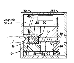
The test lnstrument 10 is ~hown as including 3 box- -^
shaped assembly which defines a finger-receiving recess 12 _.
therein. ~he assembly lncludes a body section 14 defined by
the top, bottom and elongated side walls 16, 18, 20 and 22 ~
and the back wall 24. The assembly is enclosed in a two- _


~ ~8475

piece cover or housing 25A and 25B within which the
electronic components discussed hereinafter are also
enclosed. Alternatively, the electronics can be enclosed in
a separate housing connected to the body section,- A p~ir of
first or biasing permanent magnets 26 and 28 form the top
and bottom walls 18 and 22, are positioned opposite one
another and provide the first alignins magnetic field. It
is to be noted that the poles of the respective magnets are
aligned 50 that the field is additive and provide construc-

tive lnterference, and the pole pieces or shoes shape themagnetic fleld in the finger-receiving recess 12. ~his
alignment is shown by the "X" designation, wh~ch indicates
that the magnetic field from the magnets passes through the
secess 12 in the same direction, in Fig. 2, into the paper.
A sample holder or container:34 for a standard sample
starting apparatus 30 is shown positioned in the recess.
The apparatus includes a compression blasing spring 32
pressing at one end against the back wall 24 and against the rear -
wall 30 of sample holder 34 at the other end. The holder 34 is
mounted on a post-like member 35, which is guided through an
aperture 37~ A start switch 36 is mounted to the back wall -~
offset from the member 35 so that when the sample holdRr
34 is pushed against the spring toward the back wall, the
holder will depress the start switch to start operation of
the instrument. Release of the sample holder will
release the switch. The switch may also be mounted outside,
say beneath the head 39, and operated upon movement of the
head 39.
A surface coil 38 is mounted in the hou~ing
ad~acent one of the permanent magnets 26 and 28. The coil


_ g _

47~

produces the second field and acts as a source of energy for
realignment and for sensing purposes. As seen in Fig, 1,
the second field produced by the surface coil is transverse
to the first or permanent magnet field. The surface coil
has been selected for this embodiment because the depth of
magnetization (i.e., extent of penetration of the field) is
related to the diameter of the coll and can thus be
controlled.

The surface coil 38 may be a slngle coil for both
energlzation and sensing. The co~1 can also be an assembly

in which there are multiple coils, each of which are for
energizatlon and 6ensing. Furthermore, the coil may be an,
assembly of at least two coils, where at least one is for
energization and at least one other coil is for sensing.

.
These alternatives are shown in Figs. 13, 14-and 15.
The cover,or housing 25A and 25B for the
electronics is provided with an electronic interlock system
(schematically shown as 56 in F~g.; 3 ) 80 that unauthorized

vpening or removal of the cover will disable the electronics ,-
described herelnafter, thereby prevent~ng unauthorized '--

tampering or repair of the device which could destroy ' '
calibration and result in improper usage. - -'
Physically the test is run by ~he patient inserting
his finger into the instrumen~ and pushing the sample holder
toward the back wall 24 and into engagement with the start
switch 36 to start the analysis as describsd herelnafter.

It will be noted that the finger is positioned so
that the fingernail i8 located adjacent the surface coil.
Thl~ positioning is chosen as the fingerna~l i5 dead t~ssue
but has a bed of active blood vlessels pos~tioned ~ust below

-- 10 --

s

the nail. These vessels are believ~d to provide an accurate
testing site. In many other test sites, llve body tissue or
bone must be penetrated in order to test blood in a vessel,
which means that the tissue or bone will'émlt s~gnals due to
testlng which act as noise and may interfere with analysis
of the blood for glucose concentration. The finger region
is preferable, since the nail is essentially dead material
and produces little, if any, interfering noise, thereby
increasing the signal to noise ratio. It ls believed that
other body extremities can be tested, for example, the ear
of either a human or other'~nimals. ' '''''
- ' The testing circuit 40'includes a battery power
supply 42. In'a permanent installation, such as a doctor's-
office, hosp~tal, etc., a commercial AC power supply and
battery charger may be used to supply energy to the
battery. Depression of the start switch activates the '- - - -

circuit and, thereby the microproces~or 44. The microprocessor
activates an RF generator and cyciically-operated gate 46,
,
which excites the surface coil~38 (or coil assembly) for
applying the second fleld, raising the energy state and -
realigning the nuclei.
At the appropriate time and under control of the' --
microprocessor, the RF generator is deactivated, thereby
' permittin~ the nuclei (dipolesj to relax or return to the
first alignment. The surface coil then detects the energy '' ' --
released duriny relaxation and realignment. Those sisnals
are received by receiver/gate 48, converted from analog
signals to digital signals by the ~/D converter S0 and fed
to the microprocessor 44. A rea~ only memory (ROM) 52 is
provid~d for storing the program for use with the micro-


-- 11 --

~J

~ ~f~47~rj

processor in calibrating the machine and analyzing and
displaying test results. If separate coils are used, then
the circuit ls chang0d so that the RF generator is connected
to the energizing coil and the receiver is connscted to the
sPnsing coil as shown in Fig. 12.
The ROM is continuously energized by the battery
54. A cover interlock switch 56 is provided between the ROM
52 and battery 54 to deenergize the ROM in the event the
electronics cover 25A or 25B is opened, removed or tampered
with. In such an event, the switch 56 ~s opened and the
program in the ROM is erased. In this instance, the ROM may
be selected from the well-known classes of electrically
erasable or alterable ROM's. The specific function of the
ROM-cover interlock arrangement may be selected as desired,
i.e., to generate an error message on the panel display, or
simply to disable the apparatus from operating or exhibiting
any panel display. Various other forms of electronic-type
interlocks are well-known in the computer art.
The testing circuit 40 also ~ncludes a display 58,
preferably digital, which is connectsd to the microprocessor
and a group of status lamps (read 60, calibrate 62, display
64 and error 66), which indicate the status of the system1s
operation.
The ROM 52 includes a program as represented by the
flow chart of Figs. 4a-4c, whereby operation of the tester is
controlled. In general, the operation of the tester is as
follows:
1. A finger is inserted to depress the sample
holder and activate the-start switch.
2. The ~inger ~s tested.


- 12 -

9 ~475


3. The finger test results are stored in the RAM
45.
4. The finger is released and the standard sample
moved to the test position.
5. The standard sample is tested.
6. The standard sample test results are stored in
the RAM 45.
7. The standard sample test results are compared
with predetermined calibration data previously entered in
memory to determine if the standard sample data reading is
still within preset and allowable tolerances.
8. Then the finger test results are compared with
the sample standard test result data and the finger data is
normalized and proportioned to determine glucose
concentration.
Referring now to the flow diagrsm, Figs. 4a through
4c, tha various phases of the microprocessor and ROM are
shown. These phases can considered as follows:
1. Patient reading cycle.
2. - Standard sample reading cycle.
3. Operational system check.
4. Calculation of normalized patient data and
stan~ard sample for equal ~2 peak.
5. ~alculation of glucose level.
Within each one of these broad steps are a series of smaller
steps.
Reerring first to Fig. 4a, the flow chart begins
with depression of the starting switch 36, init~ation of the


- 13 -
~J

4~i

program and activation of the read light 60. Next, a one
second homodecoupling pulse (or a plur~lity of pulses~ to
saturate the water peak is activated. A five microsecond
sample pulse is taken, and the free induction deeay output
from the A/D converter is noted. Next, th~ data points are
stored in the memory 45 and the process is repeated (i.e,,
looped) perhaps one hundred tlmes. In the right-hand
column, there is shown a serles of diagrams representing the
one second homodecoupling pulse, the five microsecond
sampling pulse, the decay, and a Fourier transformation of

the decay data points. The amplitude (Amp.) of the response
is recorded along the Y-axis. After the samplings, the read
lamp ~s deactivated, the accumulated responses are -- -
multiplied by an exponent1al decay to provlde line
broadeniny, a Fourier transformation is run, and a ~pectrum
is stored as the chemical shifts versus the peak height as
patient data.
Turning now to Fig. 4b, the standard sample reading
cycle is next activated. Here the calibrate light is turned
on, and the start switch is released. Once the switch is
released, a one second homodecoupling puls0 (or plurality of
pulses) is provided, a five mlcrosecond ~ampllng pulse is
taken, the free lnduction decay ls recorded, and the data
points are stored ln the memory 45. The system is then
repeated again,-perhaps--one hundred times.~`As `i`n ~e ; `
patient reading cycle, the accumulated responses-are- --

multiplied by an exponential decay to improve lina ~ ~-
broadening, Fourler transforms are run and the spectrum of
chemical sh~fts versus peak helght is stored as-sample~data.


- 14 -

3475


The standard sample initially contains predeter-
mined amounts of the constituent material or materials being
tested for and acts as a reference level. In order to
assure th~t there has been no significant change in these
value(s), the next step is an operational check where the
spectrum of chemical shifts versus peak height data for the
standard sample is recalled and compared to the standard
data previously taken within allowable tolerances. If the
error is not within an ac~aptable tolerance, the~- error-


display lamp 66 is lit and the operator notified. If thedata is with~n an allowable error, the system proceeds to
the next step. lt is noted that on the right-hand side of
Fig. 4c that a comparison is shown between the standard
sample data and standard sample ~pectrum showing the allow-
able shifts, peak height and frequency with amplitude
plotted along the Y-axis. ~
The next step is to normalize the patient data and
standard sample data for e~ual water heights. Here the
patient data 18 recalled and the s~andard sample data is

recalled. Next, the pati~nt data water peak height is
scal~d to match the standard sample data water peak height.
The system then executes the next step whlch is to
calculate the glucose 1PVe1. To do th~s a ratio is obtained
of the patient data glucose peak height and ths standard
sample data peak height. This ratio is then mult$plied by
the known standard sample glucose to water ratio to obtain
the patient reading and multiplied by a concentration factor
(K) from the standard samplP and expressed in milligrams per


-- 15 -

7S

deciliter or some other convenient unit. Then the patient
glucose level is displayed in relation to plasma level.
Normal 91ucose concentration is ninety milligrams per
deciliter.
This relationship is derived as follows:
1. The standard sample is prepared having a known
glucose concentration expressed, ~or example, in milligram
of glucose/deciliter of water (mg/dl) and is referred to as
K.
2. A patient is tested and the water and glucose
peak heights are obtained.
3. The standard sample ~s then tested for water
and glucose peak heights.
4. The patient's water peak height is normalized
by determining the ratio of water standard peak height/water
patient peak height. ~his ratlo can be referred to as gain.
5. The patient's glucose peak height is
normalized by multiply1ng the patient ~lucose peak height by
the gainO The result is the normallzed patient glucose
level. Expressed algebraically:

Glucose = (Water ~tandard) x glucsse pati~nt
normalized (Water patient )
6. In order to obtain the actual p~tient glucose
concentration, expressed in units such as mg/dl, the
normalized glucose now is divided by the glucose standard
and the resulting ratio ls multiplied by the concentration
factor K. In other words:

Patient glucose ~ Glucose normalized x K
concentrat1on Glucose standard


- 16 -

;,'

~ ~3847~

7. The entire expression which combines the 8tep8
of numbers 1-5 above can be stated as:

Patient glucose - ( m~ ) - K ( m~ ) -
concentration ( dl ) ( dl ) -- -
... . . . . .
(Glucose patient) (Water ~tandard)
x ( peak height ) x ( peak height
(Glucose standard) (Water patient ~
( peak height ) ~-peak height ) - - -
In Fig. 5a, a 1~ typical blood spectrum is shown

with the water (H20) and glucose peaks clearly shown. It 1s
the ratio-of the peak heights as determined from the cali-
bration and test samples that permit determination of the
test sample glucose concentration. Fig. 5a shows the work~
of Jay Block, "Analysis of Serum by High Yield NMR", Clin.
Chem. 28/9, 1983, (1982) taksn from normal blood serum. ---- -
Sample volume is 0.4 ml serum to which ~as been added D.l ml - - ---
of 2H20 for field lock. In addltion, 10 mmol/l of TSP was - ~ _
added to the 2H20 to serve as a reference to assign chemical --
shifts and peak area. The work was done on a WM 500 Bruker

spectrometer. Samples were maintained at 30C and a 1

second homodecoupling pulse was applled before the S
mill~second sample pulse (45~ notation angle) to saturate
and reduce the H20 peak. A total of 16k data points was
~eoorded in ~n ac~uisitlon time o~ 1.5 ssconds with B0 ~uch
transients averaged for each spectrum (2 min per spectrum).
~ven with the water peak suppressed, ~t is still the most
prominent feature, however, the glucose peak which is four
orders of magnitude lower i8 still easily identified. The

glucose concentration is in the normal range of 90 mgJdl as

m~asured by the conventional glucose ox$dase procedure.


- 17 -
~,

347~


Lactate was also detectable. It is also interesting to look
at the glucose peak at 5.25 in the otherwise peak free
region.
Fig. 5b is an enlarged portion of the 1H blood
spectrum of Fig. 5a, showing the ethanol and water peaks, as
also reported by Bock and showing the spectrum of serum
obtained 30 minutes after ingesting 30 ml of vodka. The
ethanol concentration measured by routine gaschromatographic
method was only 30 mg/l, while the methyl resonance of
ethanol at 1.20 ppm was detected with better -than 40:1
signal to noise ratio. The methylene resonance is buried in
the glucose region. In addition, a large peak appears at
1.93 ppm, the position of acetate, presumably derived from
the oxidation of ingested ethanol. In serum from
intoxicated patients, the ethanol resonance had a much
greater intensity and dominated the spectra.

Another smbodiment 70 of this lnvention is shown in
Fig. 6. In this embodimentj three coil pairs 72, 74 and 76,
a~e provided, whlch lie ln the same plane and are equally
spaced, that is at egually spaced 60 intervals. The coils
are arranged to provide constructive interference at the `
center of the coils where a sample ~such as a flnger or test
tube) ls to be located. These coil pa$rs act as the
energization ~r realignment coll ~nd as the sensor, in a


- 18 -

4~5

manner similar to the surface coil described hereinbefore.
This arrangement ls believed to provide better signal
discrimination by ~ncreasing the signal-to-noise ratio. The
coils are mounted in a housing similar to that shown in
Figs. 1 and 2 and are controlled by a circuit and in the
manner similar to that described in connection with Fig.
3. Physically, the standard and sample is inssrted into one
of the coils, such as the test tu~e 78 into coil 72. The

portion to be tested is located at the center of the coils
as shown.
The test sample is then tested as described above
with coils first acting as the energization or realignment
magnets and then as sensors or receivers. In other regards,
such as s$gnal pr~cess$ng and concentration analysis, this
system operates in the same manner as above.
In those cases in which it may be desirable to
implant a portion of the instrument, reference is made to
Figs. e and 9.
A third embodiment 80 is shown in Fig. 8, which is
constructed to surround a blood vessel which is internal of
or within a body, for example, a vein or artery in the body.
The test device includes the principal magnet 82,
which in this case is C-shaped and a palr o* RF coils 84.
The vein or artery 86 is positioned between the coil pa~rs
and the poles of the magnet. By so doing, blood in the vein
or artery is subjected to the first magnetic field, and the
energization or realignment field and relaxatlon is ensed
by coils 84.
In a fourth embodiment, the test instrument 90 ls
constructed for surgical implantation as shown in Fig. 9.


-- 19 --

847~

Such a device has two component parts: one part is the
internal or implanted portion 92 and the oth~r part is th~
external or power supply and sensing part 94. The two parts
are electronically coupled by transformer-like members as
described herein.
In the fourth embodiment 90 an external AC power
supply 96 is induct~vely coupled to an internal pow~r supply
98. The internal power supply 98 powers the NMR unit 100,
which is conneeted to probe snd magnet unit 102. Signals
from the probe and magnet are received by the receiver 104,
which is inductively coupled to the microprocessor 106,
through the coil element 108. The microprocessor then
provides an output to the digital display 110 of the glucose
concentration.
The magnet and probe assembly 102 is in the same
form as that in Fig. 8 and is positioned to surround an
artery. The signal processing is performed by the micro-
processor in the same manner as with the other embodiments,
particularly Fig. 3.
In a fifth embodiment, a surface blood vessel,
usually a vein, is distended and used to analyzs for glucose
concentration. Such an embodiment is shown in Figs. 10 and
lI, where a patient's arm 120 is shown surrounded by a
pressurizable cuff 122 for causing a vein 124 to protrude or
distend from the skin surface. In that situation, the NMR
unit is fitted on either side of the protruding vsssel at
the surface of the arm. In this embodiment a C-shaped
permanent magnet 126 is arranged so that its north and south
poles (~ & S) are on opposite sides ~f the vessel. A
surface coil 128, like that ln Figs. 1-2, is employed for


- 20 -

f .
i,~ j,

f347~

en0rgization and realignment and sensing. Testing circuitry
of the type shown in Fig. 3 is also employed in the embodi-
ment of Figs. lO snd 11.
A principal advantage of the test instrument shown
herein is that the device is smaller than the large ~R test
instruments now used at hospitals, etc. The reason is that
the present instruments include a large principal magnet for
surrounding the body of a patient. Here, since the tested
portion is a finger or other extremity, the principal magnet
may be smaller 50 that the instrument may be mounted on a
table top, carried in a brief case, or be even smaller. In
order to achieve such a device, the m~gnet must be small in
size, be of a comparatively light we~ght, such as one pound, - -
~
ana still exhibit an adequate field strength. ~deq~ate - --
strengths ~hould be on the order of at least five to s1x ~:
kilogauss. One particularly suitable material containing .',,7
Neodynium is manufactured by General Motors Corporation.
Fig. 12 shows the generator and gate 46 and the
receiver 46 and gate 48, respectively, connected to separate
transmit and recelve coils 38', 38". - ~--~
Fiy. 13 shows an embodiment of the coils 38' and- ---~
33" along with the field directions, including the bias ~ ~~ r'
field Ho, at 9O with respect to one another.
Figs. 14 and 15 illustrate the u~e of a plurality
of surface coils 38''', which are connected for addltive
fields, as a single transmitJreceive arrangement. --
Fig. 16 shows an alternate bias magnet, similar to
that shown in Fig. 11. The magnet 138 comprises a pair of
spaced pole pieces 132, 134, which define a gap for ---
receiving; in this example, a finger.


- 21 -


Although the invention has been described with
respect to preferred embodiments, it is not to be so
limited, as changes and modiflcations can be made which are
within the full intended scope of the invention as defined
by the appended claims.


: - - - - --


- 22 -

Representative Drawing
A single figure which represents the drawing illustrating the invention.
Administrative Status

For a clearer understanding of the status of the application/patent presented on this page, the site Disclaimer , as well as the definitions for Patent , Administrative Status , Maintenance Fee  and Payment History  should be consulted.

Administrative Status

Title Date
Forecasted Issue Date 1991-09-03
(22) Filed 1987-09-30
(45) Issued 1991-09-03
Deemed Expired 2002-09-03

Abandonment History

There is no abandonment history.

Payment History

Fee Type Anniversary Year Due Date Amount Paid Paid Date
Application Fee $0.00 1987-09-30
Registration of a document - section 124 $0.00 1988-01-15
Maintenance Fee - Patent - Old Act 2 1993-09-03 $50.00 1993-08-11
Maintenance Fee - Patent - Old Act 3 1994-09-05 $50.00 1994-09-01
Maintenance Fee - Patent - Old Act 4 1995-09-04 $50.00 1995-09-05
Maintenance Fee - Patent - Old Act 5 1996-09-03 $75.00 1996-09-03
Maintenance Fee - Patent - Old Act 6 1997-09-03 $150.00 1997-08-12
Registration of a document - section 124 $100.00 1998-06-30
Maintenance Fee - Patent - Old Act 7 1998-09-03 $150.00 1998-08-12
Registration of a document - section 124 $100.00 1999-07-29
Maintenance Fee - Patent - Old Act 8 1999-09-03 $150.00 1999-09-02
Maintenance Fee - Patent - Old Act 9 2000-09-04 $150.00 2000-07-04
Owners on Record

Note: Records showing the ownership history in alphabetical order.

Current Owners on Record
FOXBORO NMR LTD.
Past Owners on Record
ADVANCED TECHTRONICS, INC.
ELBIT-ATI (1992), LTD.
ELBIT-ATI, LTD.
PANOSH, RICHARD L.
RAPOPORT, URI
Past Owners that do not appear in the "Owners on Record" listing will appear in other documentation within the application.
Documents

To view selected files, please enter reCAPTCHA code :


To view images, click a link in the Document Description column. To download the documents, select one or more checkboxes in the first column and then click the "Download Selected in PDF format (Zip Archive)" or the "Download Selected as Single PDF" button.

List of published and non-published patent-specific documents on the CPD .

If you have any difficulty accessing content, you can call the Client Service Centre at 1-866-997-1936 or send them an e-mail at CIPO Client Service Centre.


Document
Description 
Date
(yyyy-mm-dd) 
Number of pages   Size of Image (KB) 
Representative Drawing 2000-07-27 1 14
Drawings 1993-10-23 8 218
Claims 1993-10-23 24 744
Abstract 1993-10-23 1 39
Cover Page 1993-10-23 1 13
Description 1993-10-23 21 830
Fees 1997-08-29 1 40
Fees 1998-09-01 1 46
Fees 1999-08-12 1 52
Fees 1999-09-02 1 42
Fees 1996-09-03 1 38
Fees 1995-09-05 1 37
Fees 1994-09-01 1 47
Fees 1993-08-11 1 30