Note: Descriptions are shown in the official language in which they were submitted.
sack~round of the Invent1on
This invention relates to a method and apparatus
for continuously and non-invasively measuring cutaneous
blood pressure in a small isolated flesh element. The
physiological data thus obtained are related to, but not
identical, to blood pressure measurements of a more
central arterial circulation, such as that obtained with
the conventional auscultatory method of estimating brach-
ial blood pressure.
In particular, the invention finds use as part
of a general system for measurement of blood pressure
based on repetitive evaluation of the cutaneous pressure
fluctuation patterns of minute branches of larger arter~
ies and therefore reflects the arterial blood pressure of
the general circulation. This is to be expected since the
latter is the source o~ the former.
The method and apparatus of the present inven-
tion enables continuous rnonitoring of blood pressure
patterns over extended periods of time. This is needed in
the evaluation of circulatory function and arnbulatory
monitoring of cardiac function, and is useful for hyper-
tension studies and for obtaining records of circulation
in the peripheral systems, particularly of the limbs,
fingers and toes.
In the past, various artery occlusion proce-
dures have been used stopping blood flow in radial,
brachial, dorsalis pedis, temporal and other arteries to
estimate blood pressure, particularly of the central
~ '
r~
36~3
circulatory system. Data thus obtained is by its very
nature discontinous
It has bean possible to insert pressure sensing
devices and/or catheters temporarily into the arteries of
the circulatory system for direct continuous measurements
(invasive method of measuring). While intra-arterial
catheterization may provide more precise measurements of
blood pressure than arterial occlusion devices, the
pressure measured is likely to be more related to the
central circulation, than the peripheral circulation.
Also, the blood pressure measurements and patterns thus
obtained are likely to be altered by the traumatic opera-
tion of inserting the catheter, by the drug administered
so that the catheter can be inserted, and by the presence
of a foreign body in the circulatory system.
The principal non-invasive blood pressure
measuring device used today is an auscultatory system
where a pressure is applied to occlude a major artery,
such as the brachial artery. In practice, an inflatable
encircling cuff is placed around the arm and in1ated to
occlude the major artery, e.g., brachial, to prevent flow
of blood in the artery. As the pressure in the cuff is
slowly lowered, permitting flow of blood in the artery,
Korotkoff sounds are heard. The cuff pressure at which
the first sound is heard is defined as the systollc
pressure. The pressure in the cuff is then lowered
further the pressure in the cuff at which the sound
fades is defined as the diastolic pressure.
-3-
A secol~d occluding cuff technique uses palpation
of the pulse rather than auscultation. In this palpatory
system, as the occluding cuff pressure is slowly released,
arterial pulsations are detected by palpation. The
pressure level of the cuff at which the pulsations are
first perceived is designated as systolic blood pressure.
Diastolic blood pressure cannot be detected by palpation.
Another occluding cuff system uses the maximum
and minimum oscillations o~ arterial blood pressure as
referenced to cuff pressure as indications of systolic and
diastolic blood pressure, respectively. In addition to
being an intermittent, occlusive technique, the measure-
ments thus obtained are likely influenced by the limb
volume of the limb around which the cuff is applied.
It can be generally stated that all blood
pressure measurements which are based upon arterial
occlusion are inherently discontinuous, needing to be
repeated, at best, from time to time. Such measurements
cannot resolve blood pressure patterns on a beat to beat
basis, or show the wave form of the individual beats.
Thus, although the current method of ausculta-
tory measurement of brachial blood pressure is by far the
most widely used technique for blood pressure measurement
the technique is relatively imprecise, since the observed
values vary from observer to observer and the very act of
taking blood pressure itself causes a momentary change in
blood pressure. -Additionally, since the occlusion itself
is known to have physiological and psychological ef~ects~
the measurements may be distorted.
.~ ~
A non-invasive, non--occlusive approach to the
measurement of bloocl pressure would have many advantages.
Unfortunately, prior techniques for this purpose have been
found to have disadvantages. Those directed at measuring
arterial pressure by placing a transducer directly over a
partially compressed radial or dorsalis pedis artery can,
under optimum circumstances, provide accurate records for
short periods of time. However, the required counter
pressure has to be maintained, e.g. with a pneumatic
system, and considerable difficulty is experienced in
maintaining constant mechanical coupling between the
tissue overlying the artery and pressure on the arterial
wall during even the slightest patient motion.
An e~ample of this type of measuring system is
disclosed in U.S. Patent No. 3,880,145 to E.F. Blick,
issued April 29, 1975. Blick described a system using a
strain gauge to flatten the radial artery at the inside of
the wrist. A second sensor is mounted cutaneously along-
side but off the artery. The signal from the second
sensor was subtracted from that sensor associated with the
flattened artery. In practice, the signal rom the radial
artery sensor contains arterial pulsations as well as
"noise". The noise which is measured by the cutaneous
transducer is subtraced erom the former signal, leaving a
measurement of the arterial pulsations alone. Such
systems are complex and during patient movement it is
very difficult to precisely match the "noise" component
arising from both sensors.
In tlle prior devices discussed above, the
m~jority are directed to the measurment of blood pressure
in major arteries. Elastic strain gauge techni~ues which
encircle limbs or digits have also been used. In such
devices, a finger or toe is encircled with a latex or
silastic tube which contains mercury. As the digit volume
increases with arterial inflow and decreases with venous
outflow, the change in-volume can be measured and related
to blood pressure. However, the system is occlusive in
nature and markedly decreases capillary blood flow
Hence, again it can be used only intermittently since it
causes distortion of the physiological data. It is not
possible to obtain continuous blood pressure records for
hours at a time. It is difficult to calibrate since it is
temperature sensitive, and must be calibrated off of the
body part.
Hand and thumb plethysmographs are also known
which measure changes in volume of entire digits, hands,
feet, or limbs, but they are very cumbersome and cannot be
used on an active patient, such as one jogging.
A general discussion and review of various
previously proposed systems for blood pressure monitoring
is given in the book, "The Direct and Indirect Measurement
of Blood Pressure," by L.A. Geddes (Year ~ook Medical
Publishers, Chicago, 1970) where a number of blood pres-
sure technigues are outlined (see pages 37, 71, 87 and
96).
--6--
~8~ iL3
6~75g-32
It has been clemonstratecl tha~ there i5 a hltherto
unfulfilled need for a sensitlve, continuous, non-invasive,
non-o~clusive measuring technlque for recording blood pressure
measuremants and beat to beat patterns undistorted and
unin~errupted by the measuring system, per se The method and
the apparatus of the present invention enables non-invaslve,
nonwocclusive continuous measurements over extended periods of
time. Continuous lnformation of this type is essential for
adequate evaluation of cardiac and vascular function. It is of
particular importance in the diagnosis and treatment o$
hypertension, slnce it provides detailed information concernin~
the peripheral circulation not avallable heretofore.
SummarY of the Invention
The present invention provides apparatus for
monitoring blood pressure of a body comprising isolating means
for isolating a portion o~ cutaneous tiæsue and measuring mean
for measuring chanyes in said i601ated cutaneous tissue, said
isolating means compri~ing an annular ring.
From another aspect the invention provides a method
for monitoring blood pressure of a body comprising the steps of
isola~ing a portion o~ autaneou~ tissue by pressing an annular
member on the surface of the slcin ancl measuring changes in the
pressure in the cutaneous tissue.
The apparatus of the present inventlon continuously
measures cutaneous blood pressure with a strain gauge (or
similar pressure measuring clevlce) attached to a portion of the
body, preferably on the fleshy part of the thumb.
A major use of the apparatus is to provide continuous
blood pressure measurements and patterns of blood pressure in
the fingers and toes. To maintain mechanical stability of the
platform the annular ring must be closely applied to a
~8~ 3
~ 75~-32
relatlvely hard s~able reference polnt e.g. such as t,ha bona at
the back of the ~humb whlch is covered wi~,h a small portion of
cutaneous tissua. Slnce the blood supply to the digit~: course
along their lateral aspects there ls no appreciable dimlnution
8~3
in the overall blood supply to the digits by the attach-
ment of the platform to the digit.
A small portion of cutaneous tissue is isolated
from the surrounding tissue by a ring depending Erom the
strain gauge or the platform and pressing against the
thumb. This ring projects beyond the measuring surface of
the strain gauge and serves to reduce noise emanating from
adjacent tissue and isolates the cutaneous tissue.
The blood pressure in this protruding isolated
cutaneous tissue is measured with a cylindrical strain
gauge (or other pressure measuring device) whose measurins
surface is tangentially oriented to the slightly domed
segregated cutaneous tissue. The isolating ring essen-
tially surrounds the circumference of the bottom of the
strain gauge. The long axis of the strain gauge is normal
to the upper surface of the stabilizing platform.
Together, the ring and lo~er portion of the
strain gauge form an inverted shallow dish in which the
inside circumference of the ring forms the peripheral
walls and the bottom measuring surface of the strain
gauge its bottom. Consequently, when the strain gauge
assembly i5 pressed against the isolated cutaneous tlssue,
its superEicial layers fill the space between the ring and
the strain gauge measuring surEace. The minute blood
pressure changes in this portion of the cutaneous tissue
are detected by the strain gauge and can be observed on an
oscilloscope or recorded in a conventional manner, such as
on a strip chart or magnetic tape.
--8--
~ `~
fl~
The magnitucle of the recorded blood pressure
changes are affected not only by the change within the
isolated cutanaous tissue, but also by the Eorces which
are holding the ring against the tissue. In order to keep
~hese forces sufficiently constant, the strain gauge
assembly must be attached to the stabilizing platform in a
substantially rigid, mechanical manner. This is done with
a rigid cap screw mounted to a sleeve surrounding the
gauge. Appropriate pressure is achieved by a spring
fitted between the inside surface of the cap and the
strain gauge assembly.
In practice, the initial ring pressure may be
adjusted so that the observed cutaneous blood pressure is
a given number of mms Hg. below the brachial systolic
blood pressure, if the latter is used as a reference
point. Alternatively, a predetermined known coupling
pressure may be applied to the non-active end of the
strain gauge assembly by mechanical means, such as a
calibrated spring, by pneumatic means, or directly by a
rigid rod where the coupling pressure is measured by
another strain gauge or other pressure sensing device. In
situations where known coupling pressures are used,
cutaneous blood pressure changes rnay be referenced to
them, as well as to the clinically determined brachial
blood pressure.
Since mechanical stability is highly desirable,
further stabilization may be achieved by using double-
sided adhesive materials between both surfaces of the
digit being evaluated where they meet the stabilizing
~ 2r88~3
6g7~ 32
platform and enclrcling means.
~ xternal mechanical shock to the s~rain yauge is
minimized by routing the electrical leads through the platform
bas~ where maximum s~abill~y is presen~ and placiny an all-
encompassing cover over the strain gauge assembly.
Additionally, shock absorbing materials are placed between the
transducer body and its surrounding guide -~ube.
When the foregoing non-invasive, non-occlusive
technique ls used as describedr it is possible to continuously
make cutaneous blood pressure measurements and record patterns
under most clinical clrcumstances and even during strenuous
exercise, such as jogging or running on a treadmlll.
These and other objects and features of the invention
will become apparent from the following description and claims
when taken in conjunctlon with the accompanying drawinys, of
which 5
¢~L3
6975~32
~rl~ Descxi~n~ h~
Figure 1 is a top perspa~tlve view of a first
embodiment of apparatus for cutaneou~ blood pressure measuring
apparatus.
Figure 2 is a ~ront vlew of ~he apparatus of
Figure 1.
Flgure 3 is a left side vlew ol the appara~us of
Figure 1.
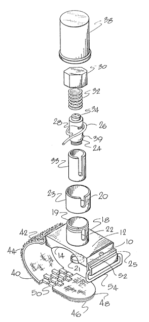
Figure 4 is an exploded top perspective view of
11 , -
6~3
the apparatus of Figure 1.
Figure 5 is a cross-sectional view of a first
embodiment of the apparatus of Figure 1.
Figure 6 is a side cross-sectional view of a
preferred embodiment of the apparatus for cutaneous blood
pressure measuring.
Figure 7 is a top partial perspective view of
the yoke assembly iri partial view.
Figure 8 is another embodiment of the present
invention.
Figure 9 is further embodiment using a calibra-
tion system employing a surrounding cuff.
Figure 10 is a partial cross-sectional view
taken along lines 11 of Figure 9.
Detailed Description of the Drawlngs
Making reference to Figures 1 through 5, the
first embodiment of the apparatus, adapted for attach-
ment to the thumb, is shown.
~ support platform plate 10 has a generally
flat rectangular top surface 12 and a concave lower
surface portion 14 adapted for conforming to the shape of
the fleshy portion of the thumb. Centrally located on
support platform 10 is a perpendicularly situated hollow
upstanding sleeve 18 having threads 19 at its outer upper
end, and extending through the support platform plate to
-12-
, r~
~2~ 3
form an opening 16 ln the support platform plate 10.
~epending below the circumference of the circular opening
16 is an isolation ring 21 that depends slightly below the
lower surface portion 14 of the support platform plate.
Within the perpendicular hollow sleeve 18 is
fitted a cylindrical pressure measuring transducer or
gauge 24 which measures pressure differentials along its
longitudinal vertical axis. The pressure measuring
transducer 24 fits slidably within shock absorbing and
guide slseve 33 fixed within the internal circumference of
the hollow sleeve 18. The diameter of the transducer 24
is approximately 1/4", and slightly smaller than the inner
diameter of the isolation ring 21. (A marking means 28 is
fixed to the external circumference of the transducer.
Electrical lead 26 from the pressure measuring
transducer 24 is fitted through a slot 25 in the support
platforrn plate 10 for stability. The electrical lead 26
is connected to a conventional recording device, not
shown.
At the upper end 35 of the transducer is a
shoulder 34 for holding one end of a coil spring 32, the
other end of which is held by an cap shoulder 36 inside
cap 30. The cap 30 has internal threads 37 corresponding
to the external threads 19 of the perpendicular hollow
sleeve 18. The coil spring 32 is thus held between the
inside of the cap 30 and the top of the transducer 24
biasing the transducer in a downward direction.
Calibration markings 20 on an outer perpendicular
-13-
~2~ 3
hollow sleeve 23 are used in a~sociation with the marking
means 28 on the hollow sleeve 1~ for setting the tension
on the coil spring 32 Erom the upwarcl force of the thumb
on the bottom surface 27 of the transc3ucer 24. The
transducer is retained in the hollow sleeve 18 by a detent
39.
The outer perpendicular hollow sleeve 23 may be
transparent or have a transparent portion Eor viewing the
marking means 28' directly. As shown in Figure 2, how-
ever, the outer hollow sleeve 23 may have a cut out
portion 22 for viewing the marking means 28.
A protective cylindrical cover 38 fits over the
cap 30 and hollow sleeve 18 and outer hollow sleeve 23,
and is held by a pressure fit against the outer periphery
of the hollow sleeve 23.
In the embodiment shown in Figures 1 through 5,
an inelastic strap 40 is connected to a first side 42.of
the lower surface 1~ of support platform plate 10. The
~h ' inelastic strap 40 has on one surface 42 a Velcro loop
portion 44, and at the end of the strap ~0 on the same
side as the Velcro loop portion 44 is a Velcro hook
portion q6. Between the Velcro loop portion q4 and Velcro
. hook portion 46 is a slightly expandable link portion 50.
Attached to the other side 52 o the lower surface 14 is
an open rectangular portion 54 for receiving the end of
the strap 40.
In an alternative embodiment shown in Figure 6,
a rigid yoke 60 is used, in place of the strap 40 shown in
-14-
cJ~ k
Figures 1 throllyh 4/ to securely attach the apparatus to
the thumb. The yoke 60 consists of a semi-circu:lar lower
portion 62 of a size and contour to fit the shape of the
back of the thumb.
A pair of screws 64 and 66 pass through openings
68 and 70 in the support platform plate 10 into screw
holes 72 and 74 in projections 76 and 78 on the sides of
the yoke 60. ~ashers 75 and 77 restrain the screws 64 and
66 from vertical rnovement.
The operation of the apparatus shown in ~igures
1 through 4 is as follows: The fleshy portion of the
patient's thumb is placed within the concave lower surface
portion 14 of the support platform plate 10. The end of
the inelastic fabric strap 40 is then passed through the
open rectangular portion 54, the end of the strap 48
having the Velcro hook portion 46 is then bent back over
itself to come into contact with the Velcro loop portion
44 on the strap 40. Prior to attaching the Velcro hooks
46 to the loops g4, the strap is subjected to tension
sufficient to stretch the expandable chain link portion 50
to its maximum extent. With the hook and loop portions 44
and 46 thus connected, the apparatus is substantially
fixed in place relative to the thurnb.
The isolating ring 21 now has a portion of
the fleshy portion of the thumb pushed into the dish
formed by the circumference of the ring 21 and the bottom
surface 27 of the transducer 24. This isolated tissue is
in contact with the lower surface 27 of the pressure
measuring transducer 24 biasing it axially upward against
-15-
~2~ 3 ' `
the tension o~ sprin~ 32 at the ~Ipper surface of the
pressure measuring transducer.
The electrical lead is connected to a conven-
tional strip recorder typically present in a doctor's
office for recording the changes in blood pressure of the
pressure measuring transducer 24. The cap 30 is then
turned until the indicator marking means 28 is approxi-
mately at the preferred position~ which should be showing
the blood pressure reading at approximately 100 rnm ~Ig.
The cap 30 would then be placed over the system. Although
not as effective, the opening 16 in the support platform
plate 10 may serve as an isolation ring, if isolating a
portion of cutaneous tissue in the opening. However, to
maintain such isolation additior.al pressure may be re-
guired of the thumb against the lower surface 14 of the
support platform plate 10 than with the use of the isola-
tion ring 21. The monitor would then provide a record of
the pressure variations on the transducer 24 corresponding
to the blood pressure in the isolated cutaneous tissue.
As a result of the isolation of the cutaneo~ls
tissue, caused by the isolation ring 21, extraneous
noise from the remainder of the digit or body itself
to the lower surface 27 of the transducer 29 is diminished
markedly or eliminated.
In the apparatus shown in Figures 6 and 7, the
apparatus is used by inserting the thumb in the opening
formed between the lower surface of yoke 62 and the
concave lower surface 14 of the support platform plate
10. The screws 64 and 66 are then rotated causing the
-16-
lower portion o~ the yoke 62 to be pulled towarcls the
concave lower surface 14 trapping the thumb between the
two surfaces. The screws 64 and 66 are turned until the
thumb is securely held in place, but there is no inter-
ference with the blood flow to the thumb. The cap 30 is
then, turned until the marking means 28 is at the desired
position. The remaining operation of the unit is the same
as that discussed in Figures 1 through 4.
Figures 8, 9 and 10 show an embodiment having a
transducer fixed in position. The pressure applied to the
isolated tissue by the lower surface of transducer is
controlled by controlling the pressure generally affixed
to the thumb.
In Figure 8 the isolation ring 21 and the
transducer 24 are fixed. The pressure is applied by the
thumb to the lower surface 27 of the transducer 24. An
expandable air sac 80 is Eitted within the concave portion
82 of the lower yoke portion 62. The air sac 80 is
inflated by use of a pump ball 84 connected through tube
86 to the air sac 80. A one way valve 88, shown diagram-
atically.
In operation, the thumb of the patient is
inserted in the opening between the air sac 80 and the
lower surface 14 of the support platform plate 10. The
screws 64 and 66 are then tightened until a pressure
meter, connected to the air sac 80 (not shown) reads the
desired pressure. Since the pressure of the air sac is
the pressure on the thumb, this is also the pressure on
the lower surface 27 of the transducer 24.
~28~ ~
In figure 9 another means of adjustiny ttle
initial pressure applied to the transducer 24 is shown.
In the embodiment cylindrical pins 90 and 92 have one
end fixedly fitted in the support platform and the other
end 94 and 96 slidably fitted within openings 98 and
100. The pins 90 and 92 can be fixed within openings 98
and 100 by locking screws 102 and l04. A pressure cuff
106, of a size of the apparatus so that inflation of the
cuff covers the compression of the support platform 10 and
the yoke 60. Once again the thumb is inserted between the
lower surface 14 and the concave surface 82 and the cuff
inflated.
When a pressure measuring device for measuring
the pressure in the cuff indicates the desired pressure,
such as 50 mm Hg then the locking screws 102 and 104 are
used to lock the pins 92 and 94 in place.
As indicated previously, the sensitivity of the
device is such that the chanyes in blood pressure to a
non-smoker may be measured when sorneone smoking a ciyar-
ette is in close proximity to the non-smoker. Such
sensitivity permits obtaining data not previously avail-
able. For example, the inventor, Dr. tlon has determined
that at the start of passive smoking (Dr. Ilon not smoking,
but having a cigarette near him), his blood pressure, as
determined by the present apparatus was approximately 100
mm of Hg. After approximately seven (7) minutes of being
in the presence of smoke his blood pressure rose to
approximately 125 mm Hg until after nine (9) minutes his
blood pressure was approximately 125 mm o~ mercury.
36~3
~fter the cigarette is removed, ~r. Hon's blood pressure
continued to rise until it ls approximately 150 mm of
mercury two (2) minutes after the cigarette has been
removed. His blood pressure remained at approximately 150
mm Hg for about seven (7) more minutes and then continued
to rise until it was approximately 165 mm Hg after eleven
(11) minutes. The blood pressure thereafter declined
steadily, but did not return to approximately normal,
until more than eighteen (18) minutes after the removal of
the smoke.
This information would not be readily discern-
able by conventional blood pressure monitoring apparatus
even of a continuous nature. Applicant is unaware of any
similar direct cardiovascular evidence of the effect of
passive smoking. In addition, other detailed cardio-
vascular effects have been determined showing the effects
of various drugs given to a patient during operations or
labor. These new data provide detailed cardiovascular
evaluation in situations where such an evaluation previ
ously was limited to relatively gross external observa-
tions.
The pressure measuring transducer 24 may of a
strain gauge type such as are available Erom Koeningsberg
Instruments, Trans ~merica Corporation or Gould, Inc.,
all of which are responsive to pressure applied to one
surface.
While a strain gauge is contemplated as the
preferred form of carrying out the present invention
it should be realized tllat many other transducers are
--19--
L3
,f~ , ~ ,
available which can measure physiological alterations in
the condition of the skin. Such conditions may be sensed
by resistive elements, by optical indicators, by linear
variable differential transformers, by frequency response
shifts in resistive capacitance systems (where the trans-
ducer is part of the capacitance of a timed circuit), by
calibrated springs, by membranes on piezo film and transis_
tors, all of which may be used in the present invention.
It will be apparent to those skilled in the art
to which this invention pertains that many adaptions and
modifications thereof may be made without departing from
its spirit and scope. For example, while the preferred
embodiment of the invention has disclosed apparatus for
particular applicability to use on the thumb, other sites
on the body may be used wherever bony eeference points or
structures are available so that a stable platform can be
established over an adjacent body part without resulting
in occlusion of the arterial flow. Examples of such
anatomic sites include fingers, toes, the entire forehead
including medial, supra-orbital and temporal areas. While
each different site may require an individualized sta-
bilizing platEorm such platform would not depart fro~ the
scope and spirit of this invention, as described.
-20-