Note: Descriptions are shown in the official language in which they were submitted.
~2~71[:~
BI NDI NG A SSAY DEV I C E
Field of the Invention
This invention relates to a device for
; performing an enzyme-labelled binding assay, the
device comprising an absorbent material and a
developing solution, wherein the absorbent material
is provided with a plurality of reagent zones
including an indicator reagent zone and is capable
of transporting the developing solution by capillary
action sequentially through each reagent zone, and
wherein the indicator reagent zone includes a
reagent capable, directly or indirectly, of
immobilising an enzyme-labelled reagent in an amount
dependent upon the assay result.
Background to the Invention
Binding assays such as immunoassaysj are in
widespread use in clinical laboratories for the
detection of substances in biological fluids.
There is however increasing interest in the
development of assays which can be performed without
~he need for complex analytical techniques and
equipment, for example, by a physician in his
consulting room or by a patient at home. Such
assays are not only more convenient but allow
savings in time and expense. Particular
applications for which convenient and simple assays
and reagent formulations are being sought are the
detection of pregnancy and of the fertile period of
the menstrual cycle
~ '
~28~90~
It is known to conduct binding assays on a
strip of material provided with a plurality of
reagent zones, in which a developing solution forms
a solvent front which passes along the strip by
capillary action piclcing up and facilitating
reaction between a sample and assay reagents located
at the reagent zones (see for example, British
patent specification GB-B-1589234). A feature of
such strips is the existence of a test location at
which, under certain conditions determined by the
assay protocol and the sample composition, a
labelled reagent becomes immobilised, giving an
indication of the assay result. In early assays,
the labelled reagent was a binding partner or
analogue of the analyte to be measured, labelled
with a radioactive isotope. Such assays require
instrumentation to detect the level of radioactive
label and may present health risk problems. A
solution to this has been the use of enzyme labels
which produce a characteristic signal (such as a
colorimetric signal) with an appropriate substrate.
A significant problem in the design of such so
called "dipstick" enzyme-labelled binding assays is
the application of the appropriate enzyme substrate
in order to produce a detectable signal. The
signal may be developed by adding substrate to the
appropriate position on the reagent strip after
allowing the assay to proceed to completion.
Alternatively, the appropriate part of the strip may
be removed and chemically analysed. All of these
represent steps which would be at least
inconvenient, if not impossible for home use of the
assay.
' ~ ~
9~
Summary of the Invention
According to the present invention, we provide a device
for perfo~ming an enzyme-labelled binding assay, the device
oomprising an absorbent material and a developing solution,
wherein the absorbent material is provided with a plurality
of reagent zones including an indicator reagent zone, and is
capable of transporting the developing solution by capillary
action sequentially through each reagent zone, and wherein the
indicator reagent zone includes a reagent capable, directly or
indirectly, of immobllising an enzyme-labelled reagent in an
amount dependent upon the assay result, characterised in that
the developiny solution includes a signal-producing substrate
for the enzyme which substrate, in use, is transported by the
developing solution slower than the enzyme--labelled reayent or
any compound of the enzyme-labelled reagent for~ed in the
assay.
The present invention, in its broadest
concept, facilitates the use of binding assays in
the home with the minimum of manipulative steps. As
the solvent front of the developing solution passes
through the absorbent material, picking up the
reagents and allowing them to react, a signal, such
as colour formation, occurs when the substrate
contacts the enzyme-labelled reagent. This may
result in a band of signal at the solvent front of
the developing solution which passes up the test
strip in the course of the assay. As the solvent
front passes through a reagent zone in which the
enzyme-labelled reagent is immobilised, a signal
separation occurs as the bound enzyme-label is
immobilised and unbound enzyme-label proceeds with
the solvent front.
This separation, whilst adequate for many
applications, may be unsatisfactory because the
~ .
~9~
separation of the signal resulting from bound and
unbound enzyme-labelled species is, under some
binding conditions unclear. In a colorimetric
assay this may result in smearing of the colour
signal and obscurity in the assay result.
Preferably, in a particularly advantageous
form of the device, the developing solution includes
a signal-producing substrate for the enzyme which
substrate, in use, is transported by the developing
solution slower than the enzyme-labelled reagent or
any compound of the enzyme-labelled reagent formed
in the assay.
The preferred form of the invention overcomes
the further problem described above because
transport of the enzyme substrate occurs slower than
transport of the enzyme-labelle~ reagent. In useS
the reactions involved in the assay take place in
the moving solvent front as the developing solution
passes through the absorbent material. The
substrate is transported slower than the
enzyme-labelled reagent and compounds formed during
the assay reactions, which remain ahead of the
substrate in the absorbent material. The assay
result is given by immobilising the enzyme-labelled
reagent, either directly or indirectly through
another species. The enzyme-labelled reagent, thus
immobilised, does not remain ahead of the substrate
which subse~uently comes into contact with any
immobilised enzyme-labelled reagent thus generating
a signal. Any enzyme-labelled reagent which is not
immobilised remains ahead of the substrate and
thereEore colour smearing does not occur. In the
absence of immobilised enzyme-labelled reagent, no
signal is generated in the immobilising region of
the absorbent material at any stage in the assay,
not even transiently as the solvent front passes
.
' ~ ,
. .
~2~39~'70
-- 5 --
through the immobilising region. This is
important~ especially when the device is intended
~or home use, to show a clear resultO The
invention provides a significant improvement to the
sensitivity and clarity of dip stick assays.
The signal-producing substrate may be a single
colour-producing compound or may be a compound
acting as a coEactor with a further compound or
compounds to produce a coloured signal in the
presence of enzyme. The ~urther compound or
compounds may be included in the developing solution
a~d may either be transported by the developing
solution with, or more slowly than, the
enzyme-labelled reagent or any compound of the
enzyme-labelled reagent formed in the assay. For
example, the substrate may be tetramethylbenzidine
which is oxidised by hydrogen peroxide in the
presence of a peroxidase to produce a coloured
signal.
The assay may be any type of enzyme-labelled
binding assay in which the amount of an enzyme-label
immobilised in an indicator reagent zone is
indicative of the result of the assay. The signal
produced in the indicator reagent zone is preferably
~ 25 colorimetric. The device is suitable for
; conducting competitive and non-competitive binding
assays in which analyte in the sample either binds
to an enzyme-labelled reagent or binds to an
enzyme-labelled reagent in competition with an
analyte analogue. Preferably the assay is an
immunoassay.
The assay may be for example a two site
immunometric assay or a dual competition assay such
as a dual analyte assay of the type described in
published British speci~ications GB-B-2029011 and
GB-B-2116318. The analyte may be any analyte which
,
~,
.
~ " ': ' ' , , .
7~
-- 6--
has a specific binding partner.
The absorbent material, which may be in the
form of an elongate strip, may be any material
capable of transporting the developing solution by
capillary action. A preferred material is a
bibulous paper, such as a glass fibre paper,
although any material exhibiting the necessary
capillary property and a low level of non-specific
binding could be used.
Differential migration of the enzyme-labelled
species and the substrate may be achieved by
selection of appropriate materials for the absorbent
rnaterial given particular combinations of
enzyme-label, substrates and buffers. Compounds
may be added to the paper to modify the differential
transport properties of the paper. ~n particular,
a compound or compounds may be added to the paper to
increase attractive interactions between the paper
and the substrate relative to interactions between
the paper and the enzyme-labelled species. For
example, where the substrate is tetramethylbenzidine
(TMB) and the paper is a borosilicate glass fibre
paper, an acrylic binder incorporated in the glass
fibre during manufacture reduces the migration rate
of TMB markedly. Conversely certain compounds such
as B-cyclodextrin interfere with the interaction and
reduce the attractive effect o~ such binders. We
have demonstrated that, on a strip of glass fibre
composed o~ pure borosilicate glass, peroxidase,
H22 and TMB migrate with the solvent front (Rf=l.0)
when an aqueous developing buffer such as 0.1 M
acetate (pH 6.0) containing 0.2% Tween 20 is
employed. However, when an acrylic binder is
included in the manufacture of the borosilicate
glass fibre, the migration f H22 and peroxidase is
unaltered but the migration rate of TMB is reduced
* TRADE MAR~
' ' ' : '
.
~2~ 7~
(Rf less than l.0). The Rf value of the TMB can
thus be controlled to the desired value by the
concentration of acrylic binder present in the
glass-fibre. The differential migration of TMB on
S glass-fibre containing an acrylic binder can also be
controlled by the use of a compound which interferes
with the interaction bet~een the TMB and the acrylic
binder, e.g. B-cyclodextrin. For example, if a
strip of Gelman AP25 extra thick glass-fibre paper
(Gelman Sciences Inc., Ann Arbour, Michigan, USA) is
placed into a solution containing O.l M acetate (pH
6.0), O.l mgml-l TMB, O.OOl~ H22 1% DMSO and 0.2%
Tween 20, the TMB migrates with an Rf value of
0.4. Inclusion of B-cyclodextrin in the developing
solution at 0.1% (w/v) increases the Rf value of TMB
to 0.55 with no effect on the H2O2. Increasing the
B-cyclodextrin concentration to 0.25% (w/v)
increases the Rf value of TMB to 0~7 with no effect
on the H2O2. In addition, B~cyclodextrin has no
effect on the migration of peroxidase which migrates
with the buf~er front (Rf=l.0~.
Alternatively, differential migration of the
enzyme-labelled species and the substrate may be
achieved by providing a substrate binding reagent
zone, capable of binding the substrate at a location
on the assay device encountered, in use, by the
developing solution prior to the enzyme-labelled
reagent zone, such that, in use, the substrate is
prevented from passing through the said binding
reagent zone until the binding reagent zone is
substantially saturated.
The reagent zones on or in the absorbent
material are arranged such that the developing
solution contacts them sequentially. The reagent
zones include the reagents for the particular assay
protocol and may be arranged on or in the material
. . .
- .
.
~39~
to allow for a predetermined incubation period
between contact with adjacent reagent zones.
The spacing of the reagent zones provides a
parameter which may be varied to set the
predetermined incubation period. Alternatively the
migration rate of the developing solution may be
controlled or modified by the inclusion of a
compound, such as a polymer, into the developing
solution. Suitable such polymers include dextran
or polyvinylpyrrolidine which cause a reduction in
the migration rate. The compound capable of
modifying the ~igration rate of the developing
solution (for example a polymer) may be provided in
a further reagent zone immediately following a given
reagent zone such thatr in use, the migration of
reagents in the solvent front will be temporarily
halted or delayed at the given reagent zone whilst
the compound at the further reagent zone is
solubilised. Thus, by halting migration of the
developing solu~ion at a given reagent zone, more
time is provided for incubation at that zone.
In the preferred form of the device, the
absorbent material is in the form of an elongate
strip with transverse reagent zones.
Preferably the cross-sectional area of the
absorbent material in at least part of the indicator
zone is smaller than the cross-sectional area of the
absorbent material in the rest of the device.
~here the absorbent material is in the orm of a
strip this may advantageously be achieved by forming
a neck in the strip o~ the indicator reagent zone.
This provides the additional advantage of
concentrating all the reagents passing up the strip
of the reagent zone, thus increasing the potential
signal intensity.
.
7~
The developing solution may be the sample
itself to which substrate is added, but is
preferably separate from the sample.
Advantageously it is contained in a rupturable sac
adjacent part of the absorbent material, suitably at
one end of a strip of absorbent material. In the
alternative, the device of the invention may be in
the form of a klt comprising separately an absorbent
material and a container of the developing solution,
as defined. Preferably, the developing solution
comprises a buffer compatible with the assay
system. A particularly preferred developing
solution for an enzyme-labelled immunoassay
comprises O.l M acetate (p~ 6) containing 0.2% (v/v)
Tween 20 and the enzyme substrate, as appropriate.
The developing solution may, in addition, include a
compound capable of modifying the migration rate of
the developing solution, such as a polymer, for
example dextran or polyvinylpyrrolidone.
The enzyme may be any enzyme capable of
producing a measurable signal in the presence of an
appropriate substrate. For example the enzyme may
be horseradish peroxidase and the developing
solution may contain tetramethylbenzidine (TMB) and
hydrogen peroxide.
The device may include a sample receiving zone
which can, if desired, be provided with a filter
member, such as a filtration pad, to remove solid
material such as cellular material and debris.
In order that the physical dimensions of the
absorbent material are not excessive, the
enzyme-labelled reagent (in its free and bound
states) should preferably exhibit an R~ value of not
less than 0.7. Preferably the Rf value of the
enzyme substrate should be in the range 50 to 9o%
that of the enzyme-labelled species (in its free and
bound states).
: . .
'
'
:~
7~)
-- 10 --
The device of the invention may include an
assay completion indicator zone comprising
immobilised enzyme to indicate completion of the
assay. Where the absorbent material is in the form
of a strip, the assay completion indicator zone is
preferably located near the end of the strip remote
from the end at which the developing solution is
applied.
The absorbent material may be enclosed within
a non-transparent covering except in the indicator
reagent zone where a transparent window may be
provided. Access to the sample zone may be
provided by removal of a resealable plug which can
be replaced after application of the sample.
Application of a sample to the device may be by way
of an applicator which delivers a predetermined
volume of the sample, for example a sampling loop.
The device may be individually packaged, but
for easy monitoring of the menstrual cycle, for
~o example for home use, a plurality of devices of the
invention may be packaged together. We further
provide there~ore a test sheet comprising a
plurality of devices of the invention.
In use, the developing solution is applied to
the absorbent material. For example, where the
absorbent material is in the form of a strip, the
developing solution is applied to one end of the
strip, advantageously by rupturing a sealed sac, for
example, by finger pressure, to release the
contents. The developlng solution advances through
the absorbent material, picking up sample applied at
the sample receiving zone, and other reagents
including an enzyme-labelled reagent. The enzyme
substrate or a cofactor included in the developing
solution, travels through the absorbent material
, ~
'~
. ~.: . :
~89~
~ 11 --
more slowly than the enzyme-labelled reagent and no
signal is therefore produced. The assay reactions
take place in the advancing solvent front of the
developing solution and after an incubation period
determined by the separation of the reagent zones,
an amount of the enzyme-labelled reagent is
immobilised in the indicator reagent zone in an
amount dependent upon the assay result. The
substrate then comes into contact with the
immobilised enzyme labelled reagent, thereby
generating a signal in the indicator reagent zone.
The result of an assay as indicated by the
device of the invention may be qualitative, read
simply by the absence or presence of a signal,
especially a coloured signal at the indicator
reagent zone. This type of result may be, for
e~ample, of considerable use where a threshold value
of a particular analyte in a sample is being
monitored (such as the level of a particular
hormone). However, the device can be employed to
provide quantitive assay results. The intensity o~
the signal produced at the indicator reagent zone
will be either proportional to or inversely
proportional to the concentration of analyte present
in the sample. Thus~ the indicator reagent zone of
the device may, following an assay, be inserted into
a reflectance spectrophotometer, or a fluorimeter
(if the signal produced is fluorescent), to measure
the intensity of the signal produced.
Alternatively, the indicator reagent zone may be
elongated in the direction of developing solution
migration or a plurality of individual indicator
reagent zones may be provided. Thus, the length of
signal produced at the indicator reagent zone or the
number of individual zones which exhibit the signal
will be quantitative and proportional to, or
~' .
~2~3~0'~61
- 12 -
inversely proportional to, the concentration of
analyte present in the sample.
Brief Description of_the Drawings
The invention is now described by way of
example with reference to the accompanying drawings
in which:
Figure 1 - shows a device for conducting
a competitive hapten assay,
Figure 2 - shows a device for conducting
a non-competitive hapten assay,
Figure 3 - shows a device for conducting
a two-site sandwich, or
immunometric assay, and
Figure 4 - shows a device for conducting
a dual analyte assay,
Figure 5 - shows a further device for
: conducting a dual analyte assay,
Figure 6 - shows test sheet comprising a
plurality of devices of the
invention arranged to monitor
the menstrual cycle~
Figure 7 - shows a device for conducting a
competitive hapten assay,
Figure 8 - shows a device for conducting a
. non-competitive hapten assay, and
Figure 9 - shows a device for conducting a
dual analyte assay.
: Embodiments of the invention are described
first generally with reference to Figures 1 to 6 and
then more specifically with reference to specific
Examples 1 to 4.
.
.
. - : . . :
.
,
~2~ t7~
- 13 -
The following description of materials and
methods applies to the general embodiments described
below and specifically to Examples 1 to 4 below.
Unless otherwise stated, all reagents were
obtained from Sigma Chemical Company, Poole, U.K.
ABSORBENT STRIP MATERIAL
Gelman AP25 extra thick glass-fibre paper
(from Gelman Sciences Inc., Ann Arbor, Michigan,
U.S.A.)
DEVELOPING SOLUTION (SUBSTRATÆ BUFFER)
.
The developing solution was prepared as
follows:
To 1000 ml of sterile DH20 was added:
2.5 g B-cyclodextrin
+ ~.2 g sodium acetate
+ 0.357 g cltric acid
+ 50 ul 30% H22
+ 10 ml of TMB in DMSO at 10 mg/ml
+ 5 g of BSA *
20 + 1 ml Tween 20
+ 5 g sodium chloride
* Sigma chemical Company No.A8647l Fraction V
SOLID-P~ASE ANTIBODY
Reagent immobilisation on the strip, may be
accomplished by physical adsorption or chemical
coupling to the strip using techniques well known in
the art (see R. Axen et al, (1967), Nature, 214,
.: . .
, ,
, .~ .
~L28~
-- 14 --
1302; S, Avrameas and T. Ternynck, (1969),
Immunochemistry, 6, 53; G.S. Bethell et al, (1979),
The Journal of Biological Chemistry~ 254, 2672;
J.M.J. Frechet, (1981), Tetrahedron, 37, 663).
S A preferred technique is however to attach the
ligand to an insoluble particle which is of the
correct size to be trapped within the framework of
the absorbent strip material and thus unable to move
with the developing solution. A suitable type of
particle is Eupergit ClZ, supplied by Rohm Pharma
GmbH, Weiterstadt, West Germany. The coupling of
antibodies to this material is described below:
Buffer A. To 1000 ml of DH2O was added:
5.96 g Na2Hpo4
+ 1.24 g NaH2PO4
+ 29.22 g NaCl
To 500 ul of the above buffer (A~ was added 3 - 4 mg
of freeze-dried antibody and 125 mg of Eupergit ClZ.
The reagents were mixed briefly and then left to
stand at room temperature for 48 hours. The
Eupergit was then resuspended in 20 ml of the
following buffer tBuffer B).
Buffer B. To 1000 ml of DH2O was added:
5.96 9 Na2HPO4
~ 1.24 g NaH2PO4
~ 3.75 y Glycine
The Eupergit ClZ was allowed to settle at 4C for 12
hours. The supernatant was then aspirated off, and
the Eupergit then resuspended in 20ml of Buffer B
and again allowed to settle. The supernatant was
then again aspirated off, and the Eupergit/antibody
resuspended in 12.5 ml of Buffer B.
:
,
:L2~9~
- 15 _
ANTIBODY-PEROXIDASE CONJUGATE
The peroxidase conjugate may be prepared
using, for example, sulfhydrylmaleimide coupling
(Ishikawa, E., (1980), Immunoassay suppl, 1, 1-16;
Duncan, R.J.S. et al, Anal. Biochem., 132, 68-72),
disulphide-thiol exchange (Carlsson, J., et al,
(1978), Biochem, J., 173, 723-737), periodate
oxidation (Nakane, P.K., et al (1974), J. Histochem.
Cytochem, 22, 1084-191) or glutaraldehyde coupling
(Avrameas, S. (1969), Immunochem., 6, 43-72;
Avrameas, S., et al, (1971), Immunochem, 8,
1175-1179).
In the preferred method, horseradish
peroxidase (HRP) was conjugated to a monoclonal
t5 antibody using an adaptation of the glutaraldehyde
method of Avrameas (loc. cit.~. 100 mg of HRP was
dissolved in 500 ul of 0.05M bicarbonate buffer (pH
9.5) to which was added 500 ul of 11% (w/v)
glutaraldehyde prepared in the same buffer. The
reaction was conducted at room temperature (20C
-25C) for two hours with gentle shaking. The
reaction mixture was then applied to a PD10 column
(Pharmacia ~td.) which had previously been
e~uilibrated with 0.05 M bicarbonate buffer (pH
9.5). Elution was achieved with the same buf~er and
those fractions containing activated HRP were
pooled. An~ibody (2-3 mg/ml) in 0.05 M bicarbonate
bu~fer (pH 9.5) was added to the activated HRP to
give a mass ratio of 6:1 of ackivated ~IRP to
antibody~ The reaction was conducted at 4C for
16-21 hours after which the antibody-HRP conjugate
was purified by gel filtration, typically on a TSK
G3000SW column (Toya Soda, Japan).
.~
. . ~ `, . -
.
.: . ,; . .
.
.
.
~L2~91~710~
-- 16 --
MIXED STEROID ANTIGEN (MSA)
Mixed steroid antigen (MSA) is a bifunctional
ligand comprising oestrone-3-glucuronide (El3G) and
pregnanediol-3-glucuronide (PD3G). The synthesis
of the compound is described in British patent
specification GB-B-2116318.
BUFFER C
To 1000 ml of sterile D~0 was added:
8.2 g sodium acetate
0.357 g citric acid
2 ml Tween 20
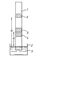
Figure l shows a device for measuring the
concentration of pregnanediol~3-glucuronide (PD3G)
in a urine sample using a competitive hapten assay
protocol. Referring to Figure l, the device
comprises a strip, l, of a bibulous paper and a
reservoir, 2, containing a developing solution, 3,
consisting of a substrate-buffer. The strip is
provided with a sample receiving zone, 4, a first
reagent zone, 5, and a second, indicator, reagent
zone, 6. The first reagent zone, 5, includes
enzyme-labelled PD3G hapten covalently attached to a
horseradish peroxidase enzyme-label, the
enzyme-labelled PD3G being impregnated into the
strip, such that, in use, it is caused to migrate
through the strip by passage of the developing
solution through the strip. The indicator zone,
6, comprises antibody to PD3G covalently bonded to
the strip. The reservoir, 2, consists of a
rupturable sac containing the substrate buffer which
includes the cofactors tetramethylbenzidine and
hydrogen peroxide. In use, a sample of urine is
applied to the sample receiving zone, 4, and the
`J: :s
':
- .' :: .
, '' "
. ~, .
. .
~iL2l3~
substrate buffer, 3, is released onto the end of the
strip by rupturing the sac, 2, with finger
pressure. The developing solution, 3, passes
through the strip by capillary action picking up the
sample and the enzyme-labelled PD3G in the solvent
front from the sample receiving zone, 4, and the
first reagent zone, 5, respectively. The enzyme
cofactor, tetramethylbenzidine, is carried through
the strip more slowly than the enzyme-labelled
PD3G. Thus, when the solvent front of the
substrate buffer, 3, reaches the first reagent zone,
5, little or no cofactor is present in the solvent
front and substantially no colour formation
occurs. Any PD3G present in the sample competes
with the enzyme-labelled PD3G for a limited number
of ~inding sites in the second, indicator, reagent
æone, 6. The amount o~ enzyme-labellea PD3G that
becomes bound to the strip in the indicator reagent
zone, 6, is therefore inversely proportional to the
concentration of PD3G in the sample. Continued
development of the strip washes any unreacted
reagents through the indicator zone, 6, and
subsequently brings the cofactor into contact with
the indicator zone where the substrate is converted
by the bound enzyme-labelled PD3G to give a coloured
product ~horseradish peroxidase catalyses the
oxidation of tetramethylbenzidine by hydrogen
peroxide). The coloured product does not migrate
further, thus giving a sharp band of colour at the
indicator æone, 6. The difference between
distances B and A as shown in Figure 1 is made
relatively small as the sample and the
enzyme-labelled PD3G do not interact until the
indicator zone, 6, is reached thus allowing a
reduction in the length of the strip. However, the
distance B must be sufficient to permit adequate
~, . . . .
.
-, ~
,
,. ,~ .
.
.;
' '
.
~2~39~
- 18 -
washing of the bound enzyme-labelled PD3G by
continued migration of the developing solution, 3,
prior to the arrival of the cofactor at the
indicator zone, 6. The duration of the competitive
reaction which occurs at the indicator zone, 6, is
equal to the time it takes for the sample and the
enzyme-labelled PD3G to pass through the indicator
zone, 6. This is determined by the physical
properties of the material from which the strip is
made.
Figure 2 shows a further device for measuring
the concentration of PD3G in a urine sample using a
non-competitive hapten assay protocol. Referring
to Figure 2, the device comprises a strip, 11, of a
bibulous paper and a reservoir, 12, containing a
developing solution, 13 consisting of a
substrate-buffer. The strip is provided with a
sample receiving zone, 14, a first reagent zone, 15, ..
and a second, indicatorj reagent zone, 16. .The ..
first reagent zone,l5, includes enzyme-labelled
anti-PD3G comprising antibody to PD3G covalently
attached to a horseradish peroxidase enzyme label,
; the enzyme-labelled anti-PD3G being impregnated into
; the strip such that, in use, it is caused to migrate
25 through the strip by passage of the ..
~ubstrate-buffer, 13, through the strip. .The
indicator zone, 16, comprises PD3G covalently bonded
to the strip. The reservoir, 12, is as described
above with reference to Figure 1.
In use, a sample of urine is applied to the
sample receiving zone, 14, and the substrate-buffer,
13, is released onto the end of the strip by .
rupturing the sac, 12, with finger pressure. The.
substrate bu~fer, 13, passes through the strip by
capillary action picking up the sample~ Any PD3G
present in the sample is bound in the first reagent
;:~ ' .
.
.
'.,~
~2~3~(;17~
-- 19 --
zone, 15, by the enzyme-labelled an~i-PD3G which is
present in excess. The incubation time for this
interaction to take place is controlled by the
difference between distances B and A as shown in
s Figure 2. As the solvent front contacts the
indicator zone, 16, PD3G covalently bonded to the
strip binds any unreacted enzyme-labelled antibody
to PD3G. Again, since the cofactor travels more
slowly through the strip than the enzyme-labelled
antibody, colour generation only occurs at the
indicator zone, 16, where the enzyme-labelled
antibody has been immobilised.
Figure 3 shows a device for measuring the
concentration of thyroid stimulating hormone ~TSH)
in a sample using a two-site sandwich assay or
immunometric assay protocol. Referring to Figure 3
the device comprises a strip 21 of a bibulous paper
and a reservoir, 22, containing a developing ~
solution, 23 consisting of a substrate-buffer. -The
strip is provided with a sample receiving zone, 24,
a first reagent zone, 25, and a second, indicator,
reagent zone, 26. The first reagent zone, 25,
includes enzyme-labelled anti-TSH comprising
antibody to TSH covalently attached to a horseradish
peroxidase enzyme-label, the enzyme-labelled
anti-TSH being impregnated into the strip, 21, such
that, in use, it is caused to migrate through the
strip by passage of the substrate-buffer, 23. ~he
indicator zone, 26, comprises a second antibody to .
TSH covalently bonded to the strip, 21. The second
antibody has specificity for a different and
non-competing epitope of TSH from that of the
enzyme-labelled antibody. In use, any TSH present
in the sample is bound by the enzyme-labelled
antibody which is present in excess as they
co-migrate through the strlp. The time allowed for
. . '
~28~
-20-
this first reaction is yoverned by the difference
between the distances B and A as shown in Figure
3.0n passing through the indicator zone~ the TSH (in
the form of an antibody complex) is bound by the
second antibody. The time of the second incubation
is governed by the speed of capillary migration of
the solvent front through the material of the
strip. Again, colour development only occurs where
the enzyme-labelled antibody is immobilised in the
indicator zone thus allowing the cofactor to be
brought into contact with it by the capillary motion
of the solvent front.
Figure 4 shows a device for measuring the
ratio of the concentrations of PD3G and
oestrone-3-glucuronide (E13G) in a urine sample.
The ratio of these two products has been shown to be
indicative of the fertile period of the female
menstrual cycle (see for example British published
specifications GB-B-2029011 and GB-B-2116318).
Referring to Figure 4, the device comprises a
strip, 31, of bibulous paper and a reservoir, 32,
containing a developing solution, 33 consisting of a
substrate-buffer. The strip is provided with a
sample receiving zone, 34, a first reagent zone, 35,
a second reagent zone, 36, a third reagent zone, 37,
a'nd a fourth, indicator, reagent zone, 38. The
first reagent zone, 35, comprises a mixed steroid
antigen (MSA) consisting of a PD3G hapten and an
E13G hapten covalently bonded to a bridging
structure. The MSA is impregnated into the strip,
31, such that, in use, it may migrate through the
strip in the advancing solvent front of the
substrate-buf~er, 33. The second reagent zone, 36,
comprises an antibody to E13G which may be free to
migrate through the strip in the advancing solvent -
front though is preferably covalently bonded to the
Y~
7~
-21-
strip. The third reagent zone, 37, comprises an
enzyme-labelled anti-E13G (antibody to E13G
covalently attached to a horseradish peroxidase
enzyme-label), impregnated into the strip, 31, such
that it may migrate through the strip with the
substrate-buffer, 33. The fourth, indicator,
reagent zone, 38, comprises antibody to PD3G
covalently bonded to the strip. The reservoir, 3~,
comprises a rupturable sac containing the
substrate-buffer.
In use, a urine sample is applied to the
sample zone, 34, and the reservoirl 32, of
developing solution~ 33, is ruptured, releasing the
substrate-buffer onto one end of the strip. The
substrate-buffer, 33, passes up the strip, 31, by
capillary action picking up sample from the sample
receiving zone, 34, and mixed steroid antigen from
the first reagent zone, 35. The sample and the MSA
co-migrate along the strip to the second reagent
zone, 36, at which the antibody to E13G is
covalently immobilised. The MSA and E13G present
in the sample compete for limited binding sites as
they pass through the second reagent zone, 36. If
the concentration of E13G in the sample is low, then
a substantial proportion of the MSA is bound in the
second reagent zone, 36, and cannot migrate
further. If, however, the sample concentration of
E13G is high, then the MSA will be free to migrate,
together with the sample, to the next reagent zone,
namely, the third reagent zone, 37, comprising
enzyme-labelled antibody to E13G. This latter
reagent is present in excess and is non-covalently
absorbed to the strip, 31. The enzyme-labelled
antibody binds to the MSA as they both migrate
together along the strip to the fourth, indicator,
reagent zone, 38, at which antibody to PD3G is
~,s'
.
- 22 -
covalently attached to the strip. At the fourth
indicator reagent zone, 38, any PD3G present in the
sample competes with the MSA/anti-El3G complex for
binding to a limited number of binding sites of the
covalently immobilised anti-PD3G antibody. The
enzyme-labelled immunocomplex will be bound at the
indicator zone, 38, only when the sample
concentration of PD3G is low. The excess reagents
are washed from the measuring location by continued
development of the strip On reaching the
indicator zone, 38, the cofactor is converted by the
enzyme-labelled, antibody-bound MSA to a coloured
product giving a clear positive signal. The assay
can be tuned to give a positive response only when a
predetermined elevated level of E13G coincides with
; a predetermined low level of PD3G. A further
reagent zone Inot shown) may be provided at a point
remote from the reservoir, 32, which comprises
covalently bound horseradish peroxidase. This
reagent zone gives an indication that substrate has
migrated through the length of the strip thus
indicating that the assay has run to completion.
Figure 5 shows a further device for measuring
the ratio of the concentrations of PD3G and El3G in
a urine sample
i Referring to Figure 5, the device comprises a
strip, 41, of bibulous paper and a reservoir, 42,
containing a developing solution. - The strip is
provided with a sample receiving zone, 43, a first
reagent zone, 44, a second reagent zone, 45, a third
reagent zone, 46, a fourth reagent zone, 47, a fifth
reagent zone, 4~, a sixth, test indicator, reagent
zone, 49 and an optional seventh, control indicator,
reagent zone, 50. The first reagent zone, 44,
comprises anti-El3G (antibody to El3G). The second
reagent zone, 45, comprises a mixed steroid antigen
7~
~ 23 -
(MSA) as described in Example 4. The third reagent
æone, 46, comprises biotin-labelled anti-PD3G
dextran-coated charcoal. The fifth reagent
zone,(antibody to PD3G covalently bonded to
biotin) The fourth reagent zone, 47, comprises
48, comprises enzyme-labelled anti~El3G (antibody to
El3G covalently bound to horseradish peroxidase).
The sixth reagent zone, 49, comprises immobilised
streptavidin. The optional seventh reagent zone,
50, comprises horseradish peroxidase.
The active components of the reagent zones are
dried onto the strip as transverse bands. The
streptavidin in the sixth, test indicator, reagent
zone, 49, is covalently attached to the strip and
the dextran-coated charcoal in the fourth reagent
zone, 47, is deposited onto the strip by adding an
aqueous suspension of microparticulate charcoal and
subsequently removing the water. All the other ~ -
reagents are soluble and are impregnated into the
strip by applying them each in solution and
subsequently drying. The soluble reagents, in use,
migrate with the solvent front of the developing
solution. The reservoir, 42, comprises a
rupturable sac containing the developing solution.
In use, a urine sample is applied to the
sample receiving zone, 43, and the reservoir 4~, of
developing solution is ruptured, releasing the
developing solution onto one end of the strip. The
developing solution passes up the strip by capillary
action, picking up sample from the sample receiving
zone, 43, and, in sequence, anti-El3G, MSA and
biotin-labelled anti-PD3G. The soluble components
of the assay pass through the strip in the advancing
solvent front, and at the same time are mixed and
allowed to react. The separation of the reagent
zones may be adjusted to facilitate optimum
~ ~ ' ' , ,
,
~2~t~
- 24 -
incubation times for reaction. Unbound, low
molecular weight species such as MSA and steroids
are removed from the solvent front by the charcoalin
the fourth reagent zone, 47. The solvent front
passes through the fifth reagent zone, ~8, picking
up enzyme-labelled anti-E13G, thus completing the
assay protocol. The presence of complexes of
enzyme-labelled anti-E13G/MSA/biotin-labelled PD3G
is indicative of a high level of E13G and a low
level of PD3G. Such complexes are immobilised in
the sixth, test indicator, reagent zone, ~9, by the
interaction of biotin with immobilised streptavidin.
The developing liquid, as previously stated,
includes a colour producing substrate for
peroxidase. If the substrate has an Rf value
substantially the same as the enzyme-labelled
species in the device, colour will develop in the
solvent front as soon as the fifth reagent zone, 48,
is reached and the solvent front will remain
coloured for the rest of its passage through the
strip. The presence of colour at the end of the
strip remote from the developing solution indicates
completion of the assay. As the solvent front
meets and passes through the sixth, test indicator,
reagent zone, 49, any coloured products and unbound
r enzyme-labelled anti-E13G will be washed clear of
the zone by incoming fresh developing solution.
However, if the complex including enzyme-labelled
anti-E13G has become immobilised, coloux generation
occurs in the sixth, test indicator, reagent zone.
Thus colour in the sixth reagent zone indicates a
positive test result. Alternatively, if the
substrate has an Rf value less than the Rf value of
the slowest moving enzyme-labelled species, colour
generation will only occur where an enzyme-labelled
species is immobilised, i.e. in the sixth reagent
~28g~7~
- 25 _
zone when a positive result is obtained. In this
alternative, which is preferred, a seventh, control
indicator, reagent zone, 50, comprising horseradish
corresponding to completion of the assay.
peroxidase may be provided to indicate arrival of
the substrate at a predetermined part of the strip,
Figure 6 shows a test sheet for monitoring the
menstrual cycle embodying a plurality of test strips
as described above with respect to Figures 4 or 5.
Referring to Figure 6, the test sheet
comprises a rigid plastics backing plate or stand,
51, supporting a plurality of test strips of the
invention, e.g. 52 (In the Figure the details of the
absorbent strips are not shown). In the embodiment
shown, fifteen test strips are provided in
side-by-side parallel arrangement. The backing
plate or stand, 51, is overlaid with a plastics
film, to cover the test strips, apart from in the
sample receiving zone, 53. The plastics film
(shown in Figure 6 as transparent for clarity) is
opaque save in the test indicator zones, 54, and in
the control indicator zones, 55. The plastics film
may be suitably masked or printed to indicate
clearly the sample receiving, test indicator and
control indicator zones. The developing solution
is contained in separate rupturable sacs, 56, one
; for each test strip.
In use, mid-stream urine is sampled using a
disposable sample loop and an ali~uot is blotted
onto the sample receiving zone, 53, of a test strip,
52. The seal of a rupturable sac, 56, of
developing solution is broken by finger pressure,
thus initiating the test. After 15 to 20 minutes,
the control indicator zone, 55, is observed and, if
coloured, the assay result is read from the test
indicator zone, 54.
,
, . .
, ' . '; ' ;
.
''
- 26 -
The presence of colour in the test zone
indicates a positive result i.e. the woman is in, or
near, her fertile period. The converse applies
with the absence of colour. For the woman who
wishes to avoid conception, it is intended that she
should test her urine once per day starting at about
day 6 or 7 of her cycle. She should continue daily
testing until a period of sustained positive results
have been observed (more than 2 days) followed by a
period of sustained negative results (more than 2
days). This would normally mean a total of lO to
15 tests in a typical cycle. Whenever a positive
result is observed, the woman should refrain Erom
intercourse and should continue to do so until two
successive daily negative results have been observed.
It is intended that colours developed in the
strips are stable, and so form a-semi-permanent
record of the woman's cyclical activity.
EXAMPLE l
A device for conducting a competitive hapten
assay similar to that described generally with
respect to Figure l was prepared.
Reerring to Figure 7, the device comprises a
;strip of Gelman glass-fibre (15 x l cm), 61, and a
reservoir, 62, containing the substrate-buffer,
63. The strip is provided with a sample receiving
zone, S~, (l.5 cm from the lower end of the strip),
a Eirst reagent zone, 65, t2.0 cm ~rom the lower end
of the strip) to which has been applied lO ul MSA
(lO00 nM in Buffer C), a second reagent zone, 66
(2.5 cm from the lower end of the strip) to which
has been applied lO ul of a monoclonal antibody to
PD3G conjugated to peroxidase (l ug ml~l in Buffer
C) and an indicator zo~e, 67, (5.0 cm from the lower
,~ '
.
~r--v~
end of the strip) to which has been applied 50 ul of
a solid phase monoclonal antibody to El3G.
In use, 20 ul of sample to be assayed for PD3G
is applied to the sample receiving zone, 64, and the
the strip is immersed. After 15 min, the indicator
lower end of the strip is placed into 2 ml of
substrate-buffer such that only the first 0.5 cm of
zone, 67, is observed. The presence of a blue
colour at the indicator zone, 67, shows that the
concentration of PD3G in the sample is less than
10000 nM. (colour at 5000 and 10000 nM: no colour at
15000, 20000 and 25000 nM)
The device operates as follows:
As the substrate-buffer migrates along the
strip, PD3G in the sample, MSA and anti-PD3G
peroxidase conjugate are transported at the buffer
front together with all the components of the
buffer-substrate exce~t TMB which exhibits a slower
rate of migration (Rf = 0.7~. During migration
along the strip, the PD3G and MSA compete for the
binding sites o~ the ant}-PD3G peroxidase
conjugate. On reaching the indicator zone, 67, the
immobilised anti-El3G antibody binds to the MSA.
If the concentration of PD3G in the sample is low,
then the MSA will also be bound by the anti-PD3G
peroxidase conjugate and when the TMB reaches the
indicator zone, 67, a blue colour will be formed.
If the concentration of PD3G in the sample is high,
then the anti-PD3G peroxidase conjugate will not
bind the MSA and no colour signal will be observed
at the indicator zone, 67.
EXAMPLE 2
A device for conducting a non-competitive
hapten assay similar to that described generally
,~ .
. ,
,
~L28~
- 28 -
with respect to Figure 2 was prepared.
Referring to Figure 8 the device comprises a
strip of Gelman glass-fibre (15 x 1 cm), 71, and a
reservoir, 72, containing the substrate-buffer,73.
The strip is provided with a sample receiving zone,
74, (1.5 cm from the lower end of the strip), a
first reagent zone, 75, (2.0 cm from the lower end
of the strip) to which has been applied 10 ul of a
monoclonal antibody to PD3G conjugated to peroxidase
(1 ug ml~l in Buffer C), a second reagent zone, 76,
(5.5 cm from the lower end of the strip) to which
has been applied 10 ul of MSA ~5,000 nM in Buffer C)
and an indicator zone, 77, (6.0 cm from the lower
end of the strip) to which has been applied 50 ul of
a solid phase monoclonal antibody to E13G.
In use, 20 ul of sample is applied to the
sample receiving zone, 74, and the lower end of the
strip is placed into 2 ml of substrate-buffer such
that only the first 0.5 cm of the strip is
immersed. ~fter 15 min, the indicator zone, 77, is
observed. The presence of a blue colour at this
zone, 77, means that the PD3G concentration in the
sample is less than 10000 nM. (colour at 5000 and
10000 nM: no colour at 15000, 2aooo and 25000 nM)
The device operates as follows:
, As the substrate-buffer migrates along the
strip, the PD3G in the sample and the anti-PD3G
peroxidase conjugate are transported at the buffer
front together with all the components of the
buffer-substrate except TMB which exhibits a slower
rate of migration (Rf = 0.7). During migration
along the strip, the anti-PD3G peroxidase binds the
PD3G. On reaching the second reagent zone, 76, the
MSA in excess binds any unreacted anti-PD3G
peroxidase conjugate, and is transported to the
indicator zone, 77, where the solid phase anti E13G
.~ .
: : '
~2~
-- 29 --
antibody binds both free and anti-PD3G peroxidase
conjugate bound MS~ . Thus, if the concentration of
PD3G present in the sample is low, most of the
anti PD3G peroxidase conjugate will be bound by the
MSA at the indicator zone, 67. When the TMB
reaches the indicator zone, 67, a blue colour will
be formed. However, if the concentration of PD3G
in the sample is high, then most of the anti-PD3G
peroxidase conjugate will be unable to bind the MSA
and thus no colour will be observed at the indicator
zone, 67,
EXAMP E 3
A device for conducting a two-site sandwich
immunoassay for thyroid stimulating hormone (TSH~
such as described generally with reference to Figure
3 was prepared.
Referring again to Figure 3, the strip, 21, is
Gelman glass-fibre (17 x 1.8 cm). At the first
reagent zone, 25, (5.5 cm from the lower end of the
strip) is applied to 20 ul of a monoclonal antibody
to TSH conjugated to peroxidase (5 u~ ml~l in Buffer
C). At the indicator zone, 26, 300 ul of a
solid-phase monoclonal antibody to TSH is applied
(7.5 cm from the lower end of the strip). In use,
50 ul of sample is applied to the sample receiving
zone, 24, and the strip is placed into 5 ml o~
substrate-buffer such that only the first 1 cm of
the strip is immersed. AEter 15 mln, the indicator
zone, 26, is observed. The presence of a blue
colour indicates that the concentration of TSH
present in the sample is greater than 200 mU/L. (No
colour at 50, 100 and 200 m~/L: colour at 250 mU/L).
'
,: ,. : .
. . .
~28~
- 30 -
EXAMPLE 4
_
In another Example of a two-site sandwich
assay of the type described generally with reference
to Figure 2, the monoclonal antibodies to TSH
described in Example 3 above (at reagent zone 25 and
indicator zone 26) were replaced with monoclonal
antibodies to human chorionic gonadotrophin. The
test can be used as a pregnancy indicator with
colour being formed when the urine sample contains
more than 200 mIU/ml hCG. (No colour at 50, lO0 and
200 mIU/ml: colour at 250, 300 and 500 mIU/ml).
EXAMPLE 5
A device for conducting a dual analyte assay
similar to that described generally with respect to
Figure 4 was prepared.
Referring to Figure 9 the device comprises of
a strip of Gelman glass-fibre (15 x l cm), 81, and a
reservoir, 82, containing the substrate-buffer,
83. The strip contains a sample receiving zone,
84, (l.5 cm from the lower end of the strip), a
first reagent zone, 85, (2.0 cm from the lower end
of the strip) to which has been applied lO ul-of a-- ~
monoclonal antibody to PD3G conjugated to peroxidase
(l ug ml~l in Buffer C), a third reagent zone, 87,
~3.0 cm from the lower end of the strip) to which is
applied lO ul of MSA ~250 nM in BuEfer C) and an
indicator zone, 88, (5.0 cm from the lower end of
~he strip) to which is applied 50 ul of a solid
phase monoclonal antibody to El3G.
In use f 20 ul of a urine sample is applied to
the sample receiving zone, 84, and the strip is
placed into 2 ml of substrate-buffer such that the
lower 0~5 cm of the strip is immersed. After 15
. . .
- 31 -
min, the indicator zoner 88, is observed. The
presence of a blue colour at this zone, 88, means
that the sample concentration of E13G is greater
than 50 nM and that the sample concentration of PD3G
is less than 10000 nM (see Table 1).
TABLE 1
PD3G (nM)
E13G ~nM) 0 1000 5000 10000 20000
O ~
- _ _ _ _
100 + ~ +
200 + + +
+ = Blue colour at indicator zone.
- - No blue colour at indicator zone. -
The device operates as follows:
As the substrate-buffer migrates along the
strip, the E13G and PD3G of the sample, anti-PD3G
peroxidase conjugate, anti-E13G antibody and MSA
migrate at the buffer front with all the components
of the buffer-substrate except TMB which exhibits a
lower rate of migration ~Rf = 0.7). If the sample
concentration of E13G is low, then the MSA will be
bound by the anti-E13G antibody during the migration
and will not be bound by the solid phase anti-E13G
antibody at the indicator zone, 88. I~ the E13G
level is high, then the MSA is free to bind at the
indicator zone, 88. If the PD3G concentration of - -
the sample is low, then the MSA will be bound by the
anti-PD3G peroxidase conjugate and thus, when the
TMB reaches the indicator zone, 88, a blue colour
will be formed. If, however, the PD3G
concentration of the sample is high, then it will
~' .
'`
:
~L2~
- 32 -
bind to the anti-PD3G peroxidase conjugate
preventing the latter from binding to the MSA and
thus no signal will be generated.
It will be understood that the invention has
been described by way of example only and
modifications of detail may be made within the scope
of the invention.
,