Note: Descriptions are shown in the official language in which they were submitted.
40906 CAN 7A
92Zi 3~
AN EXTERNAL EAR CANAL ELECTRODE TO BE
PLACED PROXIMATE THE TYMPANIC MEMBRANE AND
MET~OD OF STIMULATING/RECORDING U~ILIZING EXTERNAL
EAR CANAL ELECTRODE PLACED PROXIMATE THE
5TYMPANIC MEMBRANE
Technical Field
The present invention relates generally to
electrodes for and methods of applying/recording electrical
signals to~from the neural/neuromuscular system of a
person. The present invention more particularly relates
to such electrodes which are inserted into the external
ear canal of a person and methods therefor.
Background Art
It is well known that electrical stimulation
of the auditory system of a person can produce complex
perceptions of sounds in human subjects. Experiments
by Volta occurred in the early 1800 s. Volta inserted
metal bars in-to his external ear canals and passed current
between the bars and reported the perception of sound.
A series of studies was performed in the late 1930 s
by Jones, Stevens and Lurie which examined the relationship
between various parameters of electrical stimulatlon
and the listed auditory perceps. In these experiments,
investigators used metal electrodes placed into saline
filled external ear canals to deliver electrical current
to the auditory system.
More recently, electrodes surgica~ly implanted
into the coch~ea, are providing a sensation of hearing
in profoundly deaf individuals. The deve~opment of cochlear
imp]ants has prompted significant additional research
in the area of electrical stimulation for hearing
augmentation.
~k
:
-2~ 2Z~
Means of stimulating the auditory system which
are noninvasive, as opposed to a cochlear implant, are
desirable for several reasons. A noninvasive electrode
system for aud;tory stimulation could be used as a
Eunctional auditory prosthesis, as a tool in the diagnostic
evaluation of potentia] candidates for cochlear implants
or to record the electrical signals generated by an
auditory system stimu]ated by other means.
Several invest;gators have reported the use
of electrodes combined with a saline filled ear canal
(known as a Bremmer type electrode) to provide a noninvasive
means of auditory stimulation. This approach to the
stimu]ation of the auditory system has a major drawback.
Current passed into the sa]ine solution spreads rapidly
in a]l directions through the tissues of the ear and
head. The same electrica] current which activates the
auditory nerve can a]so stimulate cutaneous nerve fibers
thereby producing uncomfortab~e and, possibly, painEul
sensations.
Existing systems of auditory stimulation have
significant disadvantages. Cochlear implants require
surgical invasion of the body. Thus, cochlear implants
are not practical for evaluation or diagnostic purposes,
which involve temporary stimulation or recording. On
the other hand, conventiona~ external ear canal stimulation
has significant limitations in dynamic range. The e~ectrica~
current used in external ear canal stimulation must
be above an amount to exceed the hearing threshold but
must be below an amount which would produce pain within
the external ear cana] or, possibly, uncomfortab~e loud
sensations.
Disclosure of Invention
The present invention solves these problems
-3- ~2~2~S
by eliminating surgical invasion while substantially
increasing the dynamic range of the stimulation signals
which can comfortably be utilized.
The only direct physical pathway from the
external ear canal to the inner ear (aside from the
s~ull itself) is the tympanic membrane and the ossicular
chain. This tissue connection constitutes the major
pathway for current flow between the inner ear and the
external ear canal and visa versa. Electrical signa]s
which are generated inside a norma]]y functioning cochlea
travel through surrounding tissues and can be recorded
from outside of the cochlea. This technique is c]inical]y
known as electrocochleography (ECoG). The amplitude
of ECoG signals is largest at the surface of the cochlea,
the measurement of which is an invasive technique. For
noninvasive measurements, the amp]itude o~ ECoG signa]s
is greate~st at the tympanic membrane. This supports
the hypothesis that the ossic]es are the lowest resistance
pathway by which current travels between the cochlea
and the ex-ternal ear canal. The present invention provides
an electrode and a method of stimulating or recording
which takes advantage of this low resistance connection
between the inner ear and the external ear canal. The
electrode can be used to either stimulate the inner
ear with e]ectrical current or, conversely, to measure
the electrical activity originating within the inner
ear without an invasive technique.
The present invention provides a method of
stimulating the neural/neuromuscu]ar system oE a person
~ having a body and an external ear canal and ad~acent
tympanic membrane. ~ ~irst electrode is inserted into
the external ear cana] o~ the person. The ~;rst electl-ode
has an elongated flexib]e body having a proximate end
and a distal end. The ~irst electrode has a compressible
35~ material mounted at the dlstal end of the elongate flexible
::
::
` _4_ ~Z~2285
body and has an e]ect:r;ca~ly conductive ge] carried
by the compressible material. The ~irst electrode has
an electrical conductor cornmunicating with the elongate
flexible body being electrically coup]ed to the electrically
conductive gel. The first electrode is positioned with
the electrical]y conductive gel proximate the tympanic
membrane or the tympano-meatal annulus of the person.
~ second return electrode is then app]ied to the body
of the person. An e]ectrica] stimu]us signal is app]ied
to the e]ectrode pair thereby stimulating the
neural/neuromuscular system of the person without surgical]y
invasive techniques. Optiona]ly an anesthetic may be
app]ied to the area of the tympanic membrane or
tympano-meatal annu]us of the person to further increase
the dynamic range of the e]ectrica] stimu~ating signal.
This anesthetic may be incorporated into the conductive
ge] may be iontophoretical]y de]ivered or may be injected.
The present invention a]~so provides a method
of recording electrica] signals from the
neural/neuromuscular system of a person having a body
and an externa] ear canal and an adjacent tympanic membrane.
~ first electrode is inserted into the externa] ear
cana] of the person. The first electrode has an elongate
f]exible body having a proximate and a distal end. The
first e]ectrode has a compressible material mounted
at the dista] end of the e]ongate flexib]e body and
has an electrical]y conductive ge] carried by the
compressible material. The first electrode further has
an e]ectrica] conductor communicating with the e]ongate
f]exihle body being electrica]]y coupled to the e:lectrica~ly
conductive gel. The first electrode is positioned with
the e]ectrically conductive gel proximate the tympanic
memhrane or tympano-meata] annulus of the person. ~
second reference electrode i~s then applied to the body
of the person. E]ectrical ~signals generated by the coch]ea
_5_ ~ 2 ~ 2 2~
of the person are then recorded utilizing the inserted
electrocle pair.
The present invention also provides an e]ectrode
adapted to be utilized within the external ear canal
for applying/recording electrical signals to/from the
neural/neuromuscular sys-tem of a person having an external
ear cana] and adjacent tympanic membrane. The electrode
has an elongated f]exible body having a proximate end
and a distal end. ~ compressible material is mounted
at the distal end of the elongate f]exible body. ~n
electrically conductive gel is carried by the compressible
material and an electrical conduc-tor communicates with
the elongate flexible body electrically coupling the
electrically conductive ge] and adapted to be coupled
to a stimulator/recorder at the proximate end. An electrode
constructed in tl-is fashion may be p]aced in the external
ear canal with a dist~] end being proximate the tympanic
membrane or tympano-meatal annulus so electrical signals
can be applied/recorded to/from the neural/neuromuscular
system.
In an alternative embodiment, the e]ectrically
conductive gel is absorbed into the compressible material
which, in another a]ternative embodiment, may also be
resilient. The elongated flexible body is stiff enough
to enab]e it to be inserted into the external ear canal
but flexible enough to bend if the electrode is pushed
against the tympanic membrane without rupturing the
tympanic membrane. In another alternative embodiment,
an anesthetic may be carried by the compressible material.
In other alternative embodiments, a physical fixation
device is utilized for physical]y securing the e]ectrode
wil:h re~spect to the externa] ear canal. In alternativc
emhocliments, the physical fixation device may be an
inflatable cuff, radial fins, a resilient collar or
a coiled spring. In an alternative embodiment, adhesive
-6- ~Z9Z~85
tape adhesively couples the elongated flexible body
to the body of the person which in another embodiment
may be utili~ed as a return/reference electrode. In
a sti]l further alternative embodiment, the compressib]e
material may be connected to the distal end of the flexible
body by a coil.ed spring.
Brief Description of Drawings
The foregoing advantages, construction and
operation of the present invention will become more
readily apparent from the following description and
accompanying drawings in which:
Figure 1 is an illustration of an el.ectrode
of a preferred embodiment of the present invention;
F'igure 2 is a illustration of a method of
the present inventi.on;
Figure 3 is an illustration of an a]ternative
method of the present invention;
Figure 4 is an illustration of an el.ec-trode
of the present invention inserted in an external ear
canal;
Figure 5 is an illustration of an alternative
:~ embodiment of an electrode of the present invention
with an inflatable cuff;
Fi.gure 6 is an illustration of an alternative
embodiment of an electrode of the present invention
: ~ with an inflatable ring;
: ~ Fi,gure 7 is an i]lustration of an a]ternative
emhodiment of an e]ectrode of the present invention
wil:h a collar;
: 30 Figure ~ is an illustration of an alternative
: embodiment oE an e]ectrode of the present invention
with fins;
Figure 9 is an il.lustration of an alternative
embodiment of an e]ectrode of the present invention
with a speculum and an anesthetic de]ivery tube; and
::
-7~ 2~
Figure 10 is an il]ustration oE an a]ternati.ve
embodiment of an electrode of the present invention
with a spring ti.p.
Detai.led Description oE the Preferred Embodiments
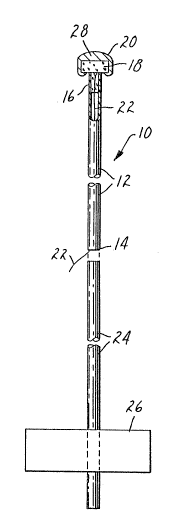
A preferred embod:iment of the electrode 10
of the present invention is illustrated in Flg~lre 1.
The electrode 10 is formed from a length of s;.licone
elastomer tubing 12 forming an elongated flexible body.
The silicone elastomer tubing 12 is of a sufficient
size and sufficient stiffness to allow the insertion
~f the electrode 10 into the external ear canal but
;.s f]exible enough so that the silicone elastomer tubing
12 wil] bend and not create damage to tl-e tympanic membrane
.shoull the electrode 10 be inserted in position proxima~e
to tympanic membrane or tympano-meatal annulus. An example
of tubing 12 which is suitable for this purpose is Dow
Corning Medical Grade ~IP Si1asticTM tubing with an outside
diameter of 0.08 inches (2.03 millimeters) and an inside
diameter of 0.077 inches (1.96 millimeters). The tubing
12 has a proximate end 14 sui-table to be grasped by
the insertion means such as the health care technician s
fingers and a distal end 16 to be placed into the external
ear canal. A compressible material 18 is mounted at
the distal end 16 of silicone elastomer tubing 12. In
a preferred embodiment the compressible material is
a sponge materia] such as an open cell compressible
foam such as a polyester/polyether sponge and is formecl
to fit snuggly within the distal end 16 of silicone
elastomer tubing 12. In a preferred embodi.ment the
compressible material 18 has an overall diameter larger
than the diameter of silicone elastomer tubin~ 12. In
a preferred embodiment the dimensions of the compressible
material 18 are about 0.25 inch (6.4 mi~limeters) in
.
-8~ 22~
diameter and about 0.125 inch (3.2 millimeters) in
compressib]e distance. It is preferred that the compressible
material also be resi]ient. The compressib]e materia]
18 cushions the tympanic membrane or tympano-meatal
annulus when the electrode 10 is inser-ted approximately
at that position. The resiliency in compressible material
18 wil] allow the electrode lO to be withdrawn and
reinserted with similar insertion characteristics on
subsequent insertions into the externa] ear cana]. ~
conductive gel is carried by the compressib]e materia]
at the dista] end 16 of si]icone elastomer tubing 12.
A preferred conductive gel is Red Dot No. 2248 Solid
Conductive E]ectrode Gel available from Minnesota Mining
and Manufacturing Company. ~n electrica] conductor,
name]y a wire, 22 is inserted through si]icone elastomer
tubing 12 from the proximate end ]4 to the dista] end
16 communicating with the conductive gel 20 carried
by compressible material 18. Electrical conductor 22
provides the mechanism for supplying the electrical
stimulus signal to the distal end 16 of the electrode
10 or to conduct electrical signals generated by the
coch]ea from the conductive gel 20 to the proximate
end 14 of the e]ectrode 10. It is important to electrica]]y
insulate electrica] conductor 22 from the wal]s of the
external ear canal and is covered with insu]ation such
as Teflon M insulation. In the preferred embodiment,
electrical conductor 22 is carried in the interior of
silicone e]astomer tubing 12. In other embodiments,
it is envisioned that the electrical conductor could
be positiorled outside of si]icone elastomer tubing ]2,
~spirally wrapped or otherwise communicating with si]icone
elastomer tubing 12 to create a conductive path between
the conductive gel 20 at the distal end 16 and the proximate
end 14 of electrode 10. A preferred e]ectrical conductor
,
::
: :~
_9_ ~ ~2~5
22 is a 0.003 inch (0.08 millimeters) diameter wire
of 90~ p]atinum ancl 10~ iridium with Tef]onTM insu]tat1On.
Those Eeatures of Figure 1 heretofor described
contain all the essential features of the electrode
10. In addition, the preferred e]ectrode 10 illustrated
in ~igure 1 has a second length oE silicone elastomer
tubing 24 affixed to the proximate end 1~ of silicone
elastomer tubing 12. One means oE securing the second
piece of si]icone elastomer ~ubing 24 to silicone elastomer
tubing 12 is to make silicone elastomer tubing 24 of
a larger diameter and of such a diameter so that a snug
fit is developed when silicone elastomeric tubing 24
is placed over or inside of the proximate end 14 of
silicone elastomer tubing 12. In a preferred embodiment,
electrical conductor 22 may exit the electrode at this
po;nt. An alternative electrode 22 could be carried
w;th si]icone elastomer tubing 24 the entire length
of the e]ectrode or exit somewhere in between. The
flexibility characteristics of silicone elastomer tubing
12 are essential for the proper placement of the electrode
10 within the external ear canal. In a preferred embodiment,
silicone elastomer tubing 24 is stiffer than and is
less flexible than silicone elastomer tubing 12. This
~silicone elastomer t~lbing ]2 may carry an adhesive tape
2fi which can be used to secure the electrode 10 to the
external ear, cheek or face of the user providing some
mechanical stability to the electrode 10 when it is
in place. Any commonly utilized adhesive tape 26 may
be utilized such as Micropore or Durapore M tape available
from Minnesota Mining and Manufacturing Company.
~ n electrocle constructe(l as clescribecl in ~igure
1 af~ords several advantac7es over existing technolo7y.
The tip, or distal end 16, of the electrode 10 is composed
o~ a compressible materia1 18, e.g., a sponge, which
is infiltrated with a conductive gel 20. The main body
I o ~L;29ZZ8s
of the electrode 10 is a soft silicone elastomer tubing
12 which allows the insertion of the electrode 10 into
the external ear canal. The flexibi]ity of the silicone
e]astomer tubing 12 combined with the compressible material
18 and conductive ge~ 20 a]lows the electrode 10 to
be positioned proximate the tympanic membrane or
tympano-meatal annulus with little or no discomfort.
The contact area of the conductive ge] 20 with the tympanic
membrane or tympano-meata] annu]us resu]ts in a low
impedance connection for recording or stimulating. It
has been found that recording with an electrode of this
design results in signals which are significantly greater
in magnitude than those obtained with a bare conductive
ball electrode and provide a more faithfu] representation
o~ the electrica] signals generated by the cochlea.
Recording of inner ear potentia]s is usefu~ in the clinical
diagnosis of ear disorders. Further, e]ectrica] stimu]ation
of the inner ear via this electrode 10 cou]d be app]ied
for prosthetic, i.e., hearing augmentation, therapeutic,
i.e., tinnitus supression, treatment of hydrops or other
inner ear disorders, or diagnostic, i.e., testing of
cochlear imp]ant candidates, purposes. The design of
the electrode 10 of the present invention affords that
current flow into the cochlea is maximized when current
is applied directly to the tympanic membrane or
tympano-meatal annu]us. Less current is ]ost into the
surrounding tissues resulting in (a) ]ower thresholds
for auditory stimulation and ~b) less stimulation oE
cutaneous pain fibers.
Several variations of the electrode 10 could
be made without departing ~rom the scope of the present
invention. ~n anesthetic 28 could be incorporated ;n
the compressib]e materia] 18. This anesthetic would
provide ]ocal anesthesia to the stimulus site to further
reduce any discomfort from the stimulation or placement
:`
.
- 1 1 - 12~Z~35
of electrode 10 proximate the tympanic membrane or
tympano-meata] annulus. A preferred anesthe-tic to be
utili~ed ;s lidocaine. Further, it is contemplated that
the size of the electrode tip at the distal end 16 could
be varied depending upon the optimal use of the electrode,
as for example, for stimulation or for recording. For
stimulation purposes, a relatively large tip wou]d be
optimal since lower current densities at the point of
contact would be obtained. ~ relative]y smaller tip
at the distal end 16 of the electrode 10 would be optimal
for recording purposes where the tip oE the electrode
10 would not load or impede -the movement of the tympanic
membrane.
To insert the electrode 10 into the external
ear canal, an insertion tool, typica]ly the health care
technician s fingers, grasps the silicone elastomeric
tubing 12 and inserts the distal end 16 into the ear
canal. The electrode 10 which contains the conductive
gel 20, resilient material 18 and, optionally, an
anesthetic, if desired, is guided down the external
ear canal until the conductive gel 20 of the electrode
10 contacts the tympanic membrane or tympano-meatal
annu]us. The electrode 10 may then be held in p]ace
or may be secured by appropriate physical fixation means,
as for exampl~e, adhesive tape 26. Electrical conductor
22 may then be connected to the appropriate instrument
for either stimulating the inner ear or recording of
signals from the inner ear.
Figure 2 illustrates a method 30 of s-timulating
the neural/neuromuscular system of a person having a
body and an external ear canal and an adjacent tympanic
mem~rane. A first electrode is inserted 32 proximate
the tympanic membrane or tympano-meatal annulus. In
order to properly localize the current density
~ 35 characteristics and to restrain the electrical current
: ~:
:: :
-12- ~2~285
to the appropr;ate conductive path and to minimize
uncomfortableness or pain sensation within the external
ear canal, the first electrode should have an elongated
flexible body with a proximate end and a distal end.
A compressible material is mounted at the distal end
of the body. A conductive gel 20 is carried by the
cornpressible material and an electrical conductor
communicates with the elongated flexible body and is
electrically coup]ed -to the electrically conductive
gel. A second electrode is then applied 34 to the body.
It is not necessary that the second electrode be of
any particular design or that it be applied at any
particular location on the body. In general, conventional
cutaneous electrodes are applied to the scalp of a person
or perhaps to the cheek. The second electrode is a return
electrode. An electrical stimulus signa] is then app~ied
36 to the electrode pair consisting of the first electrode
inserted in the external ear canal proximate the tympanic
membrane or tympano-meatal annulus and the second electrode
applied elsewhere to the body.
Similarly, Figure 3 illustrates a method of
recording 38 electrica] signa]s from the
neura]/neuromuscu]ar system oE a person having a bocly
and an external ear canal and adjacent tympanic membrane.
A first e]ectrode is inserted 40 into the external ear
canal proximate the tympanic membrane or tympano-meatal
annu]us. A first electrode similar to the elec-trode
described with respect to step 32 of Figure 2 is
appropriate. As in step 34 of ~igure 2, a second electrode
is then applied 42 to the body of a person to serve
a~s a reference electrode. Similarily, this electrode
needs to be of no .special design and maybe a conventional
cutaneous electrode applied to the scalp, cheek or other
portion of the body. The electrical conductors ~rom
the electrode pair consisting of the first electrode
~Z~2;z~15
-13-
and the second electrode are then connected to a recording
apparatus and the electrica] signals obtained from the
inner ear are recorded 44.
Figure 4 shows the electrode 10 of the present
invention inserted into the external ear canal 46 of
a patient. In particu]ar, the electrode 10 shown in
Figure 1 is illustrated. Silicone elastomer tubing 12
is st;.ff enough to be guided into the external ear canal
but flexible enough to bend once con-tact is made with
the tympanic membrane 48. ~lternatively, the electrode
10 could contact the tympano-meatal annulus 50. The
conductive gel 20 of the electrode 10 contacts the tympanic
membrane 48 or the tympano-meatal annu]us 50. Compressible
materi.a] 18 is compressed by the contact and positioning
of the electrode 10 proximate the tympanic membrane
48. Silastic tubing 24 cont;nues outside the externa]
ear canal 46 and i.s secured to the cheek of the patient
by adhesive tape 26. Electrical conductor 22 may then
be connected to the appropriate stimulating or recording
source or facility.
It can thus be seen, in reEerence to Figure
4, that the electrode 10 of the present invention, provides
a relatively localized source of stimulation and recording
at the tympanic membrane 48 or tympano-meatal annulus
50. Thus, the electrode 10 may take advantage of the
most direct current pathway via the ossicular chain
52 to or from the inner ear. The flexibility of the
silicone elastomer tubing and the compressibility of
the compressible material 18 contribute to the avoidance
of pain due to the proximity of the electrode 10 to
the tympani.c membrane 48 or tympano-meatal annulus 50.
~lso contributing to the avoidance o~ pain is the
: loca]ization of the currents such that stimulatiolls
of cutaneous nerve endings in the external ear cana]
46 are tninimized.
_~4~ 2Z~5
Once the electrode 10 has been placed in the
external ear canal 46, i-t is advantageous to be able
to physically fix the electrode 10 with respect to the
external ear canal 46. Such fixation will allow consistency
of stimulating or recordi.ng position as well as prevent
damage to the tympanic membrane 48 due to movement of
the electrode 10. Several alternative mechanical fixation
techniques are described below.
In one alternative embodiment of the electrode
10, the adhesive tape 26 may be made conductive in the
form of a cutaneous electrode and the adhesive tape
26 may form the second electrode described in the method
of stimu]ating and the method of recording at steps
3~ and 42, respective]y, in Figures 2 and 3, respectively.
Figure 5 illustrates an a]ternative embodiment
o~ an electrode 10. ~gain~ the e]ectrode 10 is formed
with an elongated fl.exible body of a si~icone elastomer
tubing 12. A compressible material 18 is carried at
the distal end 16 of the electrode 10. The compressible
material. contains a conductive ge] 20. An electrical
conductor 22 electrical1y communicates between the end
of the electrode 10. Physical fixation of this el.ectrode
].0 is achieved throu~h an inflatable cuff 54. Inflatable
cuff 54 is preferably a cylindrical. bal.loon constructed
from silicone elastomer, preferably 7 mils (0.18
millimeters) thick, fashioned in a manner similar to
that of an endotrachial tube and is affixed to silicone
elastomer tubing 12. The elec-trode 10 may be inserted
into the external ear canal with the inflatable cuff
54 in a deflated or relativel.y deflated condition ~thus
: allowing insertion of the el.ectrode. Once the electrode
: has been placed proximate the tympanic membrane or
tympano-meatal annulus, the inflatab~e cuff 54 may be
inflated through the insertion of air in-to its body
with a syrin~e. If removal of the electrode 10 or thereafter
~ desired, the inflatab~e cuff 54 could be deflated throu~h
~ ;~9Z~3S
-15-
the reverse process.
Figure 6 illustrates an electrode 10 similar]y
con~structed as the electrode 10 in Figure 5. The electrode
10 in Figure 6 contains as a mechanical fixation means
an inflatable riny 56. Inflatable ring 56 may be constructed
out of silicone elastomer material. Operating similarly
to the inflatable cuff 54 of the electrode 10 of Figure
5, inflatable ring 56 may be in a relatively deflated
condition prior to the insertion of the electrode 10
into the external ear canal. Again, once the electrode
10 is inserted into the external ear canal, inflatable
ring 56 may be inflated through conventional inflation
techniques such as, for example, the addition to the
inflatable ring 56 of air via a syringe. Inflatable
ring 56 maybe deflated through the reverse process.
One advantage of the inflatable ring 56 over the inflatable
cuff 54 is that the external ear canal is not occluded
when the electrode 10 is in place. The lack of occlusion
of the external ear canal may be advantageous in those
situations where the electrode 10 has been utilized
for recording purposes. Thus, the tympanic membrane
48 could still receive conventional auditory waveforms
through the external ear canal without the external
ear canal being obstructed.
The electrode 10 illustrated in Figure 7 contains
the silicone elastomeric -tubing 12 and compressible
material 18 and conductive gel 20 at distal end 16 as
well as electrical conductor 22. The electrode 10 of
Figure 7 for mechanical fixation carries a cone-shaped
collar 58 affixed to si]icone elastomeric tubing 12.
The collar 58 may be constructed from a transparent
silicone elastomeric material which would afford visability
; through the external ear canal by the technician while
the electrode 10 is being inserted. The narrow end of
the cone of the collar 58 is toward the distal end 16
: ,
-16- 1~ 5
of the electrode 10. Thus the electrode 10 would be
easily inserted into the ear canal and the collar 58
would take the form of the external ear canal and
mechanically fix and hold the electrode 10 in place.
The electrode 10 illustrated in Figure 8 is
similar. Again silicone elastomeric tu~ing 12 carries
electrical conductor 22 to the dis-tal end 16 where the
compressible material and conductive gel are located.
For mechanical ~ixation the electrode 10 carries
stabiliz;ng fins 60 affixed to the silicone elastomeric
tubing 12. These fins 60 may be constructed of the same
silicone elastomeric material as silicone elastomer
tubing 12. The fins are a~ixed to the silicone elastomer
tubing 12 and radiate outward ancl back away Erom distal
end 16 of electrode 10. Thus the fins 60 perform a
spring]ike effect when the e]ectrode 10 is inserted
into the external ear canal and serve to mechanically
hold it in place. Since the fins 60 do not completely
transversely surround silicone elastomer tubing 12
the external ear canal is not occluded and the health
care technician may visually observe the tympanic membrane
while the electrode 10 is being inserted and -the external
ear canal has left unoccluded fol~owing insertion.
The electrode 10 illustrated in Figure 9 shows
the silicone elastomer tubing 12 being utilized in
conjunction with a conventional speculum 62. Again
electrical conductor 22 communciates with silicone elastomer
tubing 12 to the compressible materia] and conductive
gel 20 at distal end 16 of the electrode 10. Speculum
62 is of conventional speculum design. The speculum
is slideably attached to the silicone elastomer tubing
12 at its narrowest opening. The advantages in the use
of speculum 62 with the eIectrode 10 are that conventional
ear health care specialists are very familiar with the
speculum and could use it as a guide in placing the
~ .
~ ~'
2~5
e]ectrode proximate the tympanic membrane or the
tympano-meatal annulus. Again the interior of the speculum
62 may be utilized by the health care specialist to
visualize -the external ear canal and tympanic membrane
during the insertion of the electrode 10 into the external
ear canal 46. An optional feature of the electrode 10
i]lustrated in Figure 9 is the use of auxi]iary tube
64. Auxiliary tube 64 is coupled longitudinally to silicone
elastomer tubing 12 may be utilized either in conjuntion
with speculum 62 or separately from speculum 62. Through
the use of auxialiary tube 64 once the electrode 10
is placed in the external ear canal proximate the -tympanic
membrane or tympano-meatal annulus an anesthetic may
be delivered through the auxiliary tube 64 to the
stimulation site of the e]ectrode 10 thus creating
a ~ocally anesthesized area thereby permitting the
at:tainment oE higher current densities at that location.
It is to be recognized of course that auxiliary tube
64 is shown in Figure 9 for illustrative purposes and
auxiliary tubes 64 could take many other forms and shapes
within the scope of the present invention. Auxiliary
tube 64 could alternatively be located within the interior
of silicone elastomeric tubing 12 or could take such
other forms as would facilitate the delivery of an
anesthetic to the distal end 16 of the electrode once
the electrode 10 has been inserted into the external
ear canal. In one embodiment specu~um 62 has a conductive
band 6~ on the exterior of the speculum 62 to serve
as a return/reference electrode.
Figure 10 i~lustrates an alternative embodiment
of -the electrode 10 of the present invention. Again
silicone elastomer tubing 12 comprises tlle elongated
flexible body of the electrode 10. Aga:in electrical
conductor 22 communicates ~ith silicone elastomer tubing
12 to a compressible material 18 and a conductive gel
-~8~ 85
20 at the di.stal end 16 of the electrode 10. ~s an
additional cushioning means in order to increase the
amount of comfort given by the electrode 10 of the present
invention a coiled spring 66 is provided between the
compressible material 18 and the si]icone elastomer
tubing 12 at the distal end 16 o:E the electrode 10.
As the compressible material compresses the tympanic
membrane 48 coiled spring 66 would contract giving
additional flexibility to the el.ectrode 10 and in addition
to that fl.exibility achieved by the compressible material
]8 and silicone elastomer tubing 12.