Language selection

Search

Patent 1292813 Summary

Third-party information liability

Some of the information on this Web page has been provided by external sources. The Government of Canada is not responsible for the accuracy, reliability or currency of the information supplied by external sources. Users wishing to rely upon this information should consult directly with the source of the information. Content provided by external sources is not subject to official languages, privacy and accessibility requirements.

Claims and Abstract availability

Any discrepancies in the text and image of the Claims and Abstract are due to differing posting times. Text of the Claims and Abstract are posted:

  • At the time the application is open to public inspection;
  • At the time of issue of the patent (grant).
(12) Patent: (11) CA 1292813
(21) Application Number: 1292813
(54) English Title: ASYMMETRIC X-RAY STAND
(54) French Title: SUPPORT ASYMETRIQUE DE RADIOGRAPHIE
Status: Expired and beyond the Period of Reversal
Bibliographic Data
(51) International Patent Classification (IPC):
  • H05G 01/02 (2006.01)
(72) Inventors :
  • ROW, GORDON D. (United States of America)
  • GRADY, JOHN K. (United States of America)
(73) Owners :
  • GORDON D. ROW
  • JOHN K. GRADY
(71) Applicants :
(74) Agent: SMART & BIGGAR LP
(74) Associate agent:
(45) Issued: 1991-12-03
(22) Filed Date: 1988-02-29
Availability of licence: N/A
Dedicated to the Public: N/A
(25) Language of filing: English

Patent Cooperation Treaty (PCT): No

(30) Application Priority Data:
Application No. Country/Territory Date
22,407 (United States of America) 1987-03-06

Abstracts

English Abstract


ABSTRACT
An X-ray apparatus comprises an X-ray source and X-ray
receptor mounted at the ends of two horizontal members which with
two transverse members from an adjustable parallelogram rotating
on the central axis of a support rotor. The transverse members
are located on opposite sides of a central plane coincident with
the central axis and the horizontal members are generally aligned
with the central plane but the ends of the horizontal members
which mount the source and receptor are both offset to the same
side of the central axis, thus permitting clear access by a
doctor to the patient.


Claims

Note: Claims are shown in the official language in which they were submitted.


67450-55
THE EMBODIMENTS OF THE INVENTION IN WHICH AN EXCLUSIVE
PROPERTY OR PRIVILEGE IS CLAIMED ARE DEFINED AS FOLLOWS:
1. X-ray apparatus for examination of a patient comprising:
a main support rotor mounted to turn about a central axis lying in
a central plane; an angularly adjustable parallelogram formed by
two members parallel to the central axis and two members
transverse of the central axis; and a radiation source and a
radiation receptor respectively at one end of respective parallel
members; characterized in that a first transverse member is
disposed on one side of said central plane, a second transverse
member is disposed on an opposite side of said central plane,
offset from said first transverse member, and said one end of both
parallel members is offset to the same side of the central plane.
2. Apparatus according to claim 1 wherein the radiation
source and receptor define a radiation axis intersecting the
central axis at an isocenter.
3. Apparatus according to claim 2 including means rotating
the radiation source and receptor maintain them aligned on the
radiation axis as the parallelogram is adjusted.

-6- 67450-55
4. Apparatus according to claim 1 including a counter-
weight on the parallelogram offset from the central plane oppo-
sitely to the offset of the ends of the parallel members.
5. Apparatus according to claim 4 wherein the counterweight
is in a transverse member.
6. Apparatus according to claim 4 including a first link
between one end of one transverse member and the radiation recep-
tor and a second link between the opposite end of said one trans-
verse member and the radiation source for holding the source and
receptor aligned on the radiation axis, characterized in that
the respective links are offset on opposite sides of the central
plane.

Description

Note: Descriptions are shown in the official language in which they were submitted.


l~Z~13
67450-55
BACKGROUND OF THE INVENTION
United States Patent No. 3,892,967 describes X-ray
apparatus wherein the X-ray source and X-ray receptor are mounted
at the ends of two horizontal members which with two transverse
members form an adjustable parallelogram rotating on the central
axis of a support rotor. To maintain the parallelogram stably in
balance about the axis the horizontal and transverse members are
located in a plane coincident with the central axis and the ends
of the horizontal members are offset on opposite sides of the
plane. When doctors must approach close to a patien~ lying on the
central axis one or the other of the offset ends obstructs the
doctor's access or may even strike the doctor during rotation of
the parallelogram about the central axis.
It is the object of the present invention to improve the
doctor's access to at least one side of the patient and minimize
the risk of striking him while maintaining the static balance of
the parallelogram and the X-ray source and receptor.
SUMMARY OF THE INVENTION
According to the invention there is provided X-ray
apparatus for examination of a patient comprising: a main support
rotor mounted to turn about a central axis lying in a central
plane; an angularly adjustable parallelogram formed by two members
parallel to the central axis and two members transverse of the
central axis; and a radiation source and a radiation receptor
respectively at one end of respective parallel members;
characterized in that a first transverse member is disposed on one
side of said central plane, a second transverse member is disposed
j, .,~
,~; 1
~k

2B~3
67450-55
on an opposite side of said central plane, offset from said first
transverse member, and said one end of bo~h parallel members is
offset to the same side of the central plane.
DRAWINGS
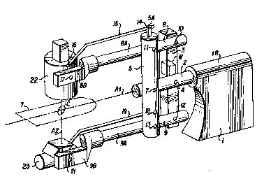
Figure 1 is an isometric vlew of a stand supporting an
X-ray source and receptor; and
Figures 2 and 3 are plan views of the source and
receptor, respectively.
DESCRIPTION
Shown in the figures is apparatus for X-ray examination
of a patient in a fixed posltion on a radiation-transparent table
T shown in Figure 1. The patient's heart, for example, would be
located at an isocenter C which is at the intersection of the
central axis of rotation A1 of the apparatus and of a radiation
axis A2 between an X-ray tube 23 and a radiation image receptor 22
which may be a radiation image intensifier 22. Subjects other
than a human organ may be examined, and the radiation receptor 22
may be a ray-sensitive film, fluorescent screen or a
scintillation counter. In each case it is desired that the
subject be at the isocenter C on the radiation axis A2 which is
coincident with the aligned axes of the X-ray tube 23 and the
lmage receptor 22. In neuro and cardiac anglography the human
organ is examined radiologically by tilting or angulating the X-
ray tube 23 and image receptor 22 through 360 of angle around the
central axis A1 of ~he system which is around the

Z~13
-3- 67450-55
longitudinal axis of the patient, and also through 90 of angle,
for example, in the head to foot direction, that is about a
secondary axis perpendicular to the central axis Al and through
the isocenter C. Angulation of the radiation axis A2 about the
isocenter is thus possible through a solid angle defined by the
360 angulation about the central axis Al and the head to foot
tilting of the radiation axis.
The apparatus producing such angulation comprises a
heavy base 1 anchored to the floor of a hospital or laboratory,
the base having a rotary bearing lB for a central rotor shaft 2
journalled in the bearing. A first transverse member 3 extending
generally vertically of the rotor 2 is pivotally attached to the
rotor at 4. A second transverse member 5 is pivotally supported
at 7 at the left end of the rotor 2. The two transverse members
3 and 5 and two other members, upper and lower arm 8 and 9, are
pivotally connected by bearing pins 10, 11, 12 and 13 to form an
angularly adjustable parallelogram (3,5,8,9) whose sides are of
a fixed length between the pivotal connections (10,11,12,13).
The two other horizontal members 8 and 9 have
extensions 8A and 9A outside the parallelogram for distances
sufficient to allow the table T and patient P to be located
close to the vertical transverse member 5. At the free ends 8A
and 8B of the extensions 8A and 9A are two pivots 17 and 21
respectively for the image intensifier 22 and the X-ray tube 23.
A short extension 5A from the transverse member 5 has a pivot
point 14 for a link 15 extending to a pivot point 16 on the image

`" 1'~9Z~13
-4- 67450-55
intensifier 22. Similarly a second link 19 is connected between
a pivot point 18 on the transverse member 5 and a pivot point
(not shown) on the X-ray tube 23.
According to the invention the transverse members 3 and
5 are disposed on opposite sides of the rotor 2 and its central
plane coincident with the radiation axis A2 and the central axis
Al, and as seen clearly in Figure 2, the end 8B of the upper
horizontal member 8 is offest to the same side of the rotor 2
and its central axis Al as the end 9B of the lower horizontal
member 9. The imbalance of the offset of the ends to the same
side is countered by the weight W in the transverse member 3
which now is on the other side of the central plane coincident
with the central axis. Since the offset of the horizontal member
ends is only on one side the other side is left unobstructed
above and below the patient table T, and there is no offset to
strike a doctor on that other side. As shown in Figure 1, the
links 15 and 19 are offset on opposite sides of the central plane.
As the parallelogram 3,5,8,9 is adjusted from the
rectangular position shown in Figure 1, axis 8 and 9 remain
parallel with one of the ends 8B, 9B extending further to the
left and the other end extending further to the right than shown
in Figure 1~ The links 15 and 19 rotate source 22 and receptor 23
about pivots 17 and 21 while parallelogram 3,5,8,9 is adjusted to
ensure that radiation axis A2 continues to pass through the
isocenter C.

Representative Drawing
A single figure which represents the drawing illustrating the invention.
Administrative Status

2024-08-01:As part of the Next Generation Patents (NGP) transition, the Canadian Patents Database (CPD) now contains a more detailed Event History, which replicates the Event Log of our new back-office solution.

Please note that "Inactive:" events refers to events no longer in use in our new back-office solution.

For a clearer understanding of the status of the application/patent presented on this page, the site Disclaimer , as well as the definitions for Patent , Event History , Maintenance Fee  and Payment History  should be consulted.

Event History

Description Date
Inactive: IPC expired 2024-01-01
Time Limit for Reversal Expired 1998-12-03
Letter Sent 1997-12-03
Grant by Issuance 1991-12-03

Abandonment History

There is no abandonment history.

Owners on Record

Note: Records showing the ownership history in alphabetical order.

Current Owners on Record
GORDON D. ROW
JOHN K. GRADY
Past Owners on Record
None
Past Owners that do not appear in the "Owners on Record" listing will appear in other documentation within the application.
Documents

To view selected files, please enter reCAPTCHA code :


To view images, click a link in the Document Description column. To download the documents, select one or more checkboxes in the first column and then click the "Download Selected in PDF format (Zip Archive)" or the "Download Selected as Single PDF" button.

List of published and non-published patent-specific documents on the CPD .

If you have any difficulty accessing content, you can call the Client Service Centre at 1-866-997-1936 or send them an e-mail at CIPO Client Service Centre.


Document
Description 
Date
(yyyy-mm-dd) 
Number of pages   Size of Image (KB) 
Claims 1993-10-22 2 40
Abstract 1993-10-22 1 14
Drawings 1993-10-22 1 21
Descriptions 1993-10-22 4 126
Representative drawing 2000-07-31 1 18
Maintenance Fee Notice 1998-01-01 1 178
Fees 1995-07-18 1 52
Fees 1993-11-29 1 41
Fees 1996-07-24 1 52
Fees 1994-07-28 1 38