Une partie des informations de ce site Web a été fournie par des sources externes. Le gouvernement du Canada n'assume aucune responsabilité concernant la précision, l'actualité ou la fiabilité des informations fournies par les sources externes. Les utilisateurs qui désirent employer cette information devraient consulter directement la source des informations. Le contenu fourni par les sources externes n'est pas assujetti aux exigences sur les langues officielles, la protection des renseignements personnels et l'accessibilité.
L'apparition de différences dans le texte et l'image des Revendications et de l'Abrégé dépend du moment auquel le document est publié. Les textes des Revendications et de l'Abrégé sont affichés :
| (12) Brevet: | (11) CA 1292813 |
|---|---|
| (21) Numéro de la demande: | 1292813 |
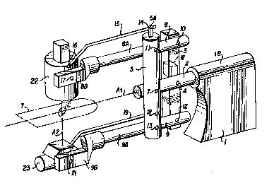
| (54) Titre français: | SUPPORT ASYMETRIQUE DE RADIOGRAPHIE |
| (54) Titre anglais: | ASYMMETRIC X-RAY STAND |
| Statut: | Périmé et au-delà du délai pour l’annulation |
| (51) Classification internationale des brevets (CIB): |
|
|---|---|
| (72) Inventeurs : |
|
| (73) Titulaires : |
|
| (71) Demandeurs : | |
| (74) Agent: | SMART & BIGGAR LP |
| (74) Co-agent: | |
| (45) Délivré: | 1991-12-03 |
| (22) Date de dépôt: | 1988-02-29 |
| Licence disponible: | S.O. |
| Cédé au domaine public: | S.O. |
| (25) Langue des documents déposés: | Anglais |
| Traité de coopération en matière de brevets (PCT): | Non |
|---|
| (30) Données de priorité de la demande: | ||||||
|---|---|---|---|---|---|---|
|
ABSTRACT
An X-ray apparatus comprises an X-ray source and X-ray
receptor mounted at the ends of two horizontal members which with
two transverse members from an adjustable parallelogram rotating
on the central axis of a support rotor. The transverse members
are located on opposite sides of a central plane coincident with
the central axis and the horizontal members are generally aligned
with the central plane but the ends of the horizontal members
which mount the source and receptor are both offset to the same
side of the central axis, thus permitting clear access by a
doctor to the patient.
Note : Les revendications sont présentées dans la langue officielle dans laquelle elles ont été soumises.
Note : Les descriptions sont présentées dans la langue officielle dans laquelle elles ont été soumises.
2024-08-01 : Dans le cadre de la transition vers les Brevets de nouvelle génération (BNG), la base de données sur les brevets canadiens (BDBC) contient désormais un Historique d'événement plus détaillé, qui reproduit le Journal des événements de notre nouvelle solution interne.
Veuillez noter que les événements débutant par « Inactive : » se réfèrent à des événements qui ne sont plus utilisés dans notre nouvelle solution interne.
Pour une meilleure compréhension de l'état de la demande ou brevet qui figure sur cette page, la rubrique Mise en garde , et les descriptions de Brevet , Historique d'événement , Taxes périodiques et Historique des paiements devraient être consultées.
| Description | Date |
|---|---|
| Inactive : CIB expirée | 2024-01-01 |
| Le délai pour l'annulation est expiré | 1998-12-03 |
| Lettre envoyée | 1997-12-03 |
| Accordé par délivrance | 1991-12-03 |
Il n'y a pas d'historique d'abandonnement
Les titulaires actuels et antérieures au dossier sont affichés en ordre alphabétique.
| Titulaires actuels au dossier |
|---|
| GORDON D. ROW |
| JOHN K. GRADY |
| Titulaires antérieures au dossier |
|---|
| S.O. |

