Note: Descriptions are shown in the official language in which they were submitted.
C)7
GLAUCOMA DRAINAGE_IN THE LACRIMAL SYSTEM
FIELD O- ~1E I~V~ oy
This invention relates to the field of ophthal-
mology, and particularly to devices and methods for
conducting fluids within and about the eye and the
lacrimal drainage system.
BACKGROUND OF THE INVENTION
Glaucoma is a disease characterized by elevated
intraocular pressure which, if not checked, may lead
to nerve damage and visual loss. Pressures in the
range of from about 15 + 3mm Hg up to about 21mm Hg
may be considered to be in the normal range for human
beings, whereas pressures substantially above that
range are considered abnormally high. If pressures in
the higher range are maintained for substantial
periods of time, damage to the optic nerve of the eye
may occur, leading to a narrowing of the field of
vision and eventually to blindness if not appropri-
ately treated. Although in certain cases glaucoma can
be treated through the administration of certain medi-
cines such as pilocarpine, epinephrine and timolol
maleate, it is often necessary to surgically provide
for the release of intraocular pressure for those
patients who do not respond to drug therapy or who
continue to lose vision under therapy.
Medical researchers have investigated a number of
methods for the surgical release of intraocular
pressure. Such surgery, in its simplest form, has
1 ~J5~()7
involved making a small, surgical incision into the
anterior chamber at or near the limbus to provide
means for releasing an overabundance of aqueous humor
from the eye into an adjacent subconjunctival space
and thus to lower the intraocular pressure. In a
modification of this procedure, a hair or other
wicking material is reported to have been placed in
the incision to provide a continuous passageway for
excess fluid to be dischar~ed from the eye. Other
researchers have implanted small tubes that extend
through the eye wall at the limbus or scleral-corneal
junction for the purpose of providing a channel
through which aqueous humor can escape.
Such surgical procedures, aithough still used to
some extent, are far from adequate. Healing of the
subconjunctival drainage space frequently results in
scarring, rendering the space non-absorbent of a~ueous
humor. When this occurs, no liquid flow through the
eye wall occurs, and the intraocular pressure may
hence rise to dangerous levels. An excellent account
of the history of glaucoma surgery is found in Bick,
"Use of Tantalum for Ocular Drainage", Archives of
Ophthalmoloqy, Vol. 42:373-388(1949).
In a recent device, the exterior end of a tube
extending through the wall of the eye is provided with
a pressure relief valve in the form of small slits
made through the wall of the tube at its end. Refer-
ence is made to Krupin, T., et al, "Valve Implants in
Filtering Surgery", Am. J._Ophthmol., Vol. 81:232-235
(1976). It is reported that fairly close control over
the pressure needed to open the valve may be ob-
tained. If the exterior or distal end of the tube is
inserted beneath a flap of conjunctiva or the like, of
course, the valved tube is subject to the same draw-
backs as the other tubes described above. Glaucoma
surgeons have discovered that when surgery fails it is
usually because the "bleb", the subconjunctival drain-
age space created by the surgeon, has become fibrosed,
causing it to shrink and become nonabsorbing.
One device that has been somewhat successful in
maintaining the fluid absorbency of the bleb during
the healing process was described by Molteno in 1969.
Molteno,"New Implant for Drainage in Glaucoma",
British Journal of Ophthalmology, Vol. 53:161 (1969).
Molteno described a device made from a "stellon" brand
acrylic monomer. The device consisted of two parts--a
flat plate fashioned to conform to the sclera and a
gutter incorporated at the point where a drainage tube
met the plate to assure an even spread of drainage
into the bleb. In 1979, Molteno disclosed a new
device that had a biconcave base plate and a long
silicone tube, which served the same function as the
first device. Reference is made to Chapter 11 of
Glaucoma SurqerY by Luntz, M.H., Harrison, R. and
Schenker, H.I. (1984) for a description of this device.
The drainage of fluid into spaces of the eye has
been unsuccessful largely due to the problem of bleb
formation. N.T. Mascati describes a different method
of drainage in "A New Surgical Approach for the
Control of a Class of Glaucomas", International
Surqery, Vol. 47:10-15 (1967). Dr. Mascati tried
inserting one end of a drainage tube into the anterior
chamber of an eye and the other end of the tube into
the nasolacrimal duct. This procedure met with only
limited success, and is not currently employed due,
presumably, to problems in ocular pressure control,
infections and related complications. Because the
Mascati device had no means for controlling liquid
flow such as a pressure relief valve there was no way
(1) to prevent collapse of the anterior chamber of the
.?5~07
eye and (2) to prevent reflux of fluid from the naso-
lacrimal drainage system into the anterior chamber of
the eye during sneezing or nose-blowing.
BRIEF DESCRIPTION OF THE INVENTION
The invention provides a device and method for
relieving high intraocular pressures associated with
glaucoma. The device includes a flexible tube having
one end extendable into the anterior chamber of the
eye and having its other end in communication with the
lacrimal drainage system of the eye. One-way valve
means are provided within the tube to restrain liquid
flow within the tube to a direction toward the lacri-
mal drainage system. Since the lacrimal drainage
system is lined with epithelial cells and is a natural
passageway for fluid flow, the scarring from fibrous
tissue usually associated with draining is avoided.
The one-way valve means also prevents back flow of
fluids from the lacrimal drainage system into the eye,
thus avoiding ascension of infection into the eye.
One embodiment of the ocular device has filter means
carried by the tube for restraining particles from
passing therethrough toward the anterior chamber of
the eye to further protect the eye from infection.
The method comprises the steps of inserting the
one end of the tube into the anterior chamber of the
eye, allowing aqueous humor to escape into the tube,
and attaching the other tube end to the eye so that
fluid will drain into the lacrimal drainage system.
The outlet end of the tube may be inserted into the
canaliculi, conjunctival cul-de-sac, lacrimal sac,
lacrimal duct, lacrimal passage, or nasal passage to
allow fluid flow outwardly from the tube into the
passages of the nasolacrimal system.
Another embodiment of the invention desirably
employs a pair of one-way valves within the tube.
lZ95907
Both one-way valves permit fluid flow only from the
anterior chamber into the lacrimal drainage system.
One one-way valve desirably is positioned at or near
the tube end (inlet end) that will be placed into the
anterior chamber. The other one-way valve desirably
is positioned at or near the tube end (outlet end)
inserted into the lacrimal drainage system.
BRIEF DESCRIPTION OF THE DRAWINGS
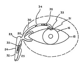
Figure 1 is a front view of an eye showing a
nasolacrimal drainage system and the device of this
invention;
Figure 2 is a cross-section of an eye showing a
tube implanted by the method of this invention;
Figure 3 is a top view of a device of this
invention; and
Figure 4 is a cross-sectional view along the line
A-A of the device of Figure 3.
DETAILED DESCRIPTION OF THE INVENTION
Figure 1 shows somewhat schematically the front
of a human eye and the lacrimal drainage system. The
lacrimal gland (not shown) continuously supplies the
eye with lacrimal fluid or tears. The lacrimal fluid
washes across the conjunctiva (11) and the cornea
(16)(Fig~ 2). Exoess lacrImal fl~id not reta~ by the eye
is commonly drained to the nasal passages, the infer-
ior nasal meatus (not shown) in particular. At times
the excess fluid is drained through a networX of
passages that commences with the puncta that appears
as a small papilla adjacent the inner canthus or the
inner corner of the eye. The fluid is collected in
the lacrimal sac (23) by a number of canaliculi (20)
connecting the puncta to the sac. The canaliculi run
inferiorly then medially to the lacrimal sac. The sac
(23) is then drained through its extension, the naso-
lacrimal duct (24~ which passes into the inferior
.... .
rI~ ~
12g5~07
nasal meatus for this purpose. This network of
passages is referred to herein as the lacrimal
drainage system (25).
A tube (30) has an inlet end portion (31) that is
shaped to be inserted through a small incision made in
the wall of the eye so that the end (31) is posi-
tioned in the interior of the eye, preferably in the
anterior chamber. The other outlet end (32) of the
tube is shaped to be inserted through a small incision
made in a portion of the lacrimal drainage system to
be positioned therein. The tube (30), desirably is on
the order of about 1 to about 8cm long and about 0.4
to about 1.5 mm in outer diameter. The end portions
may be of a comparatively rigid material such as poly-
methylmethacrylate or of a metal such as gold or other
biologically acceptable materials, and the ends may be
joined together by a more flexible length of, e g.,
silicone rubber tubing. The entire length of the tube
(30), including the end portions, is preferably made
of a flexible material such as silicone or poly-
ethylene.
In Figure 2, a cross-section of the human eye is
shown that includes the device of the invention
implanted in the anterior chamber. In that figure the
cornea is shown as (16), the iris as (17), the lens as
(18) and the limbus as (19). The inlet tube end (31)
extends into the anterior chamber (14) of the eye and
the outlet tube end (32) is positioned in the conjunc-
tival cul-de-sac to allow fluid to flow from the tube
into the lacrimal drainage system. The device may be
secured to the sclera (15) by sutures (34) or other
conventional means.
The one-way valve (3s)or valves used with this invention
(see ~igure 1) may be o~ the various types suitable
for use in the quite miniature device of the inven-
tion, and such valves often also function as pressure
~P~
12~ 7
-- 7
relief valves as well. The one-way valve employed in
this method desirably is competent to prevent back
flow of fluid even against increases of pressure such
as those that may occur within the lacrimal drainage
system when a person sneezes, blows his nose or
sneezes when the nose is plugged, e.g., 50 mm Hg. The
one-way valve preferably is also a pressure relief
valve that is adapted to open when the pressure in the
eye exceeds the pressure within the tube by a pre-set
threshold pressure, e.g. by about 8-lOmm Hg. The
pressure within the tube will be maintained at or near
atmospheric pressure (about 760mm Hg). In this way,
the valve prevents collapse of the anterior chamber.
The unidirectional flow of the fluid into the lacrimal
drainage system prevents viruses and bacteria carried
by fluids in the lacrimal drainage system from being
carried upwardly into the interior of the patient's
eye through the tube. The valves may be any of a
variety of well known designs which need not be
described in detail, but which might include by way of
example well known "duck valves".
As explained above, the one-way valve (35) may
function as a pressure relief valve, the edges of the
valve flaps pressing against one another to restrain
fluid flow until the pressure differential across the
valve increases to a level sufficient to cause the
flaps to separate slightly, permitting fluid to pass.
Reversal of the pressure gradient, as when the patient
sneezes, causes the flaps to press more tightly
together, thus restricting flow in the opposite
direction. When a tube having only one valve is used,
the valve is positioned near the inlet end of the
tube. The valve is thus more effective as a pressure
relief valve, and back flow of fluid pooling within
the tube is prevented. When two valves are used, it
1295907
-- 8
is desirable to have one valve near the inlet end and
one near the outlet end of the tube.
The outlet end of the tube is desirably surgi-
cally inserted into the eye adjacent to or in the
lacrimal drainage system. The tube end may be
inserted in the conjunctival cul-de sac, the inferior
or superior canaliculi (20), the lacrimal sac (23),
the lacrimal duct (24), the nasal passage or any of
the nasolacrimal passages or the nose. Fluid flows
from the anterior chamber (14) of the eye through the
tube (30) and into the passages of the lacrimal
drainage system (25) and ultimately into the
nasopharnyx where, if it has not been absorbed, it is
swallowed.
The ocular device shown in Figures 3 and 4
includes a microporous filter (40) that restrains
micro-organisms and other particles having a mean
diameter greater than the pore size from passing
therethrough toward the anterior chamber of the eye.
The filter may be of any known type such as a Milli-
pore filter made by the Millipore Company of Bedford,
Mass. The filter desirably has a nominal pore size in
the range of 0.1 micron to 10 microns and preferably
has a pore size in the range of 0.1 to 0.3 micron.
The filter may be carried within the tube or it may be
carried at the outlet end of the tube enclosing the
end. As shown in figures 3 and 4 the filter may be
bag-shaped and extend outwardly of the tube end. The
bag-shaped filter may comprise a pair of filter sheets
(41,42) joined at their peripheries to define an
interior space (43) communicating with the outlet end
of the tube.
One surgical technique used in practicing the
method of this invention is described below. This
description is included solely for illustrative
* Trade Mark
P
purposes; the method may be practiced using any of the
numerous known surgical techniques.
In the region to be operated upon, a rather large
limbal based flap of conjunctiva (11) is opened with
an incision of about 6 to 10 mm posterior to the
limbus. Care should be taken to provide no tears or
button-holing of the conjunctiva. If tears occur,
they should be repaired. The episcleral tissue (13)
should be cleared from the region of the limbus (19)
back for a distance (i.e., 5-8 mm) and any bleeding
controlled with gentle cautery. The posterior margin
of the conjunctiva (11) will be lifted and Tenon's
capsule (12) interrupted with combined blunt and sharp
dissection until the bare sclera of the eye is visible.
Centered at the limbus (19) a partial thickness
scleral flap is outlined, the flap desirably measuring
4-5 mm in width and approximately 4-6 mm in anterior-
posterior length. The tube (30) should be positioned
under the scleral flap and a small incision made into
the anterior chamber (14) at the limbus (19). The
tube end (31) will then be threaded into the anterior
chamber (14) until it can be visualized through the
clear cornea (16). After the tube end (31) is posi-
tioned, the limbal incision may be closed about the
tube (30) and if a flange (33) is attached to the
tube, it may be secured to the bed of the sclera (15)
with partial thickness scleral bites and through-and-
through bites through the flange (33) with a suture.
If the outlet end (32) of the tube is to be inserted
into the nasolacrimal sac (23), a false channel may be
created surgically in the posterior lateral wall of
the sac. The tube end (32) is then threaded through
that channel into the sac (23) and if desired, into
the nasolacrimal duct (24). The length of the tube
(30) must be sufficient to permit slippage of the tube
1Z~3~
-- 10 --
within the sac when the eyeball rotates without
dislodging the tube.
If the outlet end of the tube t32) is to be
inserted into the canaliculi (20) it will desirably be
threaded through the backside of the eyelid and then
into the posterior wall of the canaliculus. The out-
let end of the tube (32) may be attached to the eye so
that it is positioned in the conjunctival cul-de-sac
adjacent the lacrimal drainage system.
After the outlet tube end (32) is inserted into
the nasolacrimal system (25), it is fixed to the sur-
face of the sclera (15) under the conjunctiva (11) in
the episcleral space. The tube (30) may be fixed to
the eye using any well known surgical method such as
sutures or scleral tunnels (34).
While a preferred embodiment of the present
invention has been described, it should be under,stood
that various changes, adaptations and modifications
may be made therein without departing from the spirit
of the invention and the scope of the appended claims.
