Note: Descriptions are shown in the official language in which they were submitted.
- - ~25~77i 4
CONFOCAL TANDEM SCANNING
REFLECTED LIGHT MICROSCOPE
The present invention relates to scanned imaging; and
more particularly, it relates to devices for scanning a
field to be imaged, such as a Tandem Scanning Microscope
(TSM).
Image formation can be accomplished by confocal
imaging techniques which involve mechanical or electronic
scanning. Exemplary of these techniques is the Tandem
Scanning Reflected Light Microscope (TSRLM). The theory
of TSRLM is discussed by Petran and Hadravsky in "Tandem-
Scanning Reflected-Light Microscope," Journal of the
Optical Society of America, Vol. 58, No. 5, pp. 661-64
(May 1968). The TSRLM is further described and
illustrated by Petran, Hadravsky, and Boyde in "The Tandem
Scanning Reflected Light Microscope," SCANNING, Vol. 7,
No. 2, pp. 97-108 (1985). U.S. Patent No. 3,517,980 to
Petran et al. also describes a TSRLM device.
The scanning device in a TSRLM is a rotating disk
having holes in an annular region. The holes are arranged
on Archimedean spirals. Diametrically opposite holes are
on identical radii, and the pattern as a whole has a
central symmetry. This device is known as a Nipkow disk.
The structure of such a disk has conventionally been a
copper foil sheet stretched over a retaining ring and
having holes etched therein.
The present invention provides improvements in
scanning patterns used in imaging systems. The present
invention particularly provides improvements in the
scanning system used in a TSM.
iq
.
1 a ~ 97714
The invention in one broad aspect pertains to a
scanning device for use in an optical imaging system,
comprising an opaque disk having apertures formed therein
in an annular pattern around the circumference of the
disk, the apertures being distributed within the annular
pattern in a series of adjacent arcuate, radially-
extending arms defined by radius vectors having their
length Rn determined in accordance with the relationship:
[Rn-l + (Rn2 1 + 4K) / ]
n - 2-- -
10 where Rn 1 is the length of the preceding radius vectorand K is a constant, and their angle T of orientation
determined in accordance with the relationship:
T = COS (Rn + Rn+l2 - L2)/(2RnRn+1)
where L is the distance between the ends of the radius
15 vectors.
Further the invention pertains to a tandem
scanning microscope, comprising a rotating disk having an
array of apertures formed in a micromachined silicon
structure, means cooperating with an area of the array of
20 apertures for providing incoming light as a series of
beams which scan in a specimen field of view and
illuminate patches in a plane of focus, and means for
imaging reflected light from the illuminated patches in
the focal plane onto apertures of the array which are
25 scanning in the focal plane of an eyepiece. Preferably
the apertures provide a rotating array of light
transmissive areas in accordance with the pattern defined
above.
,~ ~,.A',`~J;~
9~714
--2--
The TSM of the present invention is similar to the
tandem scanning reflected light microscope by Mojmir
Petran and Milan Hadravsky, and described in numerous
publications. In contrast to more complex and costly
confocal laser microscopes, the present invention employs
an innovative scanning system which provides high
resolution images at high frame repetition rates. The
steady image produced is suitable for direct viewing with
the eye or TV and film cameras. The image is linearly
scanned and images may be digitized directly into a frame
buffer without correction. Illumination is provided by
standard lamps which allow rapid, convenient change-over
from visible true color to fluorescence operation using
filtered (selected) wavelength excitation.
The scanning system includes a scanning disk. In one
embodiment, this disk contains several tens of thousands
of apertures arranged in a precise pattern that is
symmetric about any diameter. The disk is placed at the
intermediate image plane of the objective. The specimen
and the image formed by the objective is viewed through
the conjugate aperture on the opposite side. As the disk
is rotated, the specimen is illuminated in small
"patchels" lying in the focal plane. As the "patchels"
are scanned across the specimen, an image of the entire
12g~
3--
field of view is created. Since only light reflected from
these illuminated "patchels" in the focal plane can pass
through the aperture holes on the viewing side, the image
contains contrast and resolution superior to conventional
light microscopes.
The discrete focal plane of the TSM of the present
invention provides an optical sectioning ability difficult
to achieve with conventional light microscopes. The TSM
provides images with unmatched clarity from the surface to
any depth within the sample, limited only by the
transparency of the material and the working distance to
the objective used. In addition, the TSM provides up to
1.28 times the resolution of conventional light
microscopes.
The TSM allows precise optical examination of
surfaces at high resolution when the surface does not
reflect enough light at normal incidence to be imaged
using conventional optics. The TSM provides a continuous
image, which means the operator can enjoy direct viewing
in true color while moving the in-focus plane through a
sample. The TSM can be used to study (with high spatial
resolution) the surface topography of reflective, opaque
specimens.
A written description setting forth the best mode
presently known for carrying out the present invention,
and of the manner of implementing and using it, is
provided by the following detailed description of a
preferred embodiment which is illustrated in the attached
drawings wherein:
FIGURE 1 is a diagram of the confocal imaging light
paths within a Tandem Scanning Microscope (TSM~;
12~77~
--4--
FIGURE 2 is an illustration of the physical
configuration of a TSM in accordance with the present
invention;
FIGURE 3 is a depiction of the aperture pattern of a
scanning disk for use in a TSM in accordance with the
present invention;
FIGURE 4 is a diagram of the mathematical
relationship of the apertures in the pattern of Pigure 3;
and
FIGURE 5 is a diagram illustrating the geometrical
relationship of the apertures in the pattern of the
scanning disk of Pigure 3.
A. The Basic Principles of the TSM
Tandem scanning microscopy (TSM) involves confocal
imaging. The light ray diagram in Figure 1 illustrates
the principles of confocal microscopy. In Figure 1,
illumination 14 enters through an illuminating aperture 10
on a scanning disk and is focused by objective lenses, 01
and 02. Only reflective light 16 from the focal plane of
the objective passes through the viewing aperture 18.
Light 17 emitted from above or light 19 from below the
focal plane 22 is not brought to focus on the viewing
aperture and is blocked by the disk. Aperture 18 is a
conjugate aperture on the observation side of the scanning
disk. In practical devices, there are thousands of
apertures on the disk.
Objects in the focal plane of the objective are
illuminated by the point source, and the light reflected
by the specimen is seen by a point detector. In practice,
the point source and point detector are obtained by
~9~7714
5--
placing apertures between a conventional source and
detector and the objective lens. Confocal imaging is
achieved when the system is precisely aligned via a system
of adjustable mirrors and a beam splitter so that rays
from the source aperture pass through the viewing
aperture. As shown, rays that emerge from objects out of
the focal plane are not focused at the viewing aperture
and are blocked from reaching the detector. The result
is a high contrast image of a small portion of the
specimen at the focal plane. To see an entire field, a
means is required to scan either the specimen or the
illumination and detector. This is accomplished by
scanning the source and detector by means of a scanning
disk having light transmissive areas.
For additional discussion of TSM theory, the
publications cited in the background are instructive.
B. Structure of the TSM
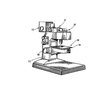
The diagram of Figure 2 illustrates a physical form
for a practical TSM instrument. The instrument 30 has a
stand 32 for placement on a planar surface such as a table
top. The stand 32 supports specimen platform 36.
~ertical adjustment of platform 36 is provided by rotation
of knobs 38. Mounted atop stand 32 is the head 40 which
includes the optical components and scanning disk. Also
included is Epi-illuminator 41 which conveys light from
the lamphouse 42 into the head. The Epi-illuminator
contains several lenses, iris diaphragms and filter
holders in order to adjust the apparent ~rightness and
emission spectrum of the light source.
For further information as to the structure for
realigning a TSM, U~S. Patent No. 3,517,980 June 1970 to
Petran et al may be referred to.
;i`. - A ~
--6--
1. The Optical Components
Eyepiece: The Ramsden type with the focal plane in
front of the lens; magnifications of 10x and 15x can be
used with the microscope.
Objectives: A standard threaded objective (RMS
thread) with tubelength (engraved on the body) of 160,
165, or 170 mm can be used. (Infinity tubelength
objectives can be used with a special correcting lens.j
Although any conventional LM objective can be used,
immersion objectives are preferred with the refraction
index of the immersion medium to be matched to that of the
specimen to be observed when studying internal structures.
Water, oil and glycerin immersion objectives are the most
commonly used. Dry objectives may be used for examination
of internal structure in translucent samples used in the
study of surfaces. Water immersion objectives are used
for living animal and plant tissues; oil for bone, tooth
and rock (fossil) samples; and glycerine or oil for
fluorescence.
Beam Splitter: The beam splitting pellicle is
extremely thin, so as not to double the image or introduce
astigmatism. As regards to the use of the TSM in the
fluorescence mode, we should note that reflection for UV
is < 50~ and the transmission of visible > 50%, thus
enhancing its characteristics in this context. Pellicles
with a variety of wavelength selective coatings to
selectively enhance transmission and reflectance for a
given application may be used.
Illumination: Practical experience has shown that
the most convenient light sources are a standard tungsten
lamp for visible and some fluorescence work, a mercury
lamp or xenon lamp for extended spectral response.
~LZ~ 4
--7--
2. The Scannlng Disk
A scanning disk pattern in accordance with a single
disk system embodiment is shown in Figure 3. This unique
disk pattern contains several tens of thousands of
apertures arranged in a precise pattern that is symmetric
along any diameter. The disk is placed at the
intermediate image plane of the objective. The specimen
image formed by the objective is viewed through the
conjugate aperture on the opposite side. As the disk is
rotated, the specimen is illuminated in small "patchels"
lying in the focal plane. As the "patchels" are scanned
across the specimen, an image of the entire field of view
is created. Since only light reflected from these
illuminated "patchels" in the focal plane can pass through
the aperture holes on the viewing side, the image contains
contrast and resolution superior to co~nventional light
microscopes.
The scanning disk has an annular pattern of apertures
around its circumference such that upon rotation, a
uniform transmission of light occurs across the entire
annulus. Confocal operation has the apertures located
across from each other along a disk diameter so as to be
precisely overlapped when viewed through the imaging
system (i.e., the pattern of apertures must be symmetric
along any diameter). These requirements are met by
locating the apertures on the arms of a plurality of
spirals and at the ends of radius vectors. The following
discussion concerning the aperture arrangement is best
understood with reference to Figures 3, 4 and 5.
The apertures are located along the arms of a
plurality of spirals. Let Ro be the radial distance from
the center of the disk to the innermost aperture on the
disk. The next aperture on the spiral is located at a
--8--
radius Rl from the center of the disk in such a way that
the increase of the radius from Ro to Rl is in an inverse
proportion to Rl as stated in Eq. 1 and 2 in which K is a
constant.
Eq. 1 1 Ro Rl/K
Eq. 2 Rl = [Ro+(Ro2+4K) /2]/2
A further requirement is that all the apertures along
a spiral be a uniform distance apart. Let this distance
be L, then the radius vectors connecting the first and
second will form an angle T at~the center of the disk as
given by Eq. 3.
Eq. 3 T = Cos [(Ro +R12-L2)/2RoRl]
Similarly with all other points on a spiral as shown
in Eq. 4 and 5 where Rn is the radius vector of point n
, 20 and Rn 1 is the radius vector of the point n-l (i.e. the
proceeding point) and K has the same value as before.
Eq. 4 Rn = [Rn_l(Rn_l +4K) / ]/2
Eq. 5 T = Cos l[(Rn 12~Rn2-L2)/2RnRn 1~
Figure 4 represents the conditions of Equations 2 and
3 where C is the center of the annulus, P0 the initial
aperture on the spiral and Pl the second aperture. Ro and
Rl are the radius vectors to these points, while T is the
angle formed by the vectors Ro and Rl. The angle A
determines the value of the constant K ~or visa versa) as
indicated in Equations 6 and 7.
77~4
g
Eq. 6 1 (Ro2+L +2RoLcosA)l/2
Eq. 7 K = R 2-R R
It should be noted that the angle A refers only to the
first cord of the spiral with the value oE Ro
The values of Ro and K are constant for all spirals.
However, in order for the apertures to overlap during the
scanning of the disk it is necessary for successive
spirals to have the initial apertures radially offset.
This offset is added to Ro to determine the actual radius
of the first point on each subsequent spiral.
Practical realization of the above-described pattern
of apertures in a disk has been achieved by micromachining
the pattern into a silicon wafer. Such a process for
producing geometrically precise silicon structures is
well-known in the art as shown in G. Kaminsky,
"Micromachining of Silicon Mechanical Structures," J. Vac.
Sci. Technol. B., 3(4), 1015-1025 (1985), which may be
referred to for further information.
Disks are fabricated in the following manner.
Silicon wafers (100 mm diameter) are thinned to the proper
thickness (.0101 inch for 60 microns holes) which in
addition to the size of the lithography mask determines
the size of the final holes in the disk. The pattern is
printed upon the wafer by normal lithographic techniques
employing a mask. The exposed wafers are etched with a
directional chemical etch to produce a series of square
- 1 0 -
holes in the desired pattern. The present disk contains
13,642 holes, each being 60 microns from side to side,
arranged along spirals in an overall annular
configuration. The inner radius of the annulus is 26 mm,
the outer radius 46 mm. Since it is desirable to keep the
transmission of the disk limited to about 1% of incident
light, hole size must be reduced if more holes are desired
in the pattern. To optimize lateral and axial resolution
in an optical microscope image, a hole size of 20 microns
0 i5 required, which determines the maximum number of holes
to be about 70,000 holes. Such a disk could be made with
the techniques described by Kaminsky. The sides of each
hole are sloped at a fifty-two degree angle. The angle is
fixed by the crystal lattices of the silicon.
Although a mechanical means for providing the
scanning disk pattern is described, other means could be
used. For example, electronic pattern generation could be
used in the TSM. One such electronic means is a Kerr cell
- 20 array. A Kerr cell is a solid state optical switch; it
transmits light in one state, and does not in the other
state. A two-dimensional array of Kerr cells could
replace the entire mechanical scanning disk, or merely
replace portions of the disk in the illumination or
viewing lens pupils. The Kerr cells would be switched
electronically so as to scan across the field of view. As
light is transmitted via an illuminating cell, the
corresponding viewing cell would also transmit reflected
light from the specimen to the eyepieces.
Another electronic means of pattern generation is CRT
illumination/image dissection. With this means,
illumination would be provided by scanning an electron
beam across the face of a CRT tube in a fixed pattern,
perhaps the spirals of our pattern. Typical spot sizes
are on the order of tens of microns. The light from the
:~L2~3)77~4
phosphor would provide the illumination for the system.
Light reflected by the sample would not be viewed directly
but detected on an image dissector tube. An image
dissector (ID) is essentially a photomultiplier Tube (PMT)
with focus and scan coils added. Internal to the
dissector is a small aperture (< 100 ~m) between the
photocathode and the dynodes. The signal from a small
area of the photocathode is focused through the aperture
at any given instant, and the entire cathode can be
scanned using the scan coils. The Image Dissector
functions as a point detector which can be scanned across
the field of view. If its scan is precisely synchronized
to the illuminating CRT scan, confocal operation is
achieved. The optical layout could be the same as in the
reflected TSM with the CRT and ID replacing the disk at
the intermediate plane of the objective. Alternatively,
transmitted light work could be done by locating the ID
below a second objective below the specimen as is done in
the laser scan confocal microscopes.
C. The Op~ration of the TSM
Basic operation of the TSM of the present invention
is similar to prior art devices. The point of distinction
concerns improvements in the image scanning performed with
the disk. In the TSM, the array of apertures provides
incoming light as a series of beams which scan the field
of view and illuminate patches in the plane of focus more
intensely than out of focus layers. Reflected light from
3~ these patches is imaged onto apertures of the array which
are scanning in the focal plane of the eyepiece. The
array of apertures which chop the illuminating beam is
identical with the array on the observation side. The
scanning, illuminating apertures are imaged by the
objective lens in the focussed-on plane. Reflected light
from the patches in this plane is imaged back into
-12-
corresponding apertures which also lie in the intermediate
image plane of the objective. All other reflected light
from out of focus layers and light scattered from optical
surfaces in the microscope is intercepted by the solid
S portions of the aperture array or by light traps in the
microscope head. Scanning of illumination and observation
is done with one device by having all the apertures
fabricated in one rotating disk. Each aperture pair in
the disk is unique, lying at a different radial distance
from the center of the disk and scanning a single line in
the image.
Both sides o~ the disk are at the same distance from
the lens which serves for both illumination (condensor)
and image formation (objective). The two functions of the
objective are separated by a very thin beam splitting,
semi-transparent mirror. The congruence (or overlap) of
both patterns of apert~ures formed by the objective in the
focussed-on plane is accomplished by a mirror system,
which converts central symmetry of the disk into
cohgruency.
Since the holes are much smaller than the distance
between them, it follows that the precision of manufacture
of the pattern of holes in the disk and of the adjustment
of the mirrors, must be exquisite. The new scanning disk
in the TSM is manufactured to high technological
standards. The mean size of the square holes is 60-80
micrometers or less, but the size can be varied as
required. The number of scanning lines is such that one
cannot observe the scanning lines in the image because
their separation is less than the resolution of the
microscope. The disk is illuminated by a strong light
source, so that a pattern of light spots is formed. The
images are created in the focussed-on layer of the
specimen. Light is reflected by the features which are to
7~14
~13-
be observed. Images of the instantaneously illuminated
structures are formed by the objective on the other side
of the scanning disk. Only the light from the focussed-on
layer can enter the eyepiece.
The disk speed is variable and can be established to
give an apparently stationary image.
The TSM described herein is an embodiment of the
present invention in which a single disk is used. Such an
embodiment requires that the disk perform two functions:
image the illumination onto the specimen and select the
resultant image point in the i~aging beam. In order to
perform these functions the disk must contain a plurality
of identical sectors. If there are two such sectors, the
disk will be symmetrical about any diameter, and the
optical axis of the illuminating and imaging beams will
intersect the disk at diametrically opposite points. If
there are 3, 4, or 5 (etc.) such sectors, then the optical
axes will intersect the disk at points whose radii from
the disk center form angles of 120, 90, and 72 (etc.)
degrees respectively.
A TSM can be constructed in which there are a
plurality of disks, for example, a disk in the
illuminating beam and one in the imaging beam. If these
disks are synchronized, the symmetry requirement no longer
applies. The disks, however, must be identical.
A TSM is also contemplated within the scope of this
invention in which there are a plurality of disks not
identical in size but geometrically similar. That is, the
aperture patterns are identical except for scale. Such a
TSM would be confocal between the objective lens and the
specimen but would contain additional optical elements in
either the illuminating or imaging beam (or both) such
-14-
that the operation effect would be that of the TSM
described here. Disk patterns which are mirror images of
any described herein are also contemplated.
The shape of the apertures may be of any desired form
including, but not limited to: circular, square,
hexagonal, octagonal, rectangular and other geometrical
forms. Furthermore, no limitation is implied concerning
the orientation of any non-circular aperture. The size
(absolute and relative) of the apertures may be varied to
fit any particular application.~ Nothing contained in the
above description shall be construed to restrict the
choice of the number of spirals that may be present or any
of the dimensions or parameters of the disk, the
apertures, or the aperture patterns.
The foregoing description of the invention has been
directed to a particular preferred embodiment for purposes
of explanation and illustration. It will be apparent,
however, to those skilled in this art that many
modifications and changes may be made without departing
from the essence of the invention. It is the applicants'
intention for the following claims to cover such
equivalent modifications and changes as fall within the
scope of the invention as defined by the following claims.