Note: Descriptions are shown in the official language in which they were submitted.
~3(~Z~S;~
- 1 -
NON-DESTRUCTIVE OPTICAL TRAP FOR BIOLOGICAL
PARTICLES AND METHOD OF DOING SAME
Tech~,l ~ield
This invention relates to trapping of particles using a single-beam
5 gradient force trap.
Rackg~ound Q~ tl~ In~Qa
Single-beam gradient force traps have been demonstrated for neutral
atoms and dielectric particles. Generally, the single-beam gradient force trap
consists only of a strongly focused laser beam having an approximately Gaussian
10 transverse intensity profile. In these traps, radiation pressure scattering and
gradient force components are combined to give a point of stable equilibrium
located close to the focus of the laser beam. Scattering force is proportional to
optical intensity and acts in the direction of the incident laser light. Gradient
force is proportional to the optical intensity gradient and points in the direction
15 of the intensity gradient.
Particles in a single-beam gradient force trap are confined transverse to
the laser beam axis by a radial component of the gradient force. Stabilizing theparticle along the axial direction of the trap is achieved by strongly focusing the
laser beam to have the axial component of gradient force dominate the
20 scattering force in the trap region.
In prior work using single-beam gradient force optical traps on dielectric
particles, trapping was demonstrated with a visible light laser source (A = 514.5
nm.) focused by a high numerical aperture lens objective. See A. Ashkin et al.,
~ti~ T ektec~, Vol. 11, p 288-~0. The dielectric particles were closely spherical
25 or spheroidal in shape and ranged in size from 10,um diameter Mie glass spheres
(a! > > )~) down to 200 Angstrom diameter Rayleigh particles (Cl! < < ~). Use ofsuch regularly shaped particles in the Mie regime was desirable as taught in this
and other articles.
For Mie particles, both the magnitude and direction of the forces depend
30 on the particle shape. This restricts trapping to fairly simple shapes such as
spheres, ellipsoids, or particles whose optical scattering varies slowly with
orientation in the trap. In the Rayleigh regime, the particle acts as a dipole and
the direction of force is independent of particle shape; only the magnitude of
' ' . . . . ..
:,.. .. . . . .
'' '
' .
`-` 13~Z753
force varies with particle orientation.
It is not an insignificant result of the prior work that silica and other dielectric
particles experienced varying amounts of irreversible optical damage ~rom the trap. While
it was suggested that the single-beam trap and the prior results would be extensible to
S biological particles, the resulting damage from exposure in the trap would destroy or
significantly incapacitate the biological particles and render them useless. Also, since prior
optical traps have been defined for quite regular-shaped, dielectric particles, their
extension to biological particles is cast in doubt because regularity of shape is not an
attribute of biological particles.
10 SummarY of the Invention
Biological particles are successfully trapped in a single-beam gradient force optical
trap incorporating an infrared light source. Reproduction of trapped particles has been
observed. After release from the trap, particles exhibit normal motility and continued
reproductivity even after trapping for several life cycles at a high laser power of 160 mW.
In one embodiment, the high numerical aperture lens objective in the single-beamgradient force trap is used for simultaneous viewing of the trapped biological particles.
Two single-beam gradient force optical traps are introduced into the same cell to
permit three-dimensional manipulation of the biological particles.
In accordance with one aspect of the invention there is provided apparatus for
20 generating a single-beam gradient force optical trap of particles, said apparatus comprising
a laser for generating a light beam at a predetermined wavelength and means for focusing
said light beam with sufficient convergence to form said optical trap in a predetermined
region, said apparatus characterized in that said predetermined wavelength is substantially
included in the infrared range of wavelengths between 0.8 ,um and 1.8 ,um inclusively, so
25 that said trap non-destructively confines at least one biological particle.
In accordance with another aspect of the invention there is provided apparatus for
generating a single-beam gradient force optical trap of particles, said apparatus being
comprised of a laser for generating a light beam at a predetermined wavelength and
means for focusing said light beam with sufficient convergence to form said optical trap in
30 a predeterrnined region, said apparatus characterized in that said predetermined
13~t;27S3
2a
wavelength is substantially included in the infrared range of wavelengths, so that said trap
non-destructively confines at least one biological particle, said apparatus further including
means for generating a second light beam substantially at the predetermined wavelength,
5 said second light beam focused by said focusing means to form a second optical trap in a
second predetermined region.
Brief Description of the Drawin .
A more complete understanding of the invention may be obtained by reading the
following description of a specific illustrative embodiment of the invention in conjunction
10 with the appended drawing in which:
FIG. 1 is a cross-sectional schematic diagram of an embodiment of the invention;FIG. 2 is a cross-sectional schematic diagram of an embodiment of the invention
employing two single-beam gradient force traps in one cell; and
FIGS. 3 th~ough 5 are schematic drawings of different modes oE operation for an
15 optical trap on particles in a cell.
,~, .. ~.. ...
.
~3~7$;;~
- 3 -
Detailed I)escription
Single-beam gradient force optical traps are useful for confining,
isolating, translating and manipulating at least one particle in a group of
particles enclosed in a cell or hanging droplet or the like. Special problems
5 surface when the particles are biological. For example, absorption of the optical
energy in the trap by the confined particle may lead to particle annihilation or a
significant loss of particle motility. Also, as the wavelength of the light beam is
varied to avoid the aforementioned problem, the intensity of the optical trap
may be sufficiently decreased so as to be rendered ineffective for the particles of
10 interest. While the wavelength selected may be sufficient for effective operation
of the optical trap, it may be at a wavelength which is absorbed by the medium
surrounding the particles and, therefore, which leads to heat generation within
the cell. Clearly, many factors must be considered when selecting the operating
wavelength for the optical trap.
In the prior optical trap experiments reported in the literature, particle
sensitivity has not been an issue. This is generally attributed to the fact thatdielectric particles have homogeneous compositions and uniformly regular
shapes 90 that it is straightforward to observe the effect of the trap on one
particle or portion of a particle and accurately predict the effect on other
20 particles or on other portions of the same dielectric particle. For biological
particles, sensitivity of the particles is extremely important. Biological particles
have heterogeneous compositions and irregular shapes. Hence, the effect of the
trap on one part of a biological particle i9 in no way determinative of the effect
in another portion of the same particle.
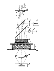
FIG. 1 shows a cross-sectional schematic diagram of apparatus for
creating a single-beam gradient force optical trap in accordance with the
principles of this invention. IR laser 10 is a standard laser emitting a coherent
light beam substantially in the infrared range of wavelengths, for example, 0.8
~m to 1.8 ~m.
Light beam 11 from IR laser 10 impinges upon a combination of optics
elements for focusing the light beam with a sufficient degree of convergence to
form a single-beam gradient force optical trap for confining biological particles
at a desired position. The combination of optics elements includes an
adjustably mounted diverging lens 12 and a high convergence lens 23.
.. ~.. .
13~2753
- 4 -
Lens 12 is adjustable in any of three dimensions (x, y, z) by manipulating
adjustable mount 13. It is important that lens 12 expand the spot size of light
beam 11 to cover a substantial area on the surface of lens 23. As shown in
FIG. 1, diverging light beam 14 impinges on a large portion of the facing surface
5 of lens 23 so that relatively high intensity of beam 14 fills the aperture of lens
23. In order to create the forces required for operation of the single-beam
gradient force optical trap, it is desirable that lens 23 be capable of focusing to a
spot size less than ~\ approaching ~/2. In an example from experimental
practice, lens 23 is a strong or high convergence water immersion microscope
10 objective lens having a numerical aperture of approximately 1.25 (measured inwater). wherein the numerical aperture is defined as the refractive index for the
medium multiplied by the sine of the half angle covered by the converging light
beam. Element 24 depicts the liquid (water or oil) in which lens 23 is immersed
for improved optical coupling into cell 25.
The optical trap is shown within cell 25 with particle 27 captured in the
trap. Particle 27 is suspended in a liquid medium such as water, for example,
which is enclosed by cell 25. Cell 25 is a transparent enclosure for enclosing the
suspended biological particles or a transparent slide from which particle
containing droplets can be hung. In one example, cell 25 has dimensions of
20 1 cm. x 3 cm. x 100 ,~m.
The position of cell 25 is adjustable in three dimensions (x, y, z) by the
use of adjustable mount 2B. In practice, mount 26 is useful in locating and
manipulating the biological particles.
Viewing of biological particles in the trap is accomplished directly or
25 through the use of a monitor. While other types of viewing such as viewing
directly in cell 25 are possible, it is an added feature of the present invention
the viewing is accomplished through the same lens objective which
simultaneously creates the optical trap.
Illumination for viewing is provided by visible light source 2~ and is
30 projected through converging lens 28 onto the particles in the field of view.High resolution viewing occurs with the aid of lens 23 through which the visiblelight passes toward either the eyepiece or the monitor 18. For direct viewing,
visible light shown as a dashed line is reflected from beam splitter 1~ to
microscope eyepiece 21. Infrared blocking filter 22 is placed in front of eyepiece
, ,, ... ~ ... .. .
:~3~ 7'S3
21 to isolate the viewing optics (viewer's eye) from back reflections from cell 25.
For monitoring, the visible light passes through beam splitter 1~ and is reflected
from beam splitter 15 toward infrared blocking filter 17 and finally monitor 18.Infrared blocking rllter 17 isolates the monitor from back reflections from cell5 25.
In FIG. 2, the apparatus shown in FIG. 1 is augmented by a second
infrared laser source and optics to create a second single-beam gradient force
optical trap in cell 25. Infrared laser source 30 generates light beam 31
impinging on adjustably mounted diverging lens 32. Lens 32 causes beam 31 to
10 emerge in a diverging pattern as light beam 34. Adjustment of lens 32 is
accomplished in three dimensions (x, y, z) via adjustable mount 33. Light beam
34 is reflected by mirror 35 which coincidently permits transmission of light
beam 14. This would occur by judiciously choosing different wavelengths of
operation for the separate laser sources. On the other hand, element 35 can be
15 realized as a beam splitter which would reflect approximately half of the light
beam incident thereon and transmit the remaining half. As shown in FIG. 2,
light beam 34 is converged by lens 23 to form a second trap in cell 25. Particle36 i9 confned in the second trap.
While not shown, it should now be apparent to those skilled in the art
20 that a second trap may be created in the cell by utilizing an additional set of
optics including another high convergence microscope. The second trap may be
created from light entering the cell on the side opposite the beam for the firsttrap or, for that matter, at any angle to the beam for the first trap.
Manipulation or orientation of particles is achieved by grabbing each end
2S of a rod-like particle, for example, and moving it at will.
In operation, it is necessary to move the trapped biological particles into
the viewing plane. This is carried out by adjusting the position of the diverging
lens or lenses. Similarly, translation, separation or isolation of the biological
particles is easily affected by adjusting mount 26 by the desired amount.
FIGs. 3 through 5 show several modes of operation for the same optical
trap. FIG. 3 shows the conventional mode of operation in which the focus of
the beam from lens 23 lies within cell 25 and the trapping action relies on the
backward gradient component of the optical force. Depending on the size of the
particles, it is possible to trap up to approximately four or five particles within
~Z~
-- 6 -
the trap at one time.
Both modes shown in FIGs. 4 and 5 require less intensity than for the
trap in FIG. 3. In FIG. 4, the bottom plate of cell 25 provides the backward
trapping force and the gradient provides the transverse trapping force. It is
5 possible to trap approximately twelve or more biological particles at one time.
In FIG. 5, the scattering force of the focused light beam provides transverse
confinement due to its inward direction; backward trapping i9 supplied by the
bottom plate of cell 25. In the latter mode of operation, it is possible to trapsignificantly greater numbers of particles than for the modes shown in FIGs. 3
10 and 4.
Various biological particles have been isolated, confined and transported
in this type of optical trap. For example, some biological particles successful
trapped are tobacco mosaic viruses (See Ashkin et al., Science, Vol. 235, pp.
1517-20 (1~87).), yeast, E. coli bacteria, blood cells containing hemoglobbin, and
15 complex cells or parts of cells containing chlorophyll structures.
In general, the biological particles investigate do not have the regular
shape of the dielectric spheres studied earlier. For example, passive, string-like
organisms were trapped wherein the organism was approximately 50 ,um long
and approximately 1 ~m in diameter. In the case of tobacco mosaic virus, the
20 particles resemble a cylinder about 200 angstroms in diameter and 3100
angstroms long.
It is a significant attribute of the present invention that particle motility
is preserved and reproductivity of the particles is maintained. Reproduction by
trapped biological particles has been observed with offspring remaining in the
25 trap. In other words, the optical trap permits non-destructive manipulation of
biological particles at optical powers approaching several hundred milliwatts.
It should be noted that the use of infrared light results in a lower
intensity trap at the focal spot for the same laser power than for traps using
visible light. However, the forces in the trap are approximately equal. Thus,
30 the infrared trap has the added benefit over visible light traps of inducing less
local heating in the focal spot.
^
...... .. .