Note: Descriptions are shown in the official language in which they were submitted.
1 3n74~8
24140/BIOT-6
BLOOD SEPARATION DEVICE
UNDER LO~ PRESSURE CONDITIONS
This invention relates to techniques and
devices for separating plasma from blood by filtration
and is particularly directed to filtration at low
pressures.
Many diagnostics are carried out in the
clinical field utilizing blood as a sample. Although
some of these techniques can be carried out on whole
blood, it i9 nece~sary ln many instances to utilize
serum or plasma as the 3ample in order to obtain an
accurate reading. For example, red blood cell~
(erythrocytes) scatter and absorb light and could
adversely affect a measurement of either reflected or
transmitted light of a diagno~tlc test relying on
either of these measurement teohniques.
Traditionally, plasma and serum have been
separated from whole blood by centrifuging either
before (for plasma) or after (for serum) clotting.
~ q
However, centrlfugation i9 time consuming and requires
equipment that ls not generally available outside the
clinical laboratory. Accordlngly, field testing of
numerous blood substances that require serum or plasma
3 is difficult.
A number of techniques have been devised to
avoid this problem. The techniques generally utilize a
filtering device capable of separating red blood cells
from plasma. Numerous materials have been used in the
past to form filters. Paper, non^woven fabric, ~heet-
like filter material composed of powders or fiber~ such
as man^made fibers or glass fibers, and membrane
~ 1 3n7448
filters having suitable pcre sizes have been
proposed. For example, U.S. Patent 4,256,693 to Kondo
et al. discloses a number of filter materials in a
multi~layered integral chemical analysis element for
ùse with blood. U.S. Patent 4,477,575 to Vogel et al.
describes a composition and process for permitting the
separation of plasma or serum from whole blood
utilizing glass fibers in combination with other
absorbent layers.
However, these prior art techniques have
proven to be unsuitable for use in applications which,
because of space and volume restraints, can only
utilize a small filter in a device in which a single
drop of blood is separated and the plasma is
transported through the device solely by means of
capillary action. Accordingly, further refine~ent in
blood separation techniques is desirable.
A device and a technique for 3eparating red
blood cells from plasma are provided ln which a whole
blood sample is applled to a filter under conditions in
which the driving force for transporting the pla~ma
from the exit face of the filter is provided solely by
capillary action. Two basic filtering techniques can
be used. The first utilizes a glass microfiber filter
and does not require the use of red cell agglutinins
talthough an agglutinin can be used if desired). The
second requires the use of agglutinins but can employ a
wide variety of filters.
The glass microfiber filter is selected in
terms of particle size retention and thickness to allow
plasma to pass more rapidly through the filter than the
red blood cells, whose pas~age through the filter is
retarded in a manner similar to that which occurs in
chromatography columns. Although the red blood cells
eventually pass through the filter, sufficient pla~Qma
1 3074~8
has separated and pa~ses by capillary action to a
reaction chamber to allow analysis of the analyte
present ln the plasma wlthout ~nterference by the red
blood cells. When agglutin1ns are u ed, the filter can
be any filter capable of separat1ng agglut1nated red
blood cells from plasma. However, both techniques are
specially adapted for use with small volumes of blood
and the low pressures available for use in transporting
blood in capillary devices.
In the drawin~s:
Figure 1 shows one embodiment of a filter-
containing device of the invention in which a number of
examples described below were carried out in which
Figure la is an expanded side view, Figure lb is a
bottom view of each of the components making up the
final devlce, and Figure 1c i9 a top view of the
assembled device.
Figure 2 shows an embodiment of a filter^
contalning device of the invention in which two or more
plastic formq are welded to form a unitary device
having internal chambers in which Figure 2a is a ~ide
view and Figure 2b ls a top view of the unitary device
after welding.
Flgure 3 is a top view of a filter-containing
device of the lnvention having multiple pathways for
the passage of separated pla~ma to a reagent chamber.
3 The present lnvention may be carried out in
the capillary flow device that is described in detail
in Canadian Patent Application Serial No. 514,890, filed
July 29, 1987.
The capillary flow device described in this earlier
application relies upon capillaries, chambers, and
orifices to pump fluids; to control measurement of
1 3074~8
fluids, reaction times, and mixing of reagents; and to
determine a detectable 3ignal. The capillaries provide
the sole driving force for the movement of liquid
through the device.
Although these devices could be utilized with
whole blood as previou~ly described, u~e with serum or
plasma required separation of red blood cells prior to
application of the serum or plasma to the device. The
present invention allows application of whole blood
directly to these devices or to any other devices which
rely on capillary action to provide the driving force
for the movement of fluids. By selecting glas~ fiber
filters or combinations of agglutinins and either glass
or non-glass filters as described in this
specification, it is possible to accomplish the desired
separation in a very small space with a minimum of cell
lysis and without requiring the application of any
additional force other than that which Ls supplied by
capillary action to move the serum or plasma to a
reaction chamber.
One useful aspect of the invention is that
separation of red blood cells from plasma can be
accomplished uti'izing a single layer of filter
material and a small volume of blood. Prior art
material~ used for blood separatiorl on a larger scale
and/or utilizing multiple-layer filter~ with absorbent
layer~ have proven not to be useful under the present
conditions for separation.
A key part of a first embodiment of the
present device is a glass fiber filter. Particularly
suLtable glass fiber filters can be prepared from
fibers of borosilicate glass, a material that contains,
in addition to silicon dioxide, approximately 10% of
boron trioxide as well a~ alkali and alkaline earth
oxides and oxides of other metals such as iron,
aluminum, and zinc. However, other glas~es can al90 be
utilized.
1 30744~
In the production of glass fiber filtering
media of the invention, microglass fiber~ are
utilized. These are ext~emely fine fibers typically
formed by blowing glass through jets as opposed to spun
glass material made from drawn gla~s filaments.
Typically, glass fiber filters are prepared from f ibers
with diameters between 0.10 and 7.0~m.
However, it i~ important to control the
di~tribution of fibers present within this diameter
range in order to prepare a glass fiber filter that
will be useful in the practice of this invention. A
narrow range of fine fibers with a minimum of large
diameter fibers should be used.
A preferred filter will have ~0~, preferably
15 80% or more, of its fibers with diameter3 from 0.10 to
1.23~m and no more than 40%, perferably no more than
20%, with diameters larger than 1.23~m. Filters with
essentially all of their fibers having diameters less
than 4.00~m are preferred.
On the other hand, the range of flber 9 izes
should not be too small within the limits outlined
above. A relatively even distribution of diameters in
the range of 0.10 to 1.23~m is preferred. An extremely
narrow range of fiber diameters (varying over a total
range of 0.14~m) has been shown to be incapable of
providing correct filter action. Accordingly, it i9
preferred to utilize a distribution of fibers of
different diameters 90 that if the 0.10 to 1.23~m range
is divided into 2-5 equal divisions, especially 3 or 4
equal divisions, approximately equal numbers of fibers
(preferably varying by no more than 10 number percent)
will fall into each division ~e.g. a 40, 30, 30;
30,40,30; or 35, 30, 35 number ratio upon division into
three ranges of diameter).
Suitable filter sheets can be prepared by
applying a mixture of glass fibers in a wet pulp in a
paper-making machine. In some cases, a small amount of
1 3074~8
a high-polymer organic binder can be utilized although
such binders are not preferred. Typical binders
include cellulo~ic or acrylic polymers.
The glass fiber filters used in the pr~ctice
of the invention are known as ~epth filters, being
composed of irregular]y filtering fibers. Separation
is cbtained mainly as a result of mechanical retention
of particles. secause of both the irregular size and
shape of the fibers, it i9 difficult to give an
absolute pore size in such a filter. The filters are
generally classified based on retention, which defines
the capacity of a filter to remove particles of a given
size from an aqueous or other solution.
In selecting glass filters, particle size
retention, composition of glass thickness, and density
shoùld be taken into consideration in order to provide
adequate flltration withoùt hemolysis. A thickness of
from 0.5 to O.9mm is preferred, with 0.50 to 0.80 being
more preferred, particularly from o.66 to 0.76mm.
Borosilicate and other glas~ that is slightly alkaline
~pH 8.0-11.0, preferably about 9.0-10.5) is
preferred. Particle size retention is preferably from
about 1.0 to 3.0 microns, more preferably from 1.4 to
3.0 microns, and most preferably from 2.3 to 3.0
microns. A density in the range of from 0.10 to
0.30g/cm3 is preferred, more preferably 0.20g/cm3 to
0.28g/cm3, and most preferably about 0.25g/cm3. Since
the approximate density of borosilicate glass is
2.61g/cm3, density can be seen to be a measure of the
3 poro~ity of the glass filter.
The numbers qet forth above are given for
borosilicate gla~ filters. Particle ~ize retention
and thicknesses would be the ~ame for other types of
glass, although the densities would vary
proporationately with the density of the respective
gla~s selected.
1 ~n7448
A number of commercially prepared glass
filters can be utilized in the practice of the
invention. F'or example, Micro Filtration Systems (MFS)
manufactures three glass fiber filters t'nat can be
utilized, identified by the manufacturing number3 GA-
200, GB-lOOR and GC-90. GB-100R and GC-90 are utilized
as doubled filters in the practice of the present
invention. GA-200 has a density of approximately
0.25g/cm3, a thickness of 0.70mm, and a retention size
of 2.3 microns when filtering liquids. A double
thickness of GB-lOOR has a density of 0.25g/cm3, a
thickness of 0.76mm, and a particle size retention of
2.0 micron. A doubled layer of GC-90 has a density of
0.30g/cm3, a thickness of 0.66mm, and a particle size
retention of 1.7 micron.
Whatman, Inc., of Clifton, New Jersey, and
Schleicher & Schuell, a West Cerman firm with a
distribution in Keene, NH, also manufacture a number of
different glass microfiber filter~. However, none of
the Whatman or Schleicher ~ Schuell filters tested
(Whatman GF/C, GF/~, GF/D, GF/F, 934-4H; S~S 3362) has
proven to be useful for the purpose of this invention,
becau~e of a difference in distribution of sizes of the
glass fibers used to manufacture their filters and the
resulting effects on red blood cell retention. Other
glass fiber filters have also been tested and have been
demonstrated not to provide adequate separation: P300,
from Nucleopore, Pleasanton, CA (with organic binder?;
HB-5341 and BG-08005, from Hollingsworth ~ Vose, East
30 Walpole, MA; glass fiber filter 111, 121, 131, 141,
151, and 161, from Eaton-Dikeman, Carlisle, PA; and
glass fiber filters 85~90F, from by Machery ~ Nagel,
Duren, West Germany.
All of the manufactured glass fibers described
above (except where noted) are prepared without organic
binders. Organic binders tend to reduce pore sizes and
otherwise interact with red blood cells as they pass
1 30744~
through filters. Accordingly, binderless gla~s filters
are preferred. However, it may be possible to utilize
binders in glass filters by selecting densities and
fibers sizes that result in equal particle size
retention. Furthermore, the strict control described
doe~ not need to be ~aintained when utilizing an
agglutinin, a~ described below.
A number of different filter types were tested
for their ability to effect the separation of plasma
from serum using a de~ice whose only motive force is
capillary action. Of all the filters tested,
binderless glass fiber filter~ having the di~tribution
of fiber diameters discussed above gave the best
separation. The pressure differential caused by
capillary action is apparently significantly lower than
that which exists either as a result of the action of
gravity on larger samples or as a result of contact of
a glass filter of the type described in U.S. Patent
4,477,575, discussed above, with an absorbant pad.
Typically, the available pre~sure is on the order of
2.5 mmHg (34 mm H20) or less.
Binderle~s glass microfiber filters having a
volume of approximately 7-10 ~l yielded about 3-4 ~l of
plasma when 25 ~1 of blood was applied. When the
filter was utilized in a device as shown in Figure l,
which i9 described in detail below, plasma appeared at
the top of the filter outlet about five second~ after
application of whole blood to the filter. Plasma
appeared in the well about twelve seconds after
3 application. Although blood cells eventually came
through the filter, $ndicating that the blood cells
were not being blocked but were being retarded,
sufficient plasma had appeared by this time in order to
conduct an adequate analysis. Filters of this type
have been shown to be useful in filtering blood with
hematocrits ranging from 33 to 60%. The ratio of
plasma obtained to filter volume can be increased by
1 3~7~8
utillzing lar~er diameter filter while maintain~ng the
~ame f1lter thlckness.
It i~ al~o pos~ible to separate pla~ma from
red blood cells in a single drop of biood in a
capillary flow device using antibodies to red blood
cell~ or other agglutinins in combination with a
filter. The filter can be either the glass fiber
filters described above (including the filters that do
not work in the absence of agg1utinins), paper, or any
other type of filter that can filter agglutinated red
blood cells. Paper, non-woven fabrics, ~heet-like
filter material composed of powders or fibers (such as
carbon or glass fibers), and membranes havlng ~uitable
pore sizes can all be utilized with antibodies and
other agglutlnins. Cellulose fibers, cotton linters,
nitrocellulo~e, wood pulp, ~-celluloqe, cellulose
nitrate, and cellulose acetate are all suitable for
manufacturing acceptable filters and/or membranes.
Agglutln1ns can be present in the filter (in
~oluble form) or can be added to the blood sample prior
to filtering tfor example, by having a whole blood
sample pass through a capillary or other chamber
containing soluble agglutlnins prlor to contacting the
fllter). Any chemlcal or blochemical agent capable of
causing agglutination of red blood cell can be used,
lncluding but not limited to antibodles and lectins.
Such agglutlnins are well known in the field of
chemical analysis. Antibodies are preferred
agglutinins, particularly for use with undiluted whole
blood. However, other soluble agglutinins are alqo
satisfactory, both for direct and indirect
agglutination of red blood cell~. See, for example,
Stites et al., Ba~ic and Clinical Immunology, 4th ed.,
Lange Medical Publications, Los Altos, CA, (1982), pp
356-359.
1 3~74~8
~o
The antibodies utilized will have binding
affinity for a determinant present on the surface of
red blood cells. If a ~pecific monoclonal antibody
that reacts with a blood antigen is u~ed, such as an
antibody that react~ with type-A antigen, it will be
neces3ary to match the blood type to the filter being
used. Antibodies reactive wit~ any antigen pre3ent on
the surface of a red blood cell can be utilized,
including but not limited to major histocompatability
antigens, cell surface proteins, cell surface
carbohydrate~, and cell surface glycoproteins.
It is preferred to utilize a source of mixed
antibodie~ that will react with all red blood cells of
the specie~ being te~ted. For example, an anti~erum
against human red blood cells can be utilized or a
mixture of monoclonal antibodies that react with all of
the major blood types. Such antibodies are available
commercially. For example, an IgG fraction of rabbit
anti-human red blood cell antibodies can be obtained
from Cooper Biomedical (Westchester, PA). The antibody
can be adsorbed onto the surface of the solid used to
prepare the filter. In the case of paper filters,
antibody can be effectively adsorbed onto paper by
merely contacting the paper with an aqueous solution
containing the antibody and ttlen removing the water by
evaporation. If desired, an antiserum can be applied
neat or it may be diluted. There is generally a
minimum a~ount of antibody that must be applied to the
filter in order for filtration to be effective. If
less than the minimum amount i9 present, red blood
cells pass too quickly through the filter. However, it
is not pos~ible to give a specific amount of an
antiserum that must be applied to the filter since
different antisera will differ in their ability to bind
red blood cells. Accordingly, the optimum amount of
antibody is determined empirically. Serial two-fold
dilutions of neat antibody-containing solution or
1 307448
"
antiserum are applied to filters in an amount
sufflcient to saturate the filter. Efficiency of
filtration, lycis of red blood cells, and amount of
plasma that passe~s t~ro~lgh the filter when a .standard
amount of whole blood is applied are measured. When
the IgG fraction of rabbit anti-human red blood cell
antibody from Cooper Biomedical was utilized, t~e
solution was reconstitlted to give 30 mg/ml of protein
and 20 mM phosphate-buffered saline at a pH of 7.3.
The minimum volume of this solution that appeared to be
necessary for good filtration was 7.5 ~1 (filter
diameter 0.l8 inch utilizing S+S GB003 paper; the
filter volume was approximately lO ~l). However, it
wa~ not necessary to apply the antibody as a neat
solution. Dilutions of 1:10 were still effective in
providing efficient filtration. Accordingly, it
appears that the volume of solution (10 ~1 in a l:10
dilution) necessary to saturate the filter is more
important than providing a high titer of antibody.
When using a filter paper disk 0.180 inch in diameter
and a volume of approximately lO ~1, at lea~t 5 ~l,
preferably at least 7.5 ~1 of solution appeared to be
necessary to saturate the disk and uniformly distribute
the antibody throughout the filter. Similar volume
ratios (0.5:l and 0.75:l) will be effective for other
filter volumes. Uniform distribution of antibody
prevents red blood cells from passing through the
filter at one location while being trapped in others.
If antibody is added to the sample prior to
contact with the filter, it is preferred to carry out
the filtration in the presence of an agent capable of
~uppressing hemolysis. Typical suppressing agents
include local anaesthetics, such as dibucaine and
lidocaine; ~-andrenergic blockers, such as propanolol;
tricyclic antidepressants, such as chlorpromazine and
anitriptreine; and 3-hydroxypyridines, such as 3-
hydroxy-6-methylpyridine.
12 1 3074~
It may be possible to utilize a filter, with
or without antibody, to control the rate of pas~age of
plasma or blood (the latter when utilizing a bare paper
filter or other material t.hat does not separate red
blood cells from plasma). Increasing the amount of
antibody on a filter increa~es the time that it takes
the plasma front to reach a 3iven location along the
capillary path. The filter and the capillary leavin~
the filter each act as a point of resistance to the
flow of fluid through the device. In effect, each acts
as a valve in a fluid stream. When passage of fluid
through the filter meets with more resistance than flow
through the caplllary, the system acts as if a first
valve is partially closed whi].e a second valve in the
fluid stream is open. However, it i~ possible to vary
the capillary flow rate so that greater re~istance is
present in the capillary. Such a ~ystem acts as if the
first valve is open while the ~econd valve is partially
closed. By varying filter thickness and density and by
~electing an appropriate capillary diameter,
con~iderable control over flow of fluid through the
system can be achieved.
The filter as described above has been
utilized in the te~t devices descrlbed in the ah~ve
25 Canadian ~at~nt i\pl lication S~rial Numb~r 514,890.
A brief
description of these devices is included here to show
how the filter is used ln combination with the
remainder of an device that utilizes (1) small volumes
of blood and (2) capillary action to cause movement of
plasma.
A test device utilized in many of the
experimental investigations described below is ~et
forth in Figure 1. The device was prepared from three
plastic pieces approximately the size and ~hape of
microocope slides and double^sided tape. Top slide 10
had a hole 12 smaller in diameter than the filter to be
1 3074~8
13
utilized drilled completely through Qlide 10 and
double-sided tape 14, which in the embodiment ~hown
does not extend the full length of the top slide but
may do so if desired. Middle slide 20 has a hole 22
drilled completely through slide 20 and double-sided
t~pe 24, which is applied to the bottom surface of
slide 20. Double-side~ tape 24 has a section 26 cut
out of the tape to provide capillary channels and
chambers when the total device is assembled. Capillary
space 26A leads from hole 2Z, which holds the filter,
to reaction chamber 26B. An additional caplllary
chamber ~6C provides a vent by extending from the
reaction chamber to the edge of the tape. Bottom slide
30 is a plain slide that forms a bottom surface of the
filter, capillary, and reagent spaces formed by middle
slide 20 and tape 24.
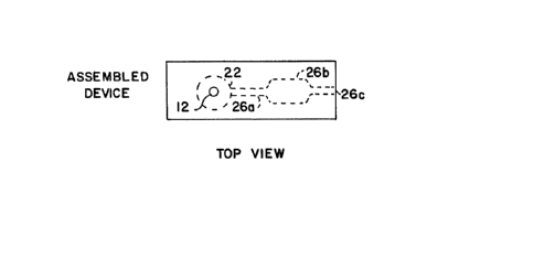
The as~embled device 1~ shown in Figure 1C in
which dotted llne~ are utilized to show the internal
chambers that have been formed. Blood i3 applied at
entry port (hole) 12, contacts the filter held ln
chamber 22, and ls separated into pla~ma while the red
blood cells are retained on the filter. Plasma passes
through capillary 26A to reaction chamber 26B while air
is vented through capillary vent 26C.
F~gure 2 ~hows a device prepared by welding
two or more plastic pieces together to form a unitary
device having internal chambers. Numerous embodiments
of this de~ice are set forth in C~rladian P~tent ~ ication
Serial Number 514,89(), referenced above.
Blood is applied to entry port 42, which is smaller in
diameter than chamber 44 which contains filter 46.
Plasma exits the bottom of the fllter into collecting
space 48 and is transported by capillary 50 to reaction
chamber 52. Vent 54 is provided for exit of air from
the device. Ridges 56 may be provlded if desired to
aid in the application of blood to the entry port.
Additional capillaries, chambers, vents, and the like
1 ~07~8
l4
~uch as are described in the incorporated patent
applications may be present in device 40 but are
ommitted in this Figure for clarity.
A whole blood sample, optionally formulated by
addition of anticQagulants or other reagents useful in
collection of blood or in undergoing a reaction with
the analyte that will be measured, is introduced into
the entry port in the receiving unit of a test
device. The receiving unit may be a capillary or a
larger cha~ber. The receiving unit may be used to
measure the particula~ sample volume or may simply
serve to receive the sample and direct the sample to
the filter. When whole blood contacts the filter, it
is separated into its components as described above.
The first component to leave the filter will be plasma
or serum, depending on the source of the sample. For
the remainder of this discussion the term plasma will
be used but ~hould be ùnderstood to represent either
plasma or serum.
The filters of the present invention typically
comprise a single layer of material rather than
multiple layers. They are intended for separation of a
single drop of blood, whlch typically has a volume of
30-50 ~1 or less. Accordingly, the volume of the
filter is also small, typically in the range of 5 to 20
~l, in order to avoid absorbing and retaining all of
the plasma. Thickness (i.e., measured in the direction
of the flow path) 19 preferably in the range of 0.2 -
1.5 mm. This range is for all filters and thus i9
somewhat broader than that expressed for glass
microfiber filters set forth above. Particle size
retention for glass microfiber filters is discussed
above. Filters used with agglutinins can be more
porous if desired but should retain agglutinated red
blood cells, which typically form clumps of cells with
apparent diameters from 6^10 ~m for a few cells to
greater than 0.1 mm (tO0 ~m) for a large number of
ce 119 .
1 30744~
~ he plasma will usually be picked up as it
leaves tne filter by one or more capillaries. When
blood is applied to the top of a filter, plasma will be
collected from the bottom. The side~ of tne filter are
5 in close contact with the walls to prevent red blood
cells from passins ar~und the ed3es of the filter.
Optionally, a sealer (usually a polymeric compound) can
be used on the sides of the filter. Plasma leavin~ the
bottom of the filter can collect in grooves or other
spaces between the filter and the surface of the device
containing the filter in closest contact with the
bottom of the filter. Capillaries will draw plasma off
from the collection space or spaces. It will be
recognized that the words top, bottom, and sides as
used here are relative terms and do not necessarily
describe orientation of the filter in relation to the
earth's surface. Capillaries will usually have
diameters in the range of about 0.01mm to 2mm. The
capillaries will vary in length but are generally
shorter than 10cm, usually not exceeding about 5cm.
The first capillary may control the rate of
flow into the chamber that will usually serve as the
reaction chamber. Thus, the capillary may aid in the
control of the time with which the plasma is in contact
with a reagent contained withln or bound to the walls
of the capillary and/or reaction chamber. However, the
flow rate of plasma through the filter is limiting in
many instances, as described above, ~o that the
capillary often i9 transporting plasma as fast as it
leaves the filter. The reagent provides a color change
or some other means of determining the amount of
analyte present in the plasma.
The capillary provides the sole driving force
for the movement of liquid through the device after
passage of the sample through the filter. The device
is normally employed with the capillaries, reaction
chambers, and other chambers being oriented in a
1 3[)7448
16
horlzontal plane so that gravity does not affect the
flow rate. The device is employed without anc~llary
motive force, such as a pump, gravity, or the like.
Accordin~ly, it i~ essential to select a filter as
described herein in order to achieve the separation
while allowing capillary forcè to transport plasma
through the device. Experimental evidence has
demonstrated that the filters described in prior art
such as U.S. Pate~ts 4,477,575 and 4,256,693, for
separating large volumes of blood aided by ~ravity or
which depend on relatively large wicking forces caused
by absorbant substances that contact the filter, are
ineffective in capillary flow devices of the type
utilized in the present invention.
Although the filters described herein can be
utilized in the same devices previously described, a
preferred configuration for use of devices with glass
fiber filters is shown in Figure 3. In this device,
whole blood is supplied to an entry port 42' situated
above a fllter, designated as a blood separater. A
number of capillaries (50') are arranged at the
periphery of the blood separater to transport plasma to
the reagent area. The capillaries may be of dlfferent
lengths and diameters but are designed to allow plasma
to reach the reagent area 52' substantially
simultaneou~ly from each capillary. Canadian Application
Serial No~5l4~89n describes sizing capillaries to
achieve this affect. This design allows for uniform
and rapid filling of the reagent area~
3 The invention will now be further described by
reference to certain specific examples which are
included for purposes of illustration only and are not
to be considered limiting of the invention unless
otherwise specified.
1 307448
1'7
EXAMPLE I
Mater1als and Methods
Blood. Whole blood in 15 USP units/ml of
lithium heparin was used in the following experiments.
Filter disks. The filter dlsks were ~ade from
commercialy available filters or other indicated
materials by usin~ a 0.180~' puncn.
Welded Cartridges. ABS (acrylamide butadiene
styrene) 31ides were welded with the Branson ultrasonic
welder at the following settings: pressure ~ 60 psi,
weld time - 0.3 sec, hold time ~ 1.5 sec, down speed -
3Ø
The essential parts of the device were a
filter chamber 33.5 mil thick with a total volume of 16
~1, a connecting chamber (wider than a normal
capillary) 3.5 mm thick, and a reaction chamber with
vent hole. The total volume of the connecting chamber
and reaction chamber wa~ 8.5 ~1.
Tape Slides. Acetate plastlc strips (6" x 1")
were washed ln SparkleenTM301ution, rinsed ln deionized
water, and then dried using llnt free towels. The
plastic strips were then cut lnto 2.5" x 1" slldes.
Plastic surfaces that contacted plasma were etched in a
plasma etcher prior to assembly. The top slide was a
clean piece of plastic with a 1" x 0.5" double stlck
tape piece stuck to the bottom of the slide. A double-
sided, 3.5 mil thick, Scotch brand tape with a pattern
that formed capillaries and other lnternal chambers cut
3 out of the tape was stuck to the bottom of what would
be the middle slide. A hole was drilled to form the
well using a #16 drill (0.1 73n) . A #25 drill was used
to make a vent hole in this cover slide. The top strip
was stuck to the top of the middle strip with the holes
carefully aligned. The filter of choice ls then placed
in the well of the middle slide, and a bottom etched
slide was stuck to the middle slide~s tape. The filter
1 307448
18
was flush against the top surface of the bottom
slide. The finished slide is shown in Figure 1.
Hemolysis Measurement. The percentage
-
hemolysis was quantitated by measuring the absorbance
of 570nm light by th~ plasma. Absorbance was measured
on a Hewlett-Pac~ard ~451 A spectrophotometer. The
readings were taken uslng cells having path length~ of
approximately 0.01cm. The 0.01 cm path length was in a
tape cartridge prepared as described above. The
absorbance was converted to percent hemolysis by
multiplication of the absorbance by a conversion
factor. The peak at 570nm was used for the 0.01cm
pathlength cell, and the conversion constant was 42Ø
_ass Fiber Filters. A number of glass fiber
fLlters were tested, including GA-200 from Micro
Flltration Systems (MFS), which is the filter used in
all examples unless another filter i9 specified. GA-
200 is a non-woven glass fiber filter containing glass
microfibers having typical diameters in the range from
20 0.5 to 1.0 micrometer. The filter i9 0.70 mm thick and
retained particles 2.3 ~m in diameter in the liquid
phase. The den~ity of the filter is 0.25g/cm3.
Density and thickness values are given prior to the
slight compression that took place during the process
of fabricating the capillary device.
Results
Blood from a patient with sickle cell anemia,
blood with artificially produced high and low
hematocrits, and normal blood were filtered through the
GA-200 filters to determine if blood with an abnormal
hematocrit would be effectively filtered.
,
1 307~4~
,9
Blood Type Filtration Time 1* (sec)_Ly3is (%)
sickle cell ~ <5 0.80
HCT - 30 + <5 ~~~~
Blood Type Filtration Time 1* Time 2* Volume**
(sec) (sec) (~l)
Fresh blood
HCT - 48.5 + 4 12.6 2.5
+ 5 13 2.5
HCT - 33.0 + 4 ~-9 5
+ 5 12.7 5
HCT - 60.0 + 4 13.2 2.5
+ 8 27 2.5
+ 7 12 2.5
* Time 1 is the time between the addition of the
blood tG the filter and the exiting of red blood
cells from the filter. Time 2 is the time for the
blood to reach the beginning of the reagent well.
Volume ~ the volume of plasma which exited the
filter before red blood cells exited the filter.
It i3 evident that the filters are as
effective in filtering the abnormal hematocrit blood as
they are with normal blood; in fact, lower hematocrit
blood appears to flow through the filters faster than
normal or high hematocrit blood.
The lower hematocrit blood was more
efficiently filtered; that is, more volume plasma per
volume of blood exited the filters before the red blood
cells~ However, sufficient plasma was separated even
in high hematocrit blood to allow plasma testing.
Comparison of Filters from MFS
A variety of filters from Micro Filtration
Systems were tested for the ability to filter RBCs from
plasma. The nomenclature of the MFS filters is based
on their physical properties. The further along the
~econd letter of the name is in the alphabet, the
tighter the weave of the filter and the slower the flow
through the filter. The numbers in the name correspond
to the thickness of the filter; that is, the higher the
1 307448
number, the thicker the filter. Three filters from the
group examined proved satisfactory: the GA-200, two
GB-lOOR ~tacked on top of each other, and two GC-90
stacked on top of each other.
Filter Time 1 Time 2 Volume ~ Ly3is *
(~ec) (sec) (~1)
GA~200 5,0 12.8 4 0.58
GB-100x2 19 32 5 0.95
GC-9Ox2 ---~ 120 S ----
1 0
* Lysis measured after removal of red blood cells by
centrifugation ~ 0.37%
Analyte Recovery After Exposure to Glas~ Fiber Filter
The purpose of this experiment was to
determine if potential analytes would be ad30rbed by
the glas.s fiber filter material. The analytes tested
were chole3terol, potassium, and total protein. The
experiment was conducted using the following protocol.
1. Serum was obtained from whole blood by drawing the
blood into glass Vacu-tainer tubes, transfering the
blood to centrifugation tubes, letting the blood
stand at room temperature for 20 minutes and then
centrifuging for 5 minutes at the blood setting on
a TRIAC centrifuge (Clay Adams).
2. The sample was then split, one sample being
contacted with the glass fiber filter material and
the other being left alone until laboratory
analysis.
3. The volume of the filter disks in the tape slides
was 12.6 ~1. Assuming 50 ~1 of blood is added to
the filter, the ratio of blood volume to filter
volume was approximately four. In the experiment,
2 ml of serum was contacted with a 24 mm diameter
di~k (depth ~ 0.7mm) with a total volume of 317
~1. The blood/filter volume ratio waY 2000/317 =
6.3 in the experiment.
1 3Q7448
4. The samples containing filters were vortexed at
medium speed for about 20 seconds and then spun in
a TRIAC centrifuge for 5 minutes to spin down the
glass fibers. The serum was drawn off using a
glass pipet. The serum was then analyzed.
Without With Fraction
filter filter recovered
CHOLESTEROL (mg/dl) 157 158 1.01
1 POTASSIUM (mEq/ml) 4.2 4.2 1.00
TOTAL PROTEIN (gm/dl) 7.2 7.1 0.99
The potassium, total protein, and cholesterol
results indicate that there was almost complete
recovery of these analytes after contact with the
filter.
All publications and patent applications cited
in the specification are indicative of the level of
skill of those skilled in the art to which this
invention pertaing. Each publication i9 individually
herein incorporated by reference to the same extent as
if each individual publication and patent application
had been incorporated by reference individually in the
location where cited.
Althou~h the foregolng invention has been
de~cribed in some detail by way of illustration and
example for purpose~ of clarity and understanding~ it
will be obvious to those ~killed in the art that
certain changes and modifications may be practiced
within the gcope of the appended claims.