Note: Descriptions are shown in the official language in which they were submitted.
J 31~6~
OPTICAL ALIGNMENT SYSTEM
BACKGROUND OF THE INVENTION
_
It is frequently desirable to be able to repeatedly
position an instrument, such as an ophthalmic instrument, in a
predetermined spatial location relative to an object. For
example, a non-contact tonometer must be carefully positioned
relative to an eye under tes-t in order to obtain an accurate
intraocular pressure reading. It is desirable that the operator
be able to position the instrument rapidly, since non-contact
tonometers are frequently used to screen a large number o*
individuals for early warning signs of glaucoma by measuring
; their intraocular pressure. Instruments, such as non-contact
tonometers, must be positioned not only relative to the eye
laterally, but also spaced a proper distance axially from the
eye. In such instruments, the patient normally places his
forehead against a rest and his chin in a cup-like support. The
operator then moves the instrument towards the eye to be tested,
while observing various indicia until the predetermined location
relative to the eye under test is achieved as indicated by the
indicia.
The alignment system of the first commercial non-
contact tonometer is disclosed in U.S. Patent No. 3,756,073,
issued September 1973 to Lavallee et al. The optical system
included a projected target, which the operator centered inside
~ 25 an aiming reticle by looking through an eyepiece, in order to
obtain correct lateral positioning. The proper distance from
~i ~
the eye under examination was achieved by moving the instrument
- 2 -
: , ~
:: .
~311 ~6~
toward the patient's eye until the image of the reflected target
was observed to be sharply focused. Since operator's were aware
that they would not be able to observe the eye through the
optical system during the alignment process, they usually per-
formed an initial positioning of the instrument by observing thelocation of the instrument relative to the eye by observing from
Gne side, while moving the instrument into an approximated
proper position. After approximated positioning, the operator
then looked through the eyepiece to obtain accurate positioning
of the instrument. This procedure avoided inadvertent contact
with the eye. A light detector was used to verify that the
operator's alignment was correct before testing.
The miniaturization of electronic components and
particularly those relating to television, i.e., cameras and
monitors, has permitted adaptation of earlier optical systems to
permit instrument operators to observe the positioning symbols
on a CRT screen. U.S. Patent No. 4,665,923, issued May 19, 1987
is an example of such an alignment system and includes three
optical subsystems. Two of the optical subsystems are
symmetrically disposed about the instrument axis and provide
visible symbols indicating the position of the instrument
relative to a predetermined location. The third optical
subsystem is used to provide the operator with a macro image of
the eye. All of the embodiments disclosed in the patent, as
well as the commercial product utilizing disclosed concepts,
present the three images to a single observation means, i.e.
,
, .
, ~' '' ' ' . '
" ~ ' , . '
,
~ 3 ~
image pickup tube 53 in Fig. 9. It is readily apparent that the
patented system has the distinct disadvantage that proper
adjustment can only be achieved by meticulous adjustment of each
component of the two symmetrically disposed systems and
manufacturing all components to very close tolerances. For
example, the first embodiment requires precise alignment of
eight reflective surfaces in the two alignment optical
subsystems, and even the simplest system, that shown in Figs. 9
and lO, requires precision alignment of five reflective
surfaces. Obviously, the dimensions of each component as well
as the mounting thereof and spacing therebetween are extremely
critical. An additional disadvantage of the disclosed optical
systems is the requirement that at least four of the reflective
elements be beam dividers. This substantially reduces the
amount of original illumination that can be presented to the
image pickup tube. A further disadvantage of the disclosed
systems is that optically presenting three separate images to a
single camera tube or CCD array causes the macro image of the
eye to be washed out or at least very faint. Similar to the
earlier system, a spot detector was used to verify correct
alignment by the operator before testing in all of the disclosed
embodiments.
The criticality in alignment is partly due to the use
o~ a "spot" detector to verify alignment. It is only after
light has passed through or been reflected by numerous elements
~; that the spot or spots are evaluated to determine how much light
l ~ - 4 -
`,
~: :
,:
13~160~
is falling on the detector. Each of these elements reduces the
amount of light available at the detector and is capable of
introducing errors because of alignment or quality. Another
factor affecting the system advsersely is the quality of the
optics required. Since the disclosed system projects a target
image that is imaged on the observation means and the detector,
the quality of the final images is controlled by the quality and
alignment of all the intervening optical components.
U. S. Patent No. 4,705,045, issued November 10, 1987,
discloses a tonometer alignment system having two oblique target
projection systems that re-image the targets through an imaging
optical system that is parallel to the discharge tube axis. Only
imaging light that is reflected from the eye parallel to the
imaging optical system axis is imaged on the detector, with both
images being superimposed when the tonometer is aligned properly.
SUMMARY OF THE INVENTION AND
BRIEF DESCRIPTION OF THE DRAWINGS
Accordingly, the present invention seeks to significantly
reduce the number of optical elements requiring critical
Z0 positioning in an optical alignment system. Further, the present
invention seeks to provide an optical alignment system presenting
a major portion of the initial illumination to the detector.
Still further, the present invention seeks to provide a
clear image of the object relative to which the instrument is
being~aligned.
~ Further still, the present invention seeks to provide an
electronic alignment of an ophthalmic instrument.
:; ~
~ - 5 -
,~ ~
.~
., ~. . ~ . .
:
3116~8
Still further, the present invention seeks to provide an
alignment verification system that does not require additional
detectors.
Still further, the present invention seeks to provide a
macro view of the eye without a light source in addition to that
used for alignment.
Still further, the present invention seeks to
automatically, i.e. electro-mechanically, align a test instrument
using information provided by at least one CCD array.
The invention in one broad aspect provides an alignment
system for an ophthalmic instrument comprising illuminating means
for illuminating an eye with diverging rays from a light source, a
first detecting means'for defining a first light detecting area,
the light detecting means including a first pinhole occluder to
pass a small bundle of reflected rays producing a first signal
identifying the XY location of reflected light on the first area,
and a second detecting means for defining a second light detecting
area, the second detecting means including a second pinhole
occluder to pass a small bundle of reflected rays and producing a
second signal identifying the XY location of reflected light on
the second area. There is means for evaluating the first and
second signals and for providing a third signal, and means for
selectlvely positioning the instrument relative to the eye
responsive to the third signal.
~ Another aspect of the invention provides an alignment
system for an ophthalmic instrument wherein there is first
illuminating means for providing light having rays from a source
dlverging to an eye from one side thersof, and second illuminsting
~ - 6 -
:
.
`-` - 13~608
means for providing light having rays from a source diverging to
an eye from the other side thereof. There is detecting means for
defining an area, the light detecting means including an occluder
having two spaced pinholes to pass two small bundles of reflected
rays, one of the bundles comprising rays from the first
illuminating means and the other of the bundles comprising rays
from the second of the illuminating means for producing first and
second signals identifying the XY location of reflected light
passing through each of the pinholes on the area. Means evaluate
the signals for providing an output signal, and means selectively
position the instrument relative to the eye responsive to the
output signal.
More particularly, the present invention includes two
light sources for reflecting spots from opposite sides of a
spherical object, such as an eye. The reflected images are
directed to at least one position sensitive detector, such as a
CCD array. The signal-produced by such a detector indicates the
location of the image on the light sensitive area. Alignment of
the optics may be achieved electronically by positioning the
lnstrument in a predetermined location relative to the spherical
ob~ect and storing the position of the spot as a reference
location. The position of the instrument at any future time can
then be presented relative to that stored reference location.
Alternatively, the system can be aligned by moving each array
normal to the light path until the spot is centered on both arrays
when the instrument is properly positioned. Inthe latter case, the
6a -
~ ,, .,, ., ,: .-:
~311 1~
signal can simply be sent to a monitor to indicate the position
of the instrument relative to the eye or other test objectO In
the former case, the signal is modified to treat the stored
location as if it were the center of the CRT screen. In both
cases, the signal sent to -the CRT can also be evaluated elec-
tronically to verify that the instrument is correctly aligned
before conducting a test.
A separate image tube or CCD array is used for a macro
image of the eye, if desired. The macro optical system is
designed to provide an image of the eye which appears sharp and
uniformly illuminated. Illumination reflected from the eye by
the alignment system is usually sufficient for the macro imaging
system when the system of the preferred embodiment is utilized.
Fig. 1 is a perspective view of an ophthalmic
instrument of a type suitable for use with the present invention;
Fig. 2 is a diagrammatical view of a first embodiment
of the present invention;
Fig. 3 is a diagrammatical view of a second embodiment
of the present invention;
; 20 Fig. 4 is a diagrammatical view of a third embodiment
of the present invention;
Fig. 5 is an optical diagram of the first embodiment of
the present invention; and
; Figs. 6 and 7 are block diagrams for explaining
electronics supporting the present invention.
- 7 -
:` ~
~"~, . . . : . . . ..
- .
;
DESCRIPTION OF THE PREFERRED EMBODIMENTS
Referring to Fig. 1, an ophthalmic instrument shown
generally at 1, has a base 2 with a frame 3 to provide a
steadying rest for the head of a patient. The test mechanism
(not shown) o instrument 1 is contained within housing 4 movably
mounted on base 2. Member 5 represents a portion of the
instrument to be positioned in a predetermined relationship to
the patient's eye. To accomplish this relationship, the operator
uses joystick 6 to move housing 4 three dimensionally on base 2,
while watching the resulting movement of symbols (not shown)
relative to reticle 7 on screen 8. When the operator has
achieved alignment by moving housing 4 until the symbols are
contained within or superimposed on reticle 7, he presses button
9 on joystick 6 to initiate the desired test.
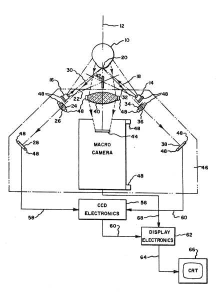
Referring to Fig. 2~ eye 10 is flooded with light on
one side of its axis 12 by light from source 14 and on the other
side of its axis with light from source 16. Sources 14 and 16
may conveniently be LEDs emitting light in the infrared region.
The advantage of infrared illumination resides in the insensi-
tivity of the human eye to the infrared region. This avoids any
discomfort, when high levels of radiation are required. Center
ray 18 from source 14 is reflected from cornea vertex 20 along
path 22 and seque~tially through pinhole occluder 24 t and lens 26
to CCD array 28. Similarly, center ray 30 of source 16 is
re~lected from cornea vertex 20 along path 32 and sequentially
through pinhole occluder 54 and lens 36 to CCD array 38. A small
8 -
~: :: : :
- : .
~::
~ ~ .
~ 3 ~ 8
bundle of rays closely adjacent to center rays 18 and 30 will
remain substantially parallel thereto and pass along with the
respective center ray through the respective pinhole occluder. A
portion of the remaining light from each of sources 14 and 16 is
reflected toward lens 40 to produce an image of eye lO on video
image detector 44. In the case of a non-contact tonometer, lens
40 is located behind air~pulse discharge tube 42.
The alignment system components may be conveniently
mounted on plate 46 which has a plurality of mounting members 48
for holding sources 14 and 16; pinhole occluders 24 and 34;
lenses 26 and 36; CCD arrays 28 and 38; objective lens 40 and
video image detector 44. One advantage of the present invention
which may be realized is that mounting members 48 do not require
precise machining to close tolerances in order to provide exact
angles and dimensions, since normal variations may be corrected
electronically rather than optically. Electronic correction is
achieved by positioning the instrument, containing the alignment
system of the present invention, in the chosen relationship to
the object such as a replica of a human eye. If -the spot
; 20 produced by occluders 24 and 34 is relatively close, e.g. .lmm,
to the center of the respective arrays, the XY location of the
spot i5 conveniently stored in a device such as an EEROM.
~; However, gross errors may be compensated for by moving the CCD to
a new position in a plane normal to the respective paths 22 and
32. The reference locations (the locations of the respective
spots on the CCD arrays) are thereafter considered to be the
: : ~
:. ,:,.
'
~.3~6~
"center" of the respective CCD array.
Referring now to Fig. 3, another embodiment of the
present invention is illustrated. Light from sources 14 and 16
is reflected back from corneal surface 120 in the same general
direction from which it came along paths 122 and 132, respec-
tively to CCD arrays 28 and 38. In other respects, this
embodiment operates in substantially the same manner as the
embodiment of Fig. 2.
Fig. 4 illustrates still another embodiment. Light
from sources 14 and 16 is reflected by the respective sides of
cornea 120 along paths 222 and 232 toward objective 20. Beam
splitter 250 diverts a portion of the light toward occluder 224
having two pinholes 252 and 254. Light passing through pinholes
252 and 254 is imaged by lens 226 on CCD array 228. In this
embodiment, sources 14 and 16 are alternately strobed in order
for CCD array 228 to identify which of sources 14 and 16 produced
the spot being observed. ;;
Referring again to Fig. 2, signals identifying the XY
location of the spots on CCD arrays 28 and 38 are delivered to
CCD evaluating electronics 56 by leads S8 and 60. Electronics 56
compares the reported XY position of the spot to the stored
reference location for each CCD array. An output from
electronics 56 representing the location of the spot relative to
~the reference location is provided to display electronics 62
whiah in turn drives CRT 66 through leads 64 to provide symbols
~ on CRT 66. The signal from video image detector 4~ is similarly
:`~
,, - 10 -
- : ~
.. . .
~3~L16~8
provided to display electronics 62 through lead 68 in order to
provide a macro image of the eye on CRT 66. The location of the
spot on a CCD array can be identified conveniently using a raster
sweep of the CCD pixel siynals. The signal and location values
of the first pixel are stored until a higher signal value is
encountered during the sweep. Each time a higher signal value is
encountered, the new pixel signal and location values are stored
replacing the values previously stored until the sweep is
complete. The location values stored at the end of the sweep
identify the center of the spot on the respective CCD array. If
a minimum signal threshold is set, artifacts, such as glare spots
that can result from illumination for the macro view, are ignored
by the system. When an optical system of the type illustrated by
; Fig. 4 is used, the timing of the raster sweep of CCD array 228
is synchronized with the strobe of sources 14 and 16 in order
that even raster sweeps relate to one source and odd raster
sweeps to the other.
.
-- 1 1 --
;~ .
~ '
~ 3 ~
A preferred optical system according to Fig. 2 is
diagrammatically presented in Fig. 5 and has the following
values:
Element Radius Thickness Spacing Index of
Refraction
14
S1=55.0
S2=60.0
24 D1=0.5 T1= 0.5
S3= 0.5
R1=30.489
26 T2= 3.00 N1=1.5168
R2= -30.489
S4=~0.0
28
.
'~ 16
S5=55.0
. 20 '
S6=60.0
~, 20 34 D2=0.5 T3= 0.5
: S7= 0.5
:'
; R3=30.489
36 T4= 3.00 N2=1.5168
R4= -30.489.
S8=60.0
38
S9=12.30
42
J' S10=87.7
:: R5=50O813
~:, 40 T5= 5.00 N3=1.5168
~-~ R6= -50.813
S11=100.0
~: 44
: - 12 -
`~' :
~'
:
-. ;. ~, , : ~
: ~
: : :
~ 3 ~
wherein, radii, Rl to R6, thicknesses, Tl to T5, spacings, Sl to
Sll, pinhole diameters, Dl and D2, are in mm; radii having their
center of curvature on the eye 10 side of the lens are indicated
by a minus (-) sign; and indexes of refraction, Nl to N3, are
absolute values. The pinhole-lens combinations can be replaced by
small diameter lenses if desired. The model Texas Instruments
TC211 CCD array is suitable for practicins this invention.
The amount of instrument movement necessary to obtain
distance (S9) of object 10 from component 42 of the instrument
being aligned can easily be calculated using the location value
related to movement in a direction parallel to the plane
containing the optical elements of the alignment system obtained
from each CCD array. For example, if a = 45 and a' = 42 and x
and x' are the relative locations in the directions indicated by
the arrows labeled x and x' in Fig. 5, ~ S9 = (x - X)-(x' - X'),
where X and X' are the reference locations for the respective CCD
array. The amount o~ movement can be presented two dimensionally,
for example on the CRT, by using one symbol for the horizonal (x)
axis and another for the vertical (y) axis. The space between the
symbols can be used to represent S9. When ~ S9 = 0, the two
symbols are superimposed. A simpler and more user-friendly
procedure is the use of a cursor which moves above the screen
center, if the instrument is too far from the object and below the
screen center, if the instrument is too close. The cursor type of
presentation is preferred because it has several advantages~ One
advantage is the ease with which the user can recognize whether
the instrument is too close or too far away. Another advantage is
- 13 -
that the x and y positions can be displayed by means that do notrequire superimposition of symbols to indicate correct
positioning. For example, a narrow vertical line can be used to
represent the relative horizonal position and a narrow horizonal
line to indicate the relative vertical position, while the cursor
indicates the relative distance from the object. Prior art
alignment systems did not permit a choice of display formats.
Referring now to Fig. 6, signals from timing generator
350 drive x/y counters 352, the raster sweep of CCD array 354 and
timing of A/D converter 358. Each pixel signal is amplified by
amplifier 356, sent to A/D converter 353, whose output is
evaluated by peak detector 360. The outputs of x/y counters 352
are stored by latches 362, each time peak detector 360 signals a
new high for the pixel signals from CCD array 354. Obviously, each
array requires an amplifier, A/D converter, peak detector and
output latches. The outputs of latches 362 are evaluated at the
end of each raster sweep by alignment and calibration electronics
364 whi~h updates operator display 366. If an automatic alignment
system is desired, motor controllers 368 and motors 370, 372 and
374 can be added as shown in Fig. 7. The value of ~z is
determined as explained previously for the determination of ~ S9,
(x - X) + (x' - X') (y - Y) + (y' - Y')
while ~x = and ~ y = --
2 2
These ~5 values are provided to motor controllers 368 by alignment
and calibration electronics 364 to position the system until all
three ~'s - 0.
- 14 -
~ '~
,
.