Note: Descriptions are shown in the official language in which they were submitted.
1 3 1 1 ~ ~ ~
IMPROVED 8UBMERGIBLE 8CR~W-TYPE DENTA~ IM_LAN~
This application is a divisional application of
Application Serial No. 561,440 filed 15 March, 1988.
Technical Field
This invention relates to dental implants and, more
particularly, to submergible screw-type implants.
Backaround Art
Screw-type implants are well known in the art. U.S.
Patent No. 3,499,222 of L. I. Linkow et al. discloses screw-type
implants which may be buried in the alveolar ridge crest bone of
a patient in an edentulous region. The implant has a threaded
lower portion which may be screwed into an opening created in the
bone after the tissue has been displaced. A coronal portion
protrudes above the bone and is used to support an artificial
dental appliance, e.g. an artificial tooth or bridge.
In more recent years submergible implants have been
created in which the threaded portions of the implants can be
completely embedded in the bone. They may then be covered with
tissue and allowed to remain in place while new bone grows around
; the implant and through vent holes in it. once it is firmly
~ anchored in new bone (3 to 6 months), the tissue is reopened and
e~, an upper post portion is screwed into the implant portion and is
.
i, used to mount the artificial dental device.
, ~:
~ It is advantageous when installing an implant portion
.: , . . . , .: - -,:,
- : - . ~ . ..
. ~ . . . .
:: :
:. : . ,,
.
1 3 ~
in the patient's bone, if the implant is self-tapping in a bore
created in the bone. This causes it to be anchored better.
Also, it would be advantageous if the bone chips created during
a self-tapping operation were deposited into the bore or opening
because these chips promote faster bone growth because of their
autogenous nature.
In order to align the artificial tooth or other dental
devices with the other teeth of the patient, it may be necessary
to have the post portion at an angle to the implant portion.
This may be accomplished by bending the post portion so that its
head is at an angle to the threaded shaft. This bending may be
accomplished before the post is threaded into the implant portion
or afterward. If the post is bent before attachment to the
implant, the proper alignment is difficult to achieve. If bent
after attachment, there is a danger that too much stress will be
put on the implant portion and it will loosen in the bone and
fail. Also bending the post may fatigue the metal of the post
and cause breakage.
, 20 Summary of the Invention
According to the invention claimed by the subject
divisional application there is provided an oral implant having
an implant portion adapted to be fitted in an opening in a bone
of a patient in the vicinity of the occlusal plane and on a
support for an artificial tooth structure attached to the implant
portion. The support includes a member on which an artificial
tooth may be mounted, the member having a bore extending
,
,,, ~ . ,
.
'
3 131194~
completely through it, one end of the bore opening into a recess
which is in the shape of half an oval. A collar has a central
opening aligned with the bore of the member, one end of the
opening being expanded into a recess which is in the shape of
half an oval in cross section. The collar is positioned relative
to the member such that the recesses are adjacent each other and
form an oval cavity. A flexible elastic washer has a hole
through the middle and is aligned with the opening when seated
in the cavity, the washer keeping the member and collar
resiliently separated from each other. A screw has a head
secured in the member and a shaft extending through the bore of
the member. The hole of the washer and the central opening of
the collar are urged into threaded engagement with the rest of
the implant. Preferably the screw has a flared portion which
engages and wedges in the collar portion so as to inhibit
unthreading of the screw.
' /
,
~, .
... .
-, :
!
,,~,;,
' ~ ' , ' :
~ :; . , '', '
- ~ ', . . - .
4 ~ ~31~ $
~rief d_scription of the_drawin~s
1 The foregoing and other features of the present
invention will be more readily apparent from the following
detailed description and drawings of illustrative embodiments
of the invention, in which:
Fig. l is a schematic cross section of the side of a
patient's-face showing the alveolar ridge crest with a screw
type implant according to the present invention installed
therein;
Fig. 2 is an enlarged view of an illustrative embodiment
of the implant portion of the device of Fig. l with an external
hex projection;
Fig. 3 is a top view of the implant portion of Fig. 2
showing the external hex portion;
Fig. 4 is a cross-sectional view through the implant
portion of Fig. 2 along line 4-4 showing the cross-sectional
shape of the channel according to the present invention;
Fig. 5 is an implant portion of a screw-type implant
according to the present invention with an internal hex recess;
Fig. 6 is a illustrative embodiment of a completed
screw-type implant with an angularly positioned threaded shaft
~ attached thereto;
;~ Fig. 7 is a cross-sectional view of a ball and socket
~; connection port4ion of an abutment according to the present
invention;
Fig. 8 illustrates a modification of the ball and socket
joint of Fig. 7;
Fig. 9 illustrates a further modification of the ball
and socket joint of Fig. 7;
Fig. lO is a ball and socket joint connection portion
with a stationary ball;
Figs. llA and llB are cross-sectional views of a unitary
inner casing and a two-part inner casing, respectively;
Fig. 12 is a side view of a healing collar according to
the present invention;
Figs 1~ and 14 are front and side sectional views of an
artificial tooth with an abutment according to Fig. 7;
,, .
.
',~
:! ~
' ~ ~ ' ' ' '
~ ~ ' ' ' .
~ ' , '
13~
Fig. 15 is a cross-sectional view of an embodiment of an
1 abutment with a shock-absorbing cushion; and
Fig. 16 is an alternative embodiment of the screw of the
shock-absorbing abutment of Fig. 15.
Descri~tion of Illustrative Embodiments
~he present invention contemplates at least a two part
screw-type dental implant, i.e, an implant portion 10 which is
buried in the bone of the patient and a post or abutmeht
portion 20 which is attached thereto and which supports an
artificial tooth structure 30. As shown in Fig. 1, an implant
screw portion 10 is located in a bore in the aveolar crest 11
at an angle that causes it to be in the center of the thickest
portion of good available bone. The abutment 20 is attached
both to the implant portion 10 and the artificial tooth 30, and
is set so that the tooth is at an angle to the implant which
causes the tooth to be in proper alignment.
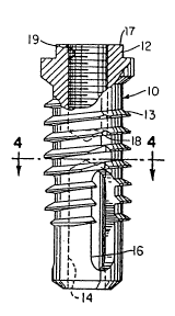
In Figs. 2 and 3 the screw implant portion 10 of Fig. 2
is illustrated in more detail. This implant portion 10
contains threads 13 which extend over the middle region of the
implant portion. These threads may have a flat bottom and be
angled up to form a ff~hristmas tree shape in cross section. The
lower half of the implant portion 10 contains a cavity 14
~shown in dotted line). Also, spaced about the lower end of
the implant are holes or vents 16, 16a and 16b, which penetrate
from its exterior to the interior cavity 14. The purpose of
these vents is to allow new bone to grow through and into the
center cavity in order to firmly anchor the implant in the
patient's bone. ~he upper surface 17 of the implant portion
defines a threaded aperture 19 which is used to connect the
abutment 20 to the implant portion 10. The projecting
structure 12 which forms surface 17 has a hexagonal shape as
shown more clearly in Fig. 3. This hexagonal shape allows a
tool, e.g. a wrench, to be used to rotate the implant portion
so as to thread it into the patient's bone.
According to the present invention a channel 18 is cut
through the threads 13 and possibly into the outer casing of
the implant portion 10. As depicted in dotted line in Fig. 3
,~,'f
,f
;; :
:: ~ : :
'' ~ '. ' ' "' '" ~" ' " ' , '
" ~ .
, ' ' "
6 ~31~ 4~
and in cross-section in Fig. 4, the channel 18 is one of three
I channels 18, 18a, 18~ in a typical implant pQrtion. These
channels are made to intersect the respective vents 16, 16a and
16b which are spaced at angles of 120- about the circumference
of the implant portion 10. The channels do not extend
completely toward the upper surface 12 in order to prevent
tissue from growing down along the channel, and to prevent the
incursion of food and bacteria. It should be particularly
noted in Fig. 4 that the channels 18 have one edqe which is at
about 90 to the circumference of the implant, i.e., surface
18', and another more obliquely shaped edge, i.e. surface 18''.
During installation of the implant, an incision is made
in the gum tissue of the patient and the underlying bone is
exposed. Then a drill or burr is used to make an opening or
bore hole in the bone which is slightly larger in diameter than
the implant portion body 10, but which is not as wide as the
threads 13. A wider counterbore may be provided to accommodate
a protection collar as explained subsequently. Next the
implant is inserted up to the first thread in the opening in
the bone. A tool, such as a wrench, is used to engage the hex
portion 12 and to rotate the implant. The threads 13 are made
to be self-tapping so that the implant portion will begin to
screw down into the patient's bone. If necessary, a bone tap
can be used to create grooves in the hard upper cortical bone
prior to insertion of the implant portion. The right angle
surface 18' of the shannel also has self-tapping properties so
as to ease the insertion of the implant, once it has reached
the depth of the channels 18. Further turning of the implant
causes the right angle surface 18' to scrape off bone as the
implant is being threaded and to push the resulting bone chips
forward. This causes the bone chips to fall through the
channels 18 and into the area of the vents 16 where they may
penetrate into the interior cavity 14. To facilitate this, the
channels 18 are made wider towards the vents 16.
; As a result of this structure, bone chips created during
the implant procedure tend to accumulate at the base of the
implant in the patient's bone. Because of the autogenous
: .
:
.
~_,.,, ,~, .. .. . . . . .
~ .
, -
7 131~9~
nature of thes~ bone chips they promote t..~ growth of new bonein the area and speed the formation of new bone around and
through the implant such that it is anchored in place more
1 rapidly;
In Fig. 5 there is shown an implant portion lo which is
nearly identical to that shown in Fig. 1. The principal
difference is that, rather than having a hexagonal projection
useful for applying torque to the implant, a hexagonal recess
12' is provided. In addition, the threaded aperture l9' is
made somewhat smaller and is located at the base of hexagonal
recess 12'. As explained previously, the threaded aperture l9'
is used for attaching the implant portion of the device to the
abutment portion. One embodiment of such an attachment is
shown in Figure 6.
In Figure 6 the upper part of the implant portion 10 is
shown partly broken away and partly in section. It is shown
partly broken away to exhibit the interior cavity 14 and the
threads 13. Towards the upper part of the implant portion it
is shown in cross section. This implant portion is like that
shown in Fig. 5 with a hexagonal recess 12' for rotating it
into position in the bone. As shown in Figure 6 the screw type
implant portion 10 is connected to an abutment portion 20 that
~ 20 includes a transitional collar 21, an angled threaded shaft 24,
$ and a tooth support cylinder 31. The threaded shaft 24 has its
lower end screwed into threaded aperture 19' in the implant
portion 10. The upper end of the threaded shaft, which is set
at an angle to the lower end, is received within a threaded
25 aperture 35 in tooth support cylinder 31. This cylinder 31
contains a recessed portion 32 which may be utilized in fixing
; on to the cylinder via cement or some other convenient and
well known method, a porcelain, plastic, or other dental tooth-
colored veneering material in the form of an artificial tooth.
The transitional collar 21 is located between the upper
j end of the implant portion lO and the cylinder 31. This collar
has an angled upper surface 25 and a perpendicular lower
surface 23. The angle of the upper surface is made to equal
the angle of the upper part of the angled shaft 24. While
. , .
: : ,
8 131:19~
collar 21 surrounds threaded shaft 24, it does not engage its
threads.
During an installation procedure the implant portion 1~
is located in the patient's bone as previously described. The
gingival tissues can then be replaced over the implant portion
and several weeks or months allowed to pass while new bone
grows around and through the implant portion. However,
alternatively the artificial tooth can be connected to the
implant immediately. Whichever manner is chosen, the
attachment is accomplished by selecting an angled shaft and
transition collar which have an angle which will cause the
artificial tooth to be correctly aligned with the other teeth
of the patient. Therefore the dentist or oral surgeon must be
provided with a variety of such shafts and collars which are at
standard angles. Also during the insertion procedure the
surgeon must appropriately angle the opening in the bone so it
penetrates a reasonably thick area of good bone. This may
require that the opening in the bone be drilled at an angle in
order to avoid penetrating a nearby sinus cavity, passing
completely through the bone, or contacting a nerve bundle.
However, in selecting the angle at which the implant is buried,
care must be ta~en to ma~e sure that this angle will
accommodate one of the standard angles available with the
,,A,, threaded shafts and collars, e.g. 10, 20 or 30 degrees, so as
to result in alignment between the new artificial tooth and the
remaining teeth of the patient.
~'Aonce the threaded shaft 24 is engaged with the implant
portion 10, the collar 21 is slipped over the free end of the
shaft. Then the shaft is rotated so that it is firmly secured
in the implant portion and is extending in the proper
direction. With the collar in place over this shaft, the
cylinder portibn 31 is threaded over the open or free end of
the shaft until it makes tight contact with the upper surface
of the collar and begins to squeeze the collar between the
~cylinder and implant portions. Notches and recesses 22 and 27
are provided in the mating surfaces such that, once the parts
are screwed together, these notches and recesses engage each
". ~ ,
~s'i' ~
, ~ ~ ~, . . . .
, . ... . . .
.
~ ' ' ' ':
: , ~
. . - .-.
.
, ,. . :, ,. ~ ,
9 ~
other and prevent unintentional unscrewing of the portions of
1 the impiant. With this firm attachment completed, the
~rtificial tooth can then be attached over the abutment
cylinder 31.
In Figure 6 the level of the patient's ~one is shown as
dotted line 70. Since the implant portion is submerged in the
bone, the line 70 intersects the lower portion of the
transitional collar 20. The gum tissue line 72 i6 towards the
upper portion of the transitional collar. As a result the
collar acts a barrier to prevent the encroachment of bacteria
and food into the interior portion of the collar and the hex
recess of the implant portion.
With the embodiment of Figure 6 fixed angles are
provided to the dentist and he must wor~ with the standard
angles and the angle which he creates for the bore in the
1~ patient's bone, in order to assure proper alignment of the
teeth. In some patients who have had serious bone disease, the
amount of available good bone is limited and the dentist has
only a limited amount of freedom in selecting the angle at
which the bore for the implant is made. Also with the
embodiment of Figure-6 it is necessary for a dentist to keep a
stock of various angled shafts and collars. The difficultly
presented by the type of implant in Figure 6 is overcome by the
implant shown in Figure 7.
In Figure 7 the angled shaft and transition collar are
replaced with a ball and socket joint which allows for the
setting of the angled relationship between the implant portion
` and the abutment portion at any selected angle within the range
of motion of the ball and socket joint, e.g. up to 30-40
, degrees. In Fig. 7 the threaded cavity 19 receives the
threaded shaft of a lower or inner abutment casing 42. This
casing has a generally Y-shape with the lower portion being the
shaft that extends into and engage the threads of cavity 19.
The upper portion of casing 42 has a hemispherical surface 45
such that it can receive a ball 46. An upper or outer casing
44 screws onto outer threads of the inner casing 42 such that
,! 35
'i '
': . : . ,:
: , .
; ~.` ~, .
lC 131194~
ball 46 is trapp~ within the abutment casin but is free to
rotate therein so as to create a ball and socket joint.
A relatively large set screw 48 penetrates the ball
1 completely. ~his set screw 48 has an internal threaded cavity
55 which passes through an upper hexagonal projection 56.
Once the implant portion 10 has been located in the bone at the
optimal angle, the ball 46 is rotated such that the center axis
5 of the set screw is at the proper angle for mounting of an
artificial tooth in line with other te~th in the patient's
mouth. Then the hexagonal portion 56 is rotated with a wrench
or other tool so the set screw comes into extreme frictional
contact with the hemispherical surface 45 of inner casing 42.
lOThis prevents further rotation of the ball and the set screw.
The artificial tooth structure in the embodiment of
Figure 7 has an interior cylinder 50, about which the
porcelain, plastic or other dental material is formed to create
the artificial tooth structure. This cylinder S0 with the
lSartificial tooth structure mounted thereon, is placed on top of
the hexagonal projection 56 and is then attached thereto by
means of a screw 52 which passes through the cylinder 50 and
into the threaded aperture 55 in set screw 48.
The bone line 70 is shown in Figure 7 as being
20 approximately mid-way through the lower abutment casing 42,
while the gum line 72 is just below the upper edge of the outer
or upper casing 44. Thus, the bone does not interfere with the
; settinq of the proper angle for the abutment and the tissue is
not likely to contact moveable adjustment parts.
The arrangement of Figure 8 is a modification of that
shown in Figure 7. In this arrangement the set screw 48, which
has a threaded recess 55 at its end in Fig. 7, is replaced with
a set screw 49 that has a further screw thread 59 on the
opposite side of the hex projection 56. This additional screw
30 thread is used to mount an artificial tooth support cylinder 53
which has an interior threaded cavity. However, this device is
essentially located and fixed in position in the same manner as
the implant of Figure 7. One difference with this implant of
Figure 8 is that the artificial tooth support cylinder 53 may
. .
`
:
11 ~.3119~
extend down to and in contact with the ~uter casing 44. This
i5 done above the gum tissue line 72 as shown in the figure.
Because of the contact between the cylinder and the casing 44,
food and baeteria are prevented from entering between these two
parts and the likelihood of infection is reduced. However,
S this arrangement allows for somewhat less range of angular
adjustment. In particular the arrangement of Fig. 7 is capable
of an angular adjustment range of approximately 37 1/2 , while
that of Fig. 8 is limited to about 30-.
As a further alternative, the set screw 48, rather than
having a projecting threaded portion located above the
hexagonal adjustment nut 56, may have a projecting cylinder
which is internally threaded (not shown). Thus either a male
or female connection of this type may be used without
difficulty.
In order to get increased angular adjustment, an
arrangement such as that shown in Fig. 9 may be used. The
abutment arrangement of Figure 9 is essentially the same as
that of Figure 7; however, the ball and socket joint are made
smaller and the ball sits higher in the soc~et joint. Further,
the set screw 54 of Fig. 9 is made to have a beveled surface 57
such that a greater a~ngular rotation may be made before it
contacts the upper part of the outer casing 44. With this
arrangement nearly 45 degrees of angular adjustment can be
achieved.
' 2 The abutment ~ylinder 50 has a recess 51 to receive the
outer end of the set screw 54. This allows for greater
stability when it is attached to the set screw by means of
attachment screw 52. The cylinder 50 is also angled in the
same manner as the surface 57 of the set screw 54 so that it
does not bind against the upper abutment casing 44 and limit
angular rotation.
In Fig. 7-9 the ball rotates with the set screw during
angular adjustment. However, as an alternative, the ball may
remain stationery and the abutment casing may rotate as shown
3 in Figure 10. In Figure 10 a threaded ball joint 60 has a
projecting threaded shaft 61 which is received in threaded
,1, .
., .
.i:
,'": , ': , '
12 13il~$
recess 19 of the implant portion 10. Various size protection
washers or collars 65 can be located about the finial part 67,
which connects the ball to the threaded shaft, in order to
cover the upper surface of whatever implant portion is used,
thereby preventing bacteria and food from entering the bore.
The opening in the bone can be countersunk as indicated by
dotted line 70 so the collar can extend out beyond the implant
portion upper surface, and bone can grow over part of the upper
surface of the collar.
A two-part casing 62, 64 is mounted on the ball 60. The
casing includes outer casing portion 62, which secures the
remote end of the ball, and an inner casing 64, which provides
the main hemispherical surface against which the outer casing
holds the ball in a rotatable manner. These two casing parts
can be threaded together or attached to each other in any
convenient manner. Their attachment, however, is such that the
casing may rotate freely on the ball.
At the opposite end of ball 60 from the screw threads is
a hexagonal recess 63, which is the means by which this
threaded ball joint is screwed into the threaded recess 19 of
the implant portion. In this arrangement the gum line 72 is
shown about 1/3 up from the base of the ball joint, but below
the lower extension of casing 62.
A hexagonal projection 66 is provided on the inner
casing 64. This projection can be used to rotate the inner
casing 64 so that th'e ball is squeezed between it and the outer
casing 62 so that swiveling can be prevented when the
arrangement is at the proper angle. A conventional cylinder 50
for a dent 1 prosthesis is attached to the inner casing 64 by
means of a screw 52. This screw 52 penetrates a threaded
aperture in the inner casing.
An enlarged view of the inner casing 64 is shown in Fig.
llA. The lower peripherial extension 64' of this casing forms
a wedge that projects between the ball 60 and the outer casing
62 as shown in Fig. 10. When the inner casing 64 is screwed
down onto ball 60, the extension 64' acts to lock the abutment
on the ball and prevents further rotation. In part this
, . . .
~ , .
..~,,..,..,, .
. `
13 1311~
loc~ing is maintained due to the fact that the diameter of the
1 extension 64~ is slightly less that the distance acro~s the
ball at its location. As a result there is an outward flaring
of the extension as shown by the arrows in Fig. llA, which
prevents the unthreading of inner casing 64.
Instead of a one piece casing as shown in Fig. llA, the
inner casing may be in two parts as shown in Fig. llB, where
the extension 64' is part of a locking ring or washer 64~.
With this arrangement the ball is surrounded by the ring 64~
and the casing 64. As the casing is threaded into contact with
10 the ball it forces the ring to wedge between the ball 60 and
the outer casing to frictionally hold the ball. The outward
flaring of the extension at the end of this compression process
tends to prevent the unthreading of the inner casing, which
prevents the abutment from becoming loose.
Installation of submergible implants is generally a two
stage procedure. During the first stage the implant portion is
buried in the bone and the tissue is restored in place over it.
Time is allowed to pass while new bone grows about, and often
over, the implant. The tissue is then reopened at the start of
20 the second stage. If bone has grown over the submerged
implant, it must be removed by a burr before the abutment can
be installed. If the bone grows into the threaded aperture for
the abutment, however, removal of this bone may be very
difficult. Consequently, it is conventional to install a
' 25 thread cap having a ~ow height into the aperture during the
first stage. However, bone also grows over this cap and it
must be removed in order to replace the cap with the abutment.
Removal of such bone may cause some loosening of the implant
portion.
With the present invention, the collar 65 is used with a
30 screw 68 as a temporary cap as shown in Fig. 12. Even if bone
grows up over the edges of the collar 65, there is no need to
remove it because it becomes part of the permanent abutment.
In particular cover screw 68 is removed during the second stage
operation, which may require the removal of a small amount of
35 bone that has grown over the screw. Then the cover screw 68
.
~ .
.j
,~
,~,
. ' .
` ~
.
14 ~3119~
is replaced with threaded shaft of abutment ball 60 which has
1 the abutment casings 62, 64 already installed. Thus the collar
65 which is anchored in bone, need not be freed from the bone
as in prior art caps, hut ~ecomes part o~ the final abutment
structure.
Figs. 13 and 14 show front and side sectional views of
an incisor of a patient ~hich is supported ~y an implant
according to the present invention. As can be seen,
particularly from Fig. ~4, the patient~s upper front jaw bone
has only a thi~ amount of good bone ll and this bone is at an
angle to the regular alignment of the other incisors in the
patient's mouth. Utilizing the present invention, implant
portion lO is located in the center of the main portion of this
bone. After this implant portion L0 is firmly anchored in good
bone, either immediately after its insertion or after several
weeks or months ha~e been a~llowed tQ pass, the abutment portion
is installed. The abutment portio~ is a ball and socket joint
like that in Fig. 7 having a set screw 48 which locks the ball
46 at the proper angle. The cylinder 50 of the artificial
tooth support is then attached to the set screw via an
attachment screw 52. As shown in cross section in Fig. 14,
cast metal 58 surrounds cylinder 50 and a porcelain or plastic
dental material 70 forms the tooth structure about the metal.
Fig. 15 illustrates a cross-sectional view of an
abutment with shock-absorbing capabilities. This abutment may
be adapted ~or use with any o~ the previously discussed
implants and angular adjustment devices.
The abutment o~ Fig. lS has a cylinder portion 70, upon
which the arti~icial tooth is mounted. In addition it has a
collar 72. The cylinder and collar are connected by a screw
74. Screw 74 also acts to connect the cylinder and collar
70,-72 to the rest of the abutment in much the same way as screw
52 connects cylinder 50 to the rest of the implants in Figs. 7,
~ 9 and lO.
b~ A flexible buna rubber washer 76, such as that used for
over dentures, is located between and separates the cylinder 70
-~ ~ and collar 72 so that the cylinder 70 may move with respect to
~!
iæ
.
1 311~
the collar 72. Typically, the artificial tooth will be mounted
1 only on the cylinder 70. As a result, some of the forces
applied to the artificial tooth during chewing or biting are
absorbed by the flexible washer 76 and are not transmitted to
the collar 72 and the rest of the implant.
In order to make it easy to install the washer 76, the
cylinder and collar parts are formed such that they define an
oval recess which seats the washer. The head 75 of the screw
74 and a peripheral flare 73 on the screw tend to keep the
washer within the oval recess.
l During installation the washer is assembled between the
cylinder 70 and collar 72. Then the screw 74 is pushed down
through the opening in the cylinder part. The flare 73
compresses the washer 76 slightly as it passes through the
washer. Then the screw 74 is passed through collar 72 and
15 threaded into the rest of the implant. At some point, the
flare 73 is drawn against the opening in collar 72. However,
the threading operation is continued in order to wedge the
flared part of screw 74 into collar 72. This acts to keep
screw 74 from unthreading after the artificial tooth is put
20 into use.
Fig. 16 shows~an alternative version of the screw 74 of
Fig. 15. In this alternative, the flared part 73 has a
triangular cross-sectional shape. Once this screw has been
s pushed through the washer 76, it cannot be withdrawn. Thus, it
25 is necessary to cut the washer to remove it.
Besides being used to mount a single tooth, the implants
according to the present invention can be used as supports for
a permanent bridge or a removable bridge. In the case of a
removable bridge the abutment cylinder is in the form of small
30 copings which can be spaced throughout the edentulous span of a
patient. These copings support a bar onto which the bridge
i~ structure may be screwed or clipped.
; While the invention has been particularly shown and
~ described with reference to preferred embodiments thereof, it
;, 35 will be understood by those skilled in the art that various
,,,
,, .
,'
,
,~ :
'
.
' ' ' .: ' .
' ~
- ~ .
16 131~4~
changes in form and details may be made thereon without
1 departing from the spirit and scope of the invention.
`
: . .
"
'
.
, '
, , . ' .. , .. ~
- . -
- ~ . '