Note: Descriptions are shown in the official language in which they were submitted.
1 3 1 ~336
MEDICAL IMAGING APPARATUS
Background of the Invention
The present invention relates generally to medical x-ray
imaging equipment and in particular to medical x-ray imaging
equipment for displaying roentgenoscopic images of a desired
; 5 target upon a monitor, selecting given images from the displayed
images, storing said selected images in memory, and recalling and
displaying the selected images on the monitor.
For diagnostic practices, medical x-ray imaging equipment is
often times employed for obtaining and recording roentgenoscopic
images of a diseased area of a patient. The images are taken
from certain predetermined angles which are necessary for
recognizing and evaluating therapeutical efficacy and determining
if further treatment is necessary.
In order to conduct the above-noted tasks using conventional
techniques, the roentgenoscopic image of the diseased part is
typically registered onto a photographic film in the form of an
x-ray image and the film is later developed. In an alternative
method, signals from a video camera are projected onto a film
while observing the transmitted images upon a TV monitor.
While a transmitted x-ray photograph can be produced
directly from the target using the above-noted film method, the
amount of x-ray dosage necessary for registering the x-ray image
upon the film generally is relatively large. Accordingly, the
amount of potential leakage of radiation is correspondingly large
resulting in a risk to both the patient and the technicians who
operate the x-ray equip~ent. It is ~urther noted that
'~
131~336
photographs produced by this method are usually unclear and
do not possess the contrask necessary for complete or
accurate diagnosis.
Directly photographing x-ray images produced on a TV
monitor have been proposed, however, the scanning periods
required by most TV systems are not compatible with those of
most x-ray systems thus making the photographic process
difficult.
Summary of the Tnvention
In order to overcome technical problems involved in most
conventional methods oE recording x-ray images on TV
monitors, it is an object of the preferred e.mbodiment of the
present invention to provide apparatus by which an x-ray
radiated image of a target can be recorded in
synchronization with the scanning signal of a TV camera to
provide instantaneous viewing and recording o~ a target
area.
In accordance with one aspect of the present invention
there is provided a system for viewing and selectively
storing x-ray images of a target that includes an x-ray
source for radiating a target, said source having a first
mode of operation whereby the target is radiated with low
intensity radiation and a second mode of operation whereby
the target is radiated with radiation of a higher intensity,
means for visually displaying x-ray image of the target,
video camera for photographing the visual x-ray images of
the target, a monitor for displaying video images of said
target, memory means for storing selected video images of
the target, control means coupled to said x-ray source, said
video camera, said monitor and said memory means for
normally holding the x-ray source in said first mode of
operation whereby low intensity x-ray images are displayed
on the monitor, and switching means associated wi-th the
- 2 -
.. .. ..
1 3 1 ~336
control means that is operable to inactivate the videocamera eor a predetermined period of time during which the
monitor screen is aleared of .residual images and said x-ray
source is switched to
said second higher mode of operation, whereupon the camera
is reactivated and the memory means is addressed for storing
a selected high intensity video image of the target.
According to one of the preferred embodiments of the
present invention, the x-ray imaginy device is constructed
o~ (1) an x-ray unit for selectively radiating a target to
be examined with one of ~wo available levels of radiation,
(2) a video camera for photographing the x-ray radiated
images obtained from the target, (3) an a/d converter for
converting the output from the video camera into digital
signal information, (4) a digital memory device for
~ producing a 1-frame memorization of said converted digital
- signals, (5) a d/a converter ~or reading out memorized
signal information and for converting this information into
an analogue siqnal for display, (6) a monitor by which the
output from the d/a converter is presented as a visual
signal and which
- 2a -
` ~ 131~336
is synchronized with the readout from said memory, ana (7) a
timing generator for synchroni~ing and controlling the various
components of the system.
¦ In operation, selected analogue electrical signals from the
1 TV camera are first converted into digital signals and memorized
in the digital memory. When selected x-ray images are to be
memorized, the following operational sequence is carried out
using the above-noted equipment. To memorize a ~iven image, the
electron beam of the TV camera is initially turned off thereby
allowing the photogene image on the monitor screerl to vanish
completely. Once the photogene image has vanished r the target is
radiated with x-rays at a second higher level and the TV camera
is once again switched on in synchronization with the vertical
and horizontal scanning signals oE th~ video system. The video
image is then sent to the memory where it is stored for viewing
at a later time.
Brief Desc iption of the Drawings
For a better understanding of these and other objects of the
present invention, preference will be made to the following
detailed description of the invention which should be read in
conjunction with the accompanying drawings, wherein:
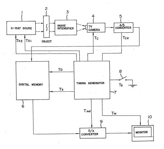
Fig. 1 is a block diagram showing a preferred embodiment of
the present invention;
Fig. 2 is a timing diagram describing the operation of the
2S apparatus shown in Fig. 1.
131~336
D iled Descri~tion of the Invention
Fig. 1 is a block diayra~ of the x-ray imaging equipment of
the present invention. The electrical components of each circuit
is constructed so that it may be independently reset. As shown
in Fig. 11 an x-ray source 1 is used to focus x-ray radiation
upon a target 2 in such a way that the x-ray images are presented
upon an imaye intensi~ier 3. The source is capable of
selectively transmitting on demand a first lower level of
radiation or a second higher level of radiation. In operation, a
TV camera 4 begins to operate automatically when the x-ray source
1 begins to irradiate the target. This, in turn, causes the
roentgenoscopic images formed on the image intensifier to ~e
projected onto the TV camera 4. The output signals from the
video camera~ which correspond to the transmitted x-ray images
formed on the image intensifier 3, are next converted from
analogue signals to digital signals by means of an a/d converter
5. The output signals from the a/d converter are applied to a
digital memory 6. In the digital memory 6, image signals for
each video frame are divided into image elements and each
corresponding imaye element is stored into a relevant memory ,
site. A one M DRAM (Dynamic Random Access Memory) may be used
for this purpose. A timing generator 7 sends a start signal Txl
to the x-ray source to initiate the imaging and storing sequence.
In addition, the timing generator 7 has added circuitry for
carrying out additional functions. The generator has the
capability of controlling the x-ray source to produce soft ~weak~
x-rays which are displayed as images on a monitor lt), as will be
-4-
" . , , .. ".. , - . ... ,-. .~ .
1314336
explained in greater detail below. The generator also controls
the synchronization of vertical and horizontal scanning signals
Tc of the video camera and converting the synchronized signals
Tcd to the a/d converter 5. The memory synchroni~ed signals Ta
and data address signals of the digital memory Tb also originate
in the timing generator.
The timing generator 7 also synchronizes the signals which
are readout from the memory 6 by sending a synchronizing signal
Tmd to the d/a converter 9 w~ich provides the input to a video
monitor lO. The timing generator sends synchronized vertical and
horizontal scanning signals Tm to the monitor lO so that the
information stored in the memory or that generated directly by
the TV camera can be displayed upon the monitor screen.
Transmitted roentgenoscopic images of the target displayed
on the image intensifier can be monitored continually using low
levels of radiation thereby exposing the patient and/or
technician to low dosages. When it becomes necessary to examine
selected images in greater detail, the exposure radiation level
is increased and the following procedures are carried out.
Initially, the ordinary operation of the camera is deactivated
for a short period o~ time by closing switch a and the photogene
image on the TV monitor is allowed to vanish completely. Closing
switch 8 also causes a signal Tx2 to be sent from the timing
generator 7 to the x-ray source thereby increasing the level
of output oi- the x-ray source. The target is radiated with
higher levels of radiation thereby causing a more defined
or sharper selective image to be produced on the image
~5-
1314336
intenslfier. The TV camera is then reactivated in
~ynchronization with the memory and monitor systems and the
selected image is passed through the a/cl converter to memory
where it is stored. Once the selected memory has been stored,
normal operations can be restoxed by opening switch 8.
The operational procedures of the above-noted apparatus will
now be described in greater detail with reference to Fig. 2.
When it is desirous to record a selected image, switch 8 is
closed at step 1 and the normal viewing load is now changed to a
photographin~ and storing mode. At step 2, the photogene
image on the TV camera is allowed to vanish completely by
blanking out at least three image frames. At step 3, the video
camera 4 iQ inactivated by means of a blanking circuit and the
x-ray source is readied to produce a higher level output. At
step 4, a signal Tx2 ~rom the timing generator is sent to the x-
ray source and the target is then exposed to higher levels of
radiation to produce a sharp image upon the image intensifier.
At step 5, the TV camera is once again turned on in
synchronization with the memory circuits and the monitor
circuits. Upon reactivation of the camera, a l-rame of the
selected image is forwarded to the memory where it is stored.
The image is simultaneously displayed on monitor lO. At this
time normal operations can be resumed by opening switch 8.
As can be seen, the above-~oted procedures makes it possible
to photograph selected images produced by x-ray radiation and
permits the selected images to be stored in memory where they can
be recalled at a later time. The transmitted images can also be
~ !314336
observed directly upon a monitor screen through means of the
video camera without passing directly through the digital memory
6. Accordingly, the x-ray signals can be monitored using low
: levels of radiation and only when a selected target is to be
memorized are higher dosages of radiation utilized. This in -turn
protects both the patient and the technicians using the equipment
while at the same time providing an important diagnostic tool to
the examining physician.
: While the present invention has been illustrated with
reference to preferred embodiments, it should be understood that
the invention is not limited to those precise embodiments, and
that many modifications and variations thereof could be carried
out by those skilled 1n the art without departure from the scope
and spirit of this invention, as defined in the appended claims.
:~:
:~