Note: Descriptions are shown in the official language in which they were submitted.
3630-TV 1 3 1 5 6 3 1
MINISCOPE
BACKGROUND OF THE INVENTION
The present invention is directed to catheters, and particularly
to steerable, miniscope catheters.
The use of miniscopes for performing diagnostic testing and for
assisting in the performance of certain types of surgery is
gaining acceptance in the f;eld of med;c;ne. M;n;scopes are
advantaseously utilized in procedures requiring passage through
very small ducts or passageways of the patient. Examples of
such procedures include the examination of the bile and
pancreatic ducts, with reference being made to "D;rect
Cholangioscopy and Pancreatoscopy at Time of Endoscopic
Retrograde Cholangiopancreatography", Richard A. Kozarek, M.D.,
The American Journal of Gastroenterology, Vol. 83, No. 1, 1988,
pages 55-57, and "Endoscopy of the Gallbladder as Control of
Gallstone Therapy with Methyl-tert-Butyl Ether", Leuschner,
~` Helstern, Birkenfeld, Leuschner, Gatzen, Kurtz and Flscher, The
American Journal of Gastroenterology, Vol. 83, No. 2, 1988,
pages 169-172.
While the use of miniscopes is increasing in diagnostic and
surgical procedures, such devices suffer intrinsic
disadvantages. Basically a miniscope is a multiple lumen
catheter e~uipped with one or more optical filaments, which
filaments may consist of a single fiber or a coherent bundle of
optical fibers. The filament is positioned within a selected
one of the catheter lumens. The optical filament used in
constructing the catheter miniscopes are selected from specific
types of op~ical filament. For example, the catheter miniscope
will include at least one optical filament, typically a coherent
bundle of glass fibers which possesses sufficient light
1 3 1 563 1
-- 2 --
transmissive properties, and while providing minimal distortion,
to function as ~he view scope. This optical filament has a lens
fitted at its distal end, that is the end which will lead the
catheter into the body. This lens will be sufficient enough to
magnify and focus the viewed object. A viewing eye piece will
be secured at the opposite, proximal end of this filament
optic. Additional optical filaments can be included for
providing illumination. These types of optical filaments need
not possess the same optical purity as the optical filament used
to function as the viewing filament.
Generally miniscopes must have a relatively narrow diameter in
order to allow access into the small conduits for which such
devices are intended. For example, min~scope catheters having
an outside diameter of seven french or less would be desirable
for atraumatic passage into the bile or pancreatic ducts. While
catheter miniscopes having this diameter or less have been
constructed, such catheter miniscopes do not possess the type of
maneuverability required to manipulate the catheter distal end
carrying the catheter optics. This ~s particularly critical
when the catheter needs to be moved into a particular duct,
which ~ntersects another duct. Specifically, the distal end of
the catheter must be deflectable to provide the doctor with
sufficient enough control to insert the catheter ~nto the proper
duct.
Catheters possess~ng the desired maneuverability do exist. Such
catheters rely upon different techniques for providing the
desired maneuverabllity. Basically, the maneuverability of the
catheter ~s provided by bending the distal tip of the catheter.
This allows the catheter to be maneuvered through tortuous
passages of the pat;ent's body during a procedure. One type of
mechanlsm for bending the catheter tip involves pre forming the
distal tip to the desired shape of the passage through which the
catheter will be positioned. While this is somewhat effective
for certain procedures, such types of catheters can not be
. . .
-- 1 31 5631
-- 3 --
satisfactorily controlled due to the lack of torque transmission
over the length of the catheter body. Another technique
involves inserting a stylet into a catheter lumen, with the
stylet being preformed. This type of procedure suffers the same
disadvantage as preforming the catheter distal end.
An early device for truly controlling the maneuverability of
catheter distal ends is taught in U.S. Patent Number 3,521,620,
~ssued to Cook on July 28, 1970. The taught device ls basically
a coil spring which is fitted about a wire. This wire is
eccentrically secured to the distal end of the coil spring. The
wire turnings at the distal end of the coil spring are spatially
separated, while the remainder of ~he coil spring ~s tightly
wound. ~hen the wire is pulled ~he coil distal end windings
become compressed on one side. The eccentric attachment of the
wire to the coil dlstal end causes a bending moment ~hich
d;fferentially compresses the coll windings. The result is an
off axis deflection of the co~l. This device can be inserted
into a lumen of a catheter. The off axis deflection of the coil
is transmitted to the distal end of the catheter, resulting ~n a
deflection of the catheter d~stal tip.
A similar approach for controlling the deflection of a cathe~er
distal end is taught in European Patent Applications 176,865,
published on September 4, 1986, and 254,885, published on March
2, 1988. The catheters taught ln these two published
applications cause the distal tip deflection by applying tension
to a wire which has been secured eccentrically at the distal end
of the catheter.
Other approaches for controlling the deflection of a catheter
distal end involve inflating an eccentric catheter balloon,
which has been constructed ln the distal end. Some catheters
may utilize short bursts of gas from out of a side vent to
deflect the catheter distal end.
1 31 5631
3630-TV
-- 4 --
Some techniques involve the construction of the catheter wall at
the distal end with portions of differing rigidity, e.y. by
varying the thickness of the wall about the catheter
circumference. This may also be accomplished by affixing a
rigid member along a portion of the catheter wall. When the
distal end is subjected to an axial compressive force the rigid
portion will not as easily constric~. This results in the
catheter bending towards the less rigld, or thinner portion of
the side wall. An example of this type of catheter is disclosed
in the Patent Cooperation Treaty Patent Application Mumber ~0
87101600, which was published on March 26, 1987.
Whlle all of the approaches taught by these references provide
for adequate control of the distal end deflec~lon, such
approaches are unavailable for the catheter min~scope. This is
because such catheter miniscopes must possess a relatively small
outside diameter, e.g. 2.8 millimeters or less. The methods
employed in the above referenced disclosures require more space
than is available with catheters of this size, particularly,
when such catheters must also include at least one working
lumen. The working lumen would have to be provided with a
sufficient size to accommodate the passage of guide wires,
electrical wiring or fluid. The space availability is also
compromised by the fact that such catheter miniscopes will be
provided with the necessary optical filaments.
There thus exists the need to construct a catheter miniscope
having the necessary optical filaments, while also being
designed to provide the necessary maneuverability to deflect the
distal end.
SUMMARY OF THE INVENTION
The present invention overcomes the above disadvantages by
providing a miniscope catheter which is constructed to allow
- 1 31 5631
-- 5
the operator to control the deflection of the catheter tip.
The catheter miniscope of the invention includes a catheter
body having at least one lumen. At least one optical
filament, which may either be an individual fiber of a
bundle of optical fibers, is positioned through this lumen
and eccentrically secured to the catheter at the catheter
distal end. In order to provide for the deflection of the
catheter tip portion the catheter miniscope further
includes a means which can be operated to apply a force
against the optical filament to longitudinally advance or
retract the optical filament alony the long catheter axis.
This longitudinally movement of the optical filament causes
a bending m~ment in the catheter tip, attributable to the
eccentric securing of the optical filament, which deflects
the tip portion. This longitudinally advancement or
retraction of the optical filament provides the ability of
deflecting the catheter distal end in at least two opposing
directions.
Specifically, the present invention in one aspect is
directed to a catheter having a catheter body with at least
three lumens. A first optical filament formed from a
bundle of fused glass fibers is positioned in one of the
lumens and eccentrically secured to the catheter at a
location proximate the catheter distal end. The mechanism
for driving the optical filament is positioned in a handle
into which the proximal end of the catheter is fitted.
This mechanism can be operated to apply the force against
the optical filament to either advance or retract this
filament longitudinally through the catheter.
Other aspec~s of this invention are as follows:
A miniscope catheter comprising: a cylindrical body
formed with a distal and proximal end and at least a first
lumen traversing through said body; at least a first
optical filament positioned in one of said lumens and
secured to said body at a location proximate said distal
'~
1 3 1 563 ~
- 5a -
end; viewing means associated with that end of said first
optical filament opposite said end secured to said body
which is formed for viewing images through said filament;
and means associated with said body which can be operated
to apply a force against said optical filament to
longitudinally drive said optical filament in at least a
first direction within said lumen.
A catheter miniscope comprising: catheter body having a
distal and proximal end and an outer diameter of no greater
than 2.8 millimeters, said body being formed with at least
three eccentrically positioned lumens; a first optical
filament formed from a fused bundle of individual glass
filaments positioned in one of said lumens and secured in
said lumen at a location proximate said cath~ter body
distal end; a second optical filament positioned through
another of said lumens; a handle means which is formed with
a cavity for receiving a portion of said catheter body
proximal end: said first and second optical filaments being
formed with exposed ends positioned in said handle cavity,
said second optical filament exposed end being positioned
to receive light; an eyepiece means coupled to said exposed
end of said first optical filament, said eyepiece means
being formed with lens to allow visual observation through
said first optical filament, said eyepiece means being
secured to said handle means; and a means located in said
housing which is formed to engage said optical filament
surface and apply a force for driving said filament in two
opposing longitudinal directions~
DESCRIPTION OF THE DRAWINGS
The present invention may be better understood and the
advantages will become apparent to khose skilled in the art
by reference to the accompanying drawings, wherein like
reference numerals refer to like elements in the several
figures, and wherein:
.~
.
1 3 1 563 1
3630-TV
-- 6 --
Figure l is a side, partially sectioned view of a miniscope
catheter in accordance with an embod;ment of the invention;
Figure 2 is an end on view of the distal end of the catheter
portion of the catheter miniscope of Figure l;
Figure 3 is an enlarged, cross-sectional view of the circled portion
of Figure 1 along lines 3-3; and
Figure 4 is an enlarged, cross-sectional view of the circled portion
of Figure l along lines 4-4.
DESCRIPTION OF THE PREFERRED EMBODIMENTS
The The present invention is directed to a mlniscope catheter,
typically having a catheter body of less than 2.8 millimeters in
outer diameter. Miniscopes having such dimensions would be useful
in performing diagnosistic procedures by the insertion of the
catheter miniscope into the smaller body ducts not presently
access~ble with ava~lable scopes. Furthermore, the miniscopes of
the invent~on may be inserted through working lumens of larger
endoscopes. The miniscopes of the ~nvention may also be formed with
at least one working lumen, 1.e. a non-obstructed lumen. Fluids may
be directed to desired body parts through th1s working lumen.
In one particular example, the min~scope of the ~nvention can be
used to v~ew the 1nterior of the gall bladder. This is possible
because of the small outer diameter of the catheter body which can
fit into the bile duct without the necessity of performing a
sphincteromtomy. The viewing capabilit;es of the miniscope allow a
surgeon to examine the inside of the bile ducts and the gall
bladder. Furthermore, if so desired a liquid, e.g. a contrast
med~um, can be directed through the catheter working lumen 1nto the
b~le duct or gall bladder. The catheter miniscopes of the invention
are useful for many other types of diagnostic or surglcal procedures.
,
3630-TV 1 31 5 6 3 ~
In order to enhance the useability of the miniscope catheter of the
invent;on, the distal tip portion of the catheter must be
steerable. This is provided by constructing the catheter miniscope
with the function of deflecting the distal tip portion in at least a
first direction away from the longitudinal ax~s of the
catheter. In this manner the surgeon operating the catheter
miniscope merely selectively deflects the distal tip to steer
the catheter miniscope in at least one directlon. Preferably
the catheter miniscope is constructed to provide for deflection
of the catheter distal tip in two or more oppos~ng directions.
0 This further enhances the ability to steer the catheter
miniscope of the invention.
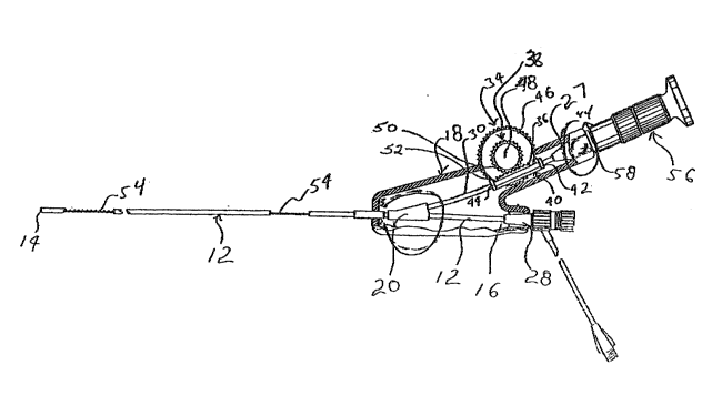
Referring now to Figure 1, a catheter miniscope in accordance
with one embodiment of the invention is seen generally at 10.
The miniscope 10 includes a catheter body 12, which has a
distal end 14 and a proximal end 16, and handle 1~. The
proximal end 16 of the catheter body 12 is partially positioned
within the handle 18. A Y-intersection connector 20 is fitted
to the catheter body 12, which connector 20 is fitted within
the hand1e 18.
The connector 20 is formed with a Y-shaped internal passageway,
illustrated in Figure 3 at 21, for rece~ving the catheter body
12. As will be described more fully herein, this passageway 21
jS formed to allow for the passage of the viewing and
~llumination opt~cal filaments through one arm 23 of the
passageway 21, while the remainder of the catheter passes down
the other arm 25 of the passageway 21.
The catheter 12 is formed by conventional procedures, e.g. by
extruding a suitable polymer, e.g. polyvinylchlor~de, through
an appropriately configured die to form one or more lumers. As
stated, the present invention will be described in relat~on to
3630-TV 1 3 1 5 6 3 1
-- 8 --
a catheter body having three lumens, with the die used to form
the catheter body 12 having the appropriate configuration to
form such a catheter body 12. The precise method of form;ng
such a catheter body 12 is not critical for the instant
invention, and will thus not be described in any deta;l
herein. In this regard, any conventional technique for forming
catheters can be used to manufacture the catheter body 12.
The handle 18 is a generally hollow structure formed to receive
the catheter body 12, connector 20 and the mechanism for
applying force against an optical filament and an optical
eyepiece, both of which will be described more fully herein.
The precise construction and shape o~ the handle 18 is not
critical to the invention, however, in accordance with the
preferred illustrated embodiment of the miniscope 10 the handle
18 is formed to be easily received within the hand of the
operator of the miniscope 10, and to allow for easy access to
the various operating components, e.g. the drive mechanism and
eyepiece, of the min~scope 10.
The miniscope 10 further includes various optical filaments,
which for the purpose of the present invention shall include
individual fibers or a bundle of fibers. These filaments, seen
generally at 30 and 32, are selectively posit~oned in
individual ones of the lumens of the catheter body 12, as
illustrated lumens 22 and 24. The optical filament 30 w~ll
function as the vlewing filament, while the filament 32 will
function as the lllumination filament.
Filaments which can be used in the practice of the invention
are those wh~ch have the characteristic of total internal
reflection and low optical attenuation. In this regard, such
filaments are typically formed wlth an lnternal core surrounded
by a cladding, with the cladding formed from a material having
a lo~er index of refractlon. ~hen such filaments are a bundle
1 31 5~31
3630-TV
_ g _
of individual fibers, such fibers are also formed with an
internal core surrounded by a cladding of a material hav~ng a
lower index of refraction. In accordance with the preferred
embodiment, the individual fibers are fused together by the
fusing of the material forming the outer cladding.
The precise materials from which the optical filaments are
formed, and the geometric shape of such filaments are not
critical to the invention so long as such materials and
geometry provide the fllaments with the desired optical
characteristics. These optical characteristics will be
dependent upon whether the filament is to function as the
viewing or illumination filament 30 or 32.
In accordance w~th a preferred embodiment, the viewing filament
w~ll have to be ~n optical filament having good light
transmissive characteristics, as depicted by an attenuation
diagram, in addition to the ~mage preservation
characteristics. The illumination filament should possess a
numerical aperture, or cone of acceptance selected to provide
illumination of the entire viewing field, which in the
preferred embodiment will have an angle of view of about 60.
This should correspond to a numerical aperture of at least
0.50. The illumination filament should also display an
attenuation below 1 dB/m in the visible range (400 to 700 nm).
In accordance with a more preferred embodiment of the
invent~on, the optical filament selected for the viewing
filament 30 is formed from glass, and even more preferably is a
filament formed from a bundle of coherent glass filaments,
which are preferably fused together. The illumination filament
32 may also be a glass fiber or bundle of glass fibers.
However, it is typically more desireable to use a polymeric
optical filament for the ~llumination filament 32, generally a
-~ 1 31 5631
3630-TV
- 10 -
filament formed with an inner core surrounded by another
polymer having a refractive index less ~han the refractive
index of the material forming the core. Typ~cal polymeric
optical filaments are formed from a polyacrylic mater~al core
coated with a fluorinated polymer cladding. The elastic moduli
of such filaments are considerably smaller than analogous glass
filaments. Accordingly, catheters employing plastic
illumination filaments will be substantially less stiff than
those employing glass illumination filaments.
As stated, the connector 20 is fixed to the catheter body 12,
with the optical filaments being passed down the arm 23 of the
passageway 21, with the remainder of the catheter body 12
passing down the arm 25. That is, the optical filaments 30 and
32 are removed from the lumens 22 and 24 by cutting open the
respective lumens 22 and 24 of the catheter body 12. The
filaments 30 and 32 are then drawn through the arm 23, while
the catheter body 12 is drawn through the arm 25. The catheter
body 12 is fixed in the connector 20 by suitable means, e.g. an
adhesive.
The exposed ends of the optical filaments 30 and 32 are fixed
in an eyepiece coupler 27. This eyepiece coupler 27 includes
two passages, not shown, for individually recelving the viewing
filament 30 and the illumination filament 32. The passage for
the viewing filament 30 will generally extend out the end of
the coupler 27, wh~le the passage for the illumination filament
32 will extend laterally through the wall of the coupler 27.
The illuminatlon fllament 32 will be exposed to the exterior of
the coupler 27 through this passage. In this regard, reference
is made to Figure 4 ~hich illustrates a portion of the handle
18 in which the eyepiece coupler 27 is mounted. As seen, a
light socket 58 extends laterally out from a hole, not shown,
formed in the side of the handle 18. This light socket 58 is
integrally formed out from the side of the coupler 27, and
3630-TV - 11 -
includes the passage through which ~he illumlnation filament 32
is passed. The exposed end of the illuminat~on filament 32 is
illuminated by any suitable source, seen generally at 60~ with
the preferred embodiment being a light source 60 which ~ncludes
a mechanism for coupling to the li~ht socket 58.
S The viewing filament 30 will extend out of the coupler 27 and
be fitted to an eyepiece 56. The eyepiece 56 may be any
suitable eyepiece, e.g. that eyepiece used in an ureterscope
manufactured by the Baxter Healthcare Corporation, a Delaware
Corporation residing in Deerfield, Illinois, which eyepiece
used in such ureterscope has a Model number GU-77. The manner
in which the viewlng filament 30 is connected in the eyepiece
56 is not critical to the invention, and will not be discussed
any further herein.
While either the viewing or illumination filaments may be used
to exert the deflection force on the distal tip of the
catheter, in accordance with the preferred embodiment, the
view;ng fllament 30 will be used. In this regard, the viewing
filament 30 is flxed to the catheter body 12 at a location
contiguous to the catheter d~stal end 14. The securing of the
filament 30 at the d~stal end 14 is generally performed by an
adhesive. As stated, the opposite end of the filament 30 is
flxed 1n the eyepiece 56. A portion of the filament 30
positioned inside the handle housing 18 is connected to a
mechanlsm, seen generally at 34, which can be operated to
either pull the filament away from or push it towards the
distal end 14. When the filament 30 is pushed towards the
distal end 14, a load is applied to the filament as a
compression force, while the load applied to the filament 30
when it is pulled away from the distal end 14 is a tensile
force. This load ~s transferred to the distal end 14, and in
particular to the point of the catheter body 12 to which the
filament 30 is secured, which causes the deflection of this end
14.
3630-TV 1 3 1 5 6 3 1
- 12 -
In order to prevent dislodging of the viewing filament from the
eye piece 56 a suff~cient amount of slack of the v;ewing
filament 30 is positioned in the interior of the handle 18.
That is, a sufficient length of the v;ewing filament is
provided in the handle housing 1~ to prevent the dislodg~ng of
the illuminat~on filament from the eye piece 56 upon applying a
load to the v;ewing filament by the mechanism 34.
The direction of the deflection will depend upon whether the
filament 30 is be~ng subjected to a compressive or tensile
load. Furthermore, the direction of the deflectlon will be
dependent upon the construction of the catheter, and whether
such catheter body 12 is reinforced in any manner. As seen in
Figure 2, that lumen 22 ~n which the vlewing filament 30 is
positioned is located off center, or to the side of the
catheter body 12 axis. When the viewing filament 30 ;s
subjected to a tensile force by pull;ng upon the f;lament a
differential compression of the catheter will occur. Thus the
deflection upon pulling the v;ew;ng filament 30 w;ll occur in
the direction indicated by the arrow A. Applying a compressive
force upon the f;lament by pushing upon the same will cause the
catheter body 12 to be deflected ;n the oppos;te d;rect;on.
The pre~erred mechan;sm 34, which is illustrated in Figure 1,
for applying the tens~le and compress;ve force upon the v~ewing
filament 30 is a rack and pinion arrangement. A rack 36 is
mounted to the f~lament 30, and a pin;on 38 is mounted for
rotat;on within the handle lB. More speci~;cally, the rack 36
is secured to the filament 30 near its proximal end, that is
the end connected to the eyep;ece 56. The rack 36 can be
affixed to the filament 30 by any suitable method, e.g.
adhesion. The rack 36 and filament 30 are mounted for a
3630-TV 1 3 1 5 6 3 1
- 13 -
sliding relationship within a frame 40. This frame 40 ;ncludes
two arms 42 and 44 which are formed with cut-outs ~hrough which
the combination of the rack 36 and filament 30 are sl~dably
received.
The pinion 38 is connected to a thumb wheel 46 by any suitable
mechanism for transferring the rotational force of the thumb
wheel 46 to the p;nion 38. In actordance with the illustrated
embodiment the pinion 38 and thumb wheel 46 are mounted
together about a shaft 48. Thus the rotation of the thumb
wheel 46 simultaneously rotates the pinion 38. However, the
pin~on 38 and thumb wheel 46 can be mounted to rotate upon two
different shafts, not shown, which are mounted in the handle
housing 18. The shaft about which the thumb wheel 46 is
mounted can be coupled to the shaft about which the pinion 38
is mounted by suitable means, e.g. pulleys or gears. This
arrangement would effect the transfer of the rotation of the
thumb wheel shaft to the pinion shaft by the pulleys or gears.
The pinion 38 ~s formed with a series of teeth about its
periphery, as seen generally at 50. These teeth 50 mate with a
series of teeth formed on the opposing surface of the rack 36,
with such teeth seen generally at 52. By rotating the pinion
38, through the operation of the ~heel 46, the teeth 50 engage
and travel along the ser1es of the teeth 52. The operation of
the rack and pinion mechanism 34 applies a load to the filament
30~ Depending upon the d~rection at which this load is applied
the filament 30 ~s subjected to a tensile or compressive load.
Since the filament 30 will be subjected to a load by the
operation of the rack and pinion mechanism 34, it is necessary
for such a filament to possess sufficient strength to resist
breakage. It has been found that the minimum ultimate strength
of a useful filament, that is a filament possessing the
3630-TV 1 3 1 5 6 3 ~
- 14 -
necessary optical characteristlcs, ~s at leas~ about seven
thousand pounds per square inch (psi). By "ultimate strength"
it is meant the stress at which the ultimate failure (breakage)
of the filament occurs. Furthermore, while mechanism 34 may
apply the force to a plastic filament, it has been found that
the most preferred ~ilament is a glass fllament. The glass
filament will not plastically distort as easily as a polymeric
filament, and thus not suffer as much distortion in light
transmission characteristics. Generally, in glass filament the
difference between the elasticity of the mater~als forming the
fllament core and the cladding is closer than with plastic
0 filaments. Thus the application of a load to the glass
f;lament will not as likely cause a separation between the
cladding and the core as will occur with the plastic filaments.
An even more preferred embodiment is the use of a bundle of
fused glass fibers as that filament to which the load is
applied by the mechanism 34. An unfused bundle of glass fibers
was found to have an uneven distribution of stress when load
was applied in comparison to a bundle of fused fibers. As a
result, a few of the indivldual fibers of the bundle carried an
inordinate proportion of the load, and would easily break.
- This would result in a distortion of the light transmission
characteristics of the viewing bundle fiber 30.
In order to promote the direction of deflection of the distal
end 14 the catheter miniscope 10 can include a stiffening
element. This stiffening element, seen generally in phantom at
54, will provide for different regions of stiffness about the
viewing filament 30. The stiffer regions will only bend, or
deflect at a higher load. Thus by the appropriate positioning
of the different regions of stiffness, the positioning of the
deflection is controlled to the desired location along the
catheter body 12. Since it is usually desireable to provide
for the most deflection at the distal end 14, it is the region
at this location which has the least degree of stiffness.
Conversely, the remain;ng regions are more stiff.
1 31 56~1
3630-TV
- 15 -
While any suitable structure can function as the stiffening
element 54, preferably the st;ffening element 54 1s a wire
coil, or metal spring, through which is pos1tioned the viewing
filament 30. The different regions of stiffness are provided
by manipulating the spacing between adjacent windings of the
coil. The normal spring windings are closely ~ound. At those
locations at which the overall stiffness is to be lessened,
these windings are moved apart. Thus the spring at these
locat1Ons will more easily bend in response to a tensile load
in the filament, ~hat 1s become compressed, and thus allow for
a greater degree of deflection.
In this regard, reference is made to the teachings of U.S.
Patent Number 3,521,620, 1ssued to Cook on July 28, 1970,
perta;ning to physical arrangement of the coil spring. In
part1cular, reference is made to the spatial separation of the
distal end coil windings, and the effect of this separation
when the spring is subjected to a compressive force.
Accordingly, these teachings are ~ncorporated herein by
reference.
- While the man1pulation of the coil spr1ng 54 is usually
provided at the distal end 14, it should be noted that such
manipulation may also be provided at a mid-point along the
spring 54, or even at different locations along the spring 54.
Furthermore, while the invention 1s being descr~bed and
illustrated as using a spring 54 as the stiffening element,
other embodiments are also envisioned. For example, the
catheter body 12 can be formed w1th a wire mesh, or similar
material, wrapped about the body up to a predetermined location
3~ from the distal end 14. This will provlde the catheter body 12
with one region of stiffness along which the wire mesh is
3630-TV 1 31 5 6 31
- 16 -
situated, with a second, lesser region of stiffness at the
distal end. The method of winding a wire mesh about a catheter
body is known and not critical to the invention.
It may also be suitable to form the catheter body 12 from two
types of polymeric material. That is, a body section can be
formed from a relatively rigid polymeric material, whlle a tip
portion can be formed from a soft pslymeric material. This
will provide for the two regions of differing stiffness. The
methods of fabricating such types of catheters are well known
in the art and not critical to the invention.
While the described and illustrated embodiment uses only a
single optical filament for causing the deflect;ng of the
catheter distal end 14, it should be noted that more than one
opt~cal filament can be used for the same purpose. In this
regard, a second mechanism, e.g. rack and pinion mechanism 34
~s included in the handle 18, w~th the second rack affixed to
the other opt~cal filament, ~.e. illumination filament 32. If
such an embodiment is desired, then the optical filament used
for illuminat~on filament 32 should be able to bear a tenslle
load of at least two pounds. Typically slzed filaments which
are useful for the practice of the ~nvention, that are those
filaments which are small enough to fit within the
described catheter body 12, should have a minimum strength of
2~ at least seven thousand psi. Greater safety can be achieved by
using an even larger filament, however the overall size
constraints of the catheter body 12 must be followed in order
to provide a min~scope with the des~red outer diameter.
Furthermore, a bundle of fused glass ~ilaments may be used as
the illumination filament 32, w~th this filament being used
alone or in combination with the viewing filament 30 to caùse
deflection of the catheter distal end 14.
' . ' '
1 31 5631
3630-TV
- 17
~h~le the preferred embodiments have been descr~bed, various
modifications and substitutions may be made thereto without
departing from the scope of the invention. Accordingly, ~t is
to be understood that the invention has been descr~bed by way
of illustration and not limitation.
. - . . ;. ,,
., ~ '