Note: Descriptions are shown in the official language in which they were submitted.
1 31 563~
` 1
~CH~NICALLY ~OC~ING BLOO~ C~OT ~ILT~R
BAC~GROUND OF T~ INY~NTION
Field of the Inventio~
The pre~ent invention relates generally to devices or
methods for filtering blood clot~ from blood ve~sels, and
more particularly to vena cava blood clot filter~ and
methods and apparatu~ for in~erting such vena cava blood
clot filters transvenously.
Back~round of the Invention
~t i8 e~timated that each year, approximately 750,000
patient~ in the United States suffer pulmonary emboli~m or
passage of blood clots to the lungs. O$ the~e,
approximately 150,000 patients die each year from such
pulmonary embolism. Most commonly, these clots originate
in the veins of the pelvis or lower limbs. While most
patients can be treated with blood thinning medications,
these medications can jeopardize the wellbein~ o~ some
patients becau~e of other, co-existing medical problems.
Other patients exhibit recurrent embolism even while being
treated with these medications. In these situations, a
mechanical barrier is neces~ary to prevent such blood clots
from travellin~ through the inferior vena sava to the heart
and lun~s.
Initially, surgical procedures were devised to form ~uch
~5 a ~echanical barrier. These procedures consis~ed of either
tying a ligature around the inferior vena cava or placing a
special clip around it. The sur~ery necessary to perform
this procedure is exten~îve and requires a general
ane~thetic. Moreover, such ~urgical procedure~
si~nificantly ~urther jeopardize the health of an already
ill patient.
Over the last ~i~teen years, several devices have been
used to place a filterin~ device into the inferior vena
cava usin~ a tran~venous route, commonly originatin~ from
2 131 5h3~
the ri~ht jugul~r vein or from either femoral vei~. For
example, the method disclo~ed in U.S. Pat~nt No. 3,834,394
to ~unter, et ~1., use~ a detachable bnlloon which i~
deliver2d to the inferior ven~ cava at ~he end o a
~atheter. The balloon and cathete~ are inserted into one
of the veins in the neck u~ing a ~urgical incision a~d
passed to the lower inferior vena cava ~here the balloon is
inflated. Once det~ched, the ~alloon occludes the inferior
vena cava entirely, thereby preventin~ any flow o~ blood or
blood clots to ~he heart. While insertion of ~his device
avoid~ major abdominal surgery, it Ctill requires a ~mall
surgical procedure to be performed in order to 0xpo~e a
neck vein. The balloon occlude~ the inferior vena cava
compleSely, resulting in s~elling of the lower extremitie3
until collateral circulation develops around the balloon.
With time, these collateral Ghannel~ may beco~e large
enough to permit life threatening emboli to pass to the
lung.
Another device for preventing pulmonary embolism but
which does not require total occlusion of the inferior vena
cava i5 an implantable cone-shaped filter device consisting
of six spoke~ with sharpened points at the end and
connected together at the other end by a central hub. A
thin membrane ~ith 4 ~m. holes covers the device. The
umbrella-like device is folded into a cylindrical capsule
connected to the end of a catheter. This device is
described in U.S. Patent No. 3,540,431, to Mobin-Uddin.
Thi3 device also requires a ~uryical cutdown on a major
rig~t neck vein for acce~s to the venous system. The
device and delivery cap3ule are po~itioned in the inferior
vena cava and released by pu~hing the device out of the
capsule. While the device acts as an efficient filter,
approximately 60% of the patient~ U5i~ the Mobin-Uddin
filter develop occlusion o~ the i~ferior vena cava,
sometimes resulti~g in severe s~ellin~ of the legs.
Furthermore, in~tances of mi~ration of the filter to the
3 1 3 1 56~
heart have been reporte~; ~uch ln~tance~ pre~ent a hi~h
mortal~ty ri~k.
The Hunter balloon and the Mobin-Uddin umbrella su~er
from ~imilsr disa~vanta~e~ in that they require a ~ur~ic~l
procedure on the neck for expo~ure of a vein into which the
filter may be pa~ed. Furthermore, morbidity from
occlusion of the inferior vena cava could be ~evere. A
device which coul~ be ea~ily in~erted from the ~emoral
approach u~ing ~tandard angiographic technique~, and
thereby avoid 3urgery, ~ould be de~irable. Ideally, the
device ~hould not totally occlude the inPerior vena cava or
be thrombogenic. It ~hould alao be ~ecurely ~nchore~
within the inferior vena cava to prevent migration.
U.S. Patent No. 3,952,747, to ~immel, di~closes a blood
ve~sel filter and filter insertion in~trument which
overcome some of the di~advantage~ of the previous two
devices. The Rimmel patent de~cribe~ device which may be
inserted either from the jugular or femoral approach u~ing
a ~urgical exposure of a major vein. The conical ~haped
ao device consists of 8iX strands of wire each connected to a
hub at one end and having recurved hooks on the other end.
~he aevice i8 loaded into a cylindrical delivery capsule
which is connected to a catheter. The ~elivery cap~ule
measures 6 ~m. in diameter and 5 cm. in length. Becau~e of
its 3ize, a surgical exposure of the vein is necessary ~or
introduction of the delivery capsule into the vascular
system. Nore recently, the delivery cap~ule ha~ been
introduced into the vascular sy~tem through a large
catheter usin~ angio~raphic technique~. However, this
technique has been ~hown to signi~icantly injure the vein
at the introduction site. Sometimes it may not be po~ible
to pas~ the cap~ule from below through tortuou3 pelvic
vein~ into the inferior vena cava because of the
inflexibility of the ~apsule. The filter engages the ~all
of the vein at one end and there~ore often tilt~ to one
side. It is very di~ficult to deliver the filter in a
13 1 5G3~
manner that maintain~ the longitudinal axis of the fllt~r
centere~ alon~ the lon~itudin~l ~xi~ o the vena cava. A
tilte~ filter ha~ been ~ho~n to be le3~ efficient at
capturing blood clots. ~igration of the filter ha~ not
been A problem.
Another method of preventin~ pulmonary emboli from
reachlng the lun~ i8 a device di~clo~ed in U.5. Patent No.
4,425,908, to Simon. Thi~ device uses the thermal ~hape
memory proper~ie~ of Ni~inol to deploy the filter following
delivery. The filter consi3~ of ~even wire3 banded at one
end and al80 in the middle. The wire~ bet~een the~e two
points form a predetermined filter me~h derived from the
thermal memory. The free-ends of the ~ires ~orm anchorin~
points which radially engage the inferior vena cava. The
device may be inserted through a jugular or femoral vein
approach usiny standard angio~raphic catheters. The device
reli@s on the thermal shape memory properti0~ of the
Nitinol ~ire to form an effective filter following
delivery. It i nok yet clear whether the filter di closed
in the Simon patent will be biocompatible in humans or if
it will be thrombogenic. Concern exi~t regarding its
reliability when ~tored at different temperature~ and also
whether the ~aterial can be manufactured with the ~ame
consi~tency.
U.S. Patent No. 4,494,531, to Gianturco, also disclo3e~
a blood ~es~el ~ilter which can be inserted through
angiographic catheter~. The device con~ist~ of a number of
strands of wire which ~re interconnected and wadded
together to form a curly wire mesh. The filter includes a
number of projection~ which serve as an anchorinq means for
anchorin~ the filter at a ~uitable body location within the
inferior vena cava. Problem~ with the device include
migration and demonstration invitro of filteriny
inefficiency. The random nature of the filtering mesh
make~ it difficult to as~es~ the overall efficacy.
Perforation of the anchoring limb~ throu~h the vena cava
- 1 31 5634
ha~ al~o been de~crib~d.
A devi~e de~cribed by Gunth~r e~ al. in a 1985 tech~ical
article con~i~t~ of a helic~l ba~ket made oP a number of
~ire3 and radially placed legs. Originally, it ~a~
intended to be implanted te~porarily in the in~erior vena
cava until the ~atient'~ risk o~ ~ulmonary embolism had
passed. Limited clinical experience i~ aqailable.
The blood clot filter device and related delivery
apparatu~ disclosed in the pre~ent invention, overcome the
disadvantagea associated with the prior art by employin~ a
nonocclusive filter ~hich i~ de3igned to be inserted into
the vena cava using normal percutaneous catheterization
techniques through a femoral or jugular approach. Thus the
need for surgery i~ totally eliminated. The device is
~elf-centering and has a positive mechanical locking
system. This sy~tem does not require the patient to b2 at
a ~iven temperature in order for the filter to ~orm it~
~hape. Moreover, it i~ made of metal~ which have been
shown to be biocompatible ~hen used in other devices such
as pacemakers and inferior vena cava filters. This is not
true of the filter di~clo~ed by Simon. The filter
configuration i8 predetermined and not random as described
by Gianturco.
Accordingly, it i5 an object of the present invention to
provide a blood clot filter ~hich may be implanted using
normal percutaneous an~io~raphic catheter techniques
through either a femoral or ju~ular approach.
It i~ a further object of the pre~ent invention to
provide a blood clot filter ~hich i5 designed to be placed
~ithin the inferior ~ena cava below the renal veins.
It is yet a further object of the pre~ent invention to
provide a blood clot filter ~hich does not obstruct blood
flow ~ithin th~ blood vessel at any time.
It is ~till a further object of the present invention to
provide a blood clot filtsr ~hich ~ill not cause thrombus
formation or emboli after implantation.
6 l 31 563~
An additioDal obj~ct of the pre~ent invention iu to
provide a blood clot filter ~hlch i~ capable of being
aecurely anchored within the blood ve~sel.
It i~ a further object of the pre~ent invention to
provide a blood clot filter which forms it~ ~hape u~ing
mechanically induced conver~ion of ~traight wireR into a
filter mesh ~hich may accommodate vena cava~ of varying
sizes.
It i9 another object of the pre3ent invention to provide
such a blood clot filter which uses well-known
biocompatible materials and which avoids reliance upon
thermal memory shape characteri~tics, thereby providing a
reliable and less expensive filter.
Sum~arv of the Invention
--
~ ~ Ihè present~invention provides a blood clot
filter which includes a central core wire extending ~long a
central longitudinal axis and surrounded b~ a number of
peripheral wires evenly spaced about the central core wire.
A first connector connects the peripheral wires together at
one end of the central core wire at a fir~t fixed
connection point. A second connector connects the
peripheral wires together at a second connection point
spaced apart from a the first connection point, the second
~5 connection point surrounding the central core wire and
being slidably secured thereto. The blood clot filter
includes a one-way lock device permitting the second
connector to slide along the central core wire toward the
first fixed connector from a first ~osition remote from the
first connector to a second position proximate the first
connector. However, the lock device prevents the second
connector from returning from the second proximate position
back to the first remote position. The p~rtions of the
peripheral wires extending between the first and second
connectors initially extend generally along the central
L' r~
- 7 l 31 563~
core ~ire. A~ the ~econd connector i8 advance~ from the
fir~t re~ot~ po~ition to the aecond proxl~ate positio~, the
por~lo~ of khe peripheral ~ire~ extending between the
firat and oecond connector~ ~ov~ radially away from the
central core wire to a deployed po~ition ~or forming a
~ilter mesh.
In a prePerred embodiment of the pre~ent invention, the
peripheral wire~ include leg portion~ ~hich exten~ beyond
either the ~ir~t or ~econd ~onnector~ The leg portions are
biased a~ay from the cen~ral lo~gitudinal axi~ of the blood
clot filter and terminate in hooked feet adapted to enya~e
the walls of a blood vessel for anchorin~ the blood clot
filter at a desired location therein. The leg portions
provide a second ~ilterin~ component in addition to the
flattened fil~er mesh, and the leg portion~, in conjunction
~ith the ~lattened filter me~h, automatically center the
blood clot filter ~ithin a blood vessel and prevent the
Bame from tilting away ~rom the central longitudinal axi~.
The aforementioned ~econd connector may be in the form
of a tubular sleeve which slide~ over the central core
wire. ~ach of the peripheral ~ires may be attached, a~ by
weldin~, to the exterior ~all of the tubular ~leeve. In
another embodiment of the present invention, the ~econd
connector i8 in the form of a collar through which each of
the peripheral wires pa~es, the ~ollar 3erving to collect
and connect the peripheral wires to permit the ~ame to
~lide along the central core wire.
A first embodiment of the prs~ent inYention i9 primarily
desi~ned for delivery u~ing a tran~femoral approach. In
this embodiment, the a$orementioned fir~t connector join~
first ends of the peripheral wire~ together a~d fixedly
~ecure~ the ~ame to a first end of ~he core wire. The
~econd connector join3 central portions of the peripheral
wire~ to one another for ~liding alo~g the central core
wire. The second ends o~ the peripheral wire~ extend from
the ~econd connector to provide the aforementioned le~
~ - 1 31 5634
portion~. The central core ~ire extend~ beyon~ the aecond
connector and beyond the ~acon~ ~nds of the ~2ri~heral
wires for being retracted to ~e~loy the flattened filter
me~h.
A ~econd embodiment of the present invention i~ adapted
for deli~ery using a tran~jugular approach. In this
embodiment, the fir~t connector join~ the central portion~
of ths peripheral ~ire~ together and fixedly ~ecure3 the
~ame to the first end o the central core ~ire. The -~econd
connector join~ first end~ of the peripheral wires to one
another and ~lidin~ly secure~ the same about the central
core wire. The second end~ of the peripheral wires extend
from the first connec~or to form the le~ portions. The
central core wire extends through and beyond the ~econd
connector in a direction oppo~ite to which the leg portion~
extend. The second end of ~he central core ~ire is again
adapted to be retracted for deploying the filter me~h.
The pre~ent invention al80 contemplate~ a filter
delivery apparatus for use in conjunction with a blood clot
ao filter of the type ~ummarized above. The filter delivery
apparatus includes a delivery catheter having a distal end
for percutaneous introduction into A blood ve~sel, the
distal end of the delivery catheter being adapted to
deliver the blood clot filter ~ithin the blood vessel. A
as pusher catheter i8 ~lidin~ly received ~ithin the delivery
catheter through the proximal end thereof. The distal end
of the pu~her eatheter i~ advanced into the delivery
catheter until it abuts the blood clot filter. Retraction
of the delivery catheter, ~hile maintaining the pusher
catheter en~ayed ~ith the blood clot filter cause~ the
leading portion of the blood clot filter to be delivered
from the distal end of the delivery catheter.
The filter delivery apparatus further includes a
retractor cable which ~lidinyly extend~ throu~h the pusher
catheter and which i~ relea~ably coupled to the re~raction
end of the central core ~ire. By pulling back on the
9 1 31 563~
retractor ~ble ~hile main-tainin~ the pu~her catheter in
abutment ~ith the bloo~ clot filt~r, the u~er force~ the
filter me~h to become locked in its ~e~loyed configuration~
Further retraction of th~ ~livery catheter while
maintaining the pusher catheter fixed release~ the blood
clot filter entirely out of ~he di~tal end of the delivery
catheter, permitting the leg portions to ~pring outwardly
and engage the ~all~ of the blood ve~sel. Retraction of
both the delivery catheter and pu3her catheter then permit~
the retractor cable to be disen~aged from the retraction
end of the central core wire. The delivery apparatus may
then be removed, leaving the blood clot filter in the
desired location.
Brief Description o~ the Drawin~s
Fig. 1 is a side view of a blood clot filter con~tructed
in accordance ~ith the teachin~ of the present invention
and de~i~ned for percutaneou~ introduction and delivery
usiny a transfemoral approach.
Fig. 2 is a frontal view as viewed through the plane
indicated b~ line~ 2-2 in Fig. 1.
Fig. 3 is a ~ectioned view o~ the filter device ~hown in
Fig. 1, as viewed from the plane indicated by lines 3-3 in
Fig. 1 and illustrating six anchoring legs f or anchoring
the blood clot filter at a desired location in a blood
ve~sel.
Figs. 4A, 4B, 4C, AD, 4E and 4F illustrate the deliv~ry
and deployment of the blood clot filter shown in Fig. 1
using a novel filter delivery apparatus.
Figs. 5A and 5B are per~pective and cro~ ectional
vie~s, respectively, of one form of releasable couplin~ for
releasably connecting a retraction end of a central core
wire within the blood clot filter to a retractor cable
~ithin the delivery apparatu~.
Fig~. 6A and 6B are per~pective and top views,
re~pectiv01y, of a ~econd form of releas~ble coupling for
~ 1 31 563~
relea~ably connectin~ the retrAction end oP the central
core ~ire ~ithi~ the bloo~ clot filter to the retractor
cable.
~ig. 7 i~ a side vie~ of a blood clot filter ~imilar to
that ~hown in Fig. 1, but wherein the anchoring leg~ are
curved rather than ~traight, and are o~ different le~gth3.
~ ig~. 8A, 8B, and 8C ~re ~artially cut a~ay ~etailed
Vie~B of a lock device for lockin~ the filter me~h sf the
blood clot filter in a deployed po~ition and ~imultaneouYly
~preading the anchoring legs to more firmly anchor the
~lood clot filter ~ithin the blood vessel.
Fig . 9 i5 an alternate form of lock device in the form
of a one-~ay wa~her which slide~ in one direction along the
central core wire, but not in the opposite direc~ion.
Fi~s. lOA, lOB, and lOC are cross-~ectional side ~iew~
of an alternate form of blood clot filter intended ~or
percutaneou~ introduction and delivery using a tran~jugular
approach, together ~ith a delivery apparatus for
introducing such blood clot filter.
Fig. 11 i~ an al~ernate ~orm of delivery apparatus
including a delivery catheter in ~hich the blood clot
f ilter may be pre-loaded at the di~tal end thereof, and
further including an outer catheter into which the delivery
catheter may be in~erted for gaininSI access to the blood
ves~el.
Fig~. 12, 13, 14 and 15 illu~trate variou~ stages in the
delivery of the blood clot filter using the filter delivery
apparatu~ shown in Fi~. 11.
Fig. 16 i~ a sectional vie~ of the slidable connector
shown in Fig. 1 as viewed through the plane indicated by
lines 15-16 in Fig. 1, wherein the ~lidable connector i~ in
the form of a collar encircling th~ peripheral wire~ o~ the
blood clot f ilter .
Fig. 17 is a sectional view of a ~lidable connector in
the form of a tubular ~lee~e ~urrounding the central core
wire and having a circular exterior wall to ~hich the
1 31 5~3ll,
11
peripheral ~ir~ are nttached.
Fig~. 18A, 18~, and 18C are sectioned vie~ of an
alternate orm o~ lock device for ~e~hanicall~ lockin~ the
filter ~e~h of the blood clot filter in the deployed,
flattened confi~uration.
Fi~. 19 show~ the proximal end of a delivery cathet~r,
as well as a filter ~torage tube in ~hich the blood clot
filter may be preloaded.
Fig. 20 illu~trate~ a ~ire sha~ing jig ~hich may be u~ed
to form the peripheral ~ires that are u~e~ to conatruct the
blood clot filter.
Fig. 21 disclo~es an assembly ji~ ~hich may be u~ed
during assembly of the blood clot filter in order to hold
the central core ~ire and peripheral ~ire~ in place during
a~sembly.
Detailed DescriPtion of the Preferred ~m~odimsnt~
In Fig. 1, a blood clot filter of the type intende~ for
percutaneous introduction and delivery u~in~ a tran~femoral
approach i3 ~hown and iB designated generally by reference
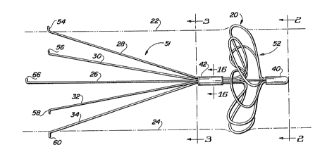
numeral 20. Within Fig. 1, dashed lines 22 and 24 indicate
the outline of an interior wall of a blood v~ssel, ~uch as
tha inferior vena cava. Blood clot ~ilter 20 con~ist
essentially of a central core wire 26 which extends
generally along the central lonsitudinal axis of blood clot
filter 20, as well as 3ix peripheral ~ires 28, 30, 32, 34,
36 and 38 spaced equiangularly about central core wire 26.
Peripheral wires 36 and 38 are hidden from view in Fig. 1
by peripheral wires 30 and 32, respectively; however,
peripheral wires 36 and 38 are vi~ible in Fig~. 2 and 3.
In Fi~. 1, a first connector 40 is 3hown forming a nose
of blood clot ~ilter 20. Connector 40 serve~ to connect
together a first end of each of peripheral wires 28-38, and
attaches such peripheral ~ires about the firqt end of
central core ~ire 26 at a fir~t connection point.
1~ 1 31 563~
Connactor 40 i8 ~elded, crimped or otherwiae attached to
the f~rst an~ of cantral core ~ire 26 ~n~ ~o the f irat en~
of peripheral wires 2B-38 ~o that a $ixed ~onnection i~
achieved between the central core wire an~ the ~ix
peripheral wires.
Still referring to Fi~. 1, ths ix peripheral ~ires 28-
38 are again joined along their central portions by a
second connector 42. As ~ho~n best in Fi~. 16, connector
42 i in the for~ of a tubular collar havin~ a central
opening defining an interior wall 44. ~ach of the
peripheral wires 28-38 passes throu~h tu~ular collar 42 and
is ~ecured to interior ~all 44 thereof, as by ~elding or by
other means of attachment. Thu~, ce~ond connector 42
serves to connect toge~her peripheral ~ires 28-38 at a
second connection point spaced apart from the fir~t
connection point at first connector 40. Referring again to
Fig. 16, it ~ill be noted that central core wire 26 passes
freely through the interior space de~ined by tubular collar
42 and the peripheral wire ~ecured therein, thereby
allowin~ 3econd connector 42 to ~lide along ~entral core
wire 2G.
Referring briefly to Fig. 17, ~n alternate form of
second connector i~ shown designated by re~erence numeral
42'. Second connector 4~' includes a tubular sleeve 46
having a central bore 48 through which central core wire 26
extends. Tubular ~leeve 46 includes an exterior circular
wall 50 to which each of peripheral wires 28-38 are
attached, as by welding. Like connector 42 shown in Fig.
16, connector 42' sho~n in Fig. 17 slidingly secures the
central portions of peripheral wires ~8-38 about central
core wire 26.
Within Fig. 1, second connector 4~ is shown after having
been advanced to a position relatively proximate to nose
40. The portions of peripheral ~ire~ a8-38 lying between
fir~t connector 40 and ~econd connector 42 are shown as
formin~ a ~lattened filter mesh, desi~nated generally by
- `
.
~3 l 31 563~
reference numeral 52. B~ch of the portion~ of peri~heral
wires 28-38 l~ing bet~een fir~t connector 40 ana ~econd
connector 42 rotate~ through an an~le of a~proximately 90-
120. A~ ~econd connector 42 ~ advanced to~ard fir~t
connector 40, the portions of peripheral wires 28-38 lying
between fir~t connector 40 and ~econd connector 42 extend
radially away from central core wire 26, to a flattened,
deployed po~ition sho~n in Fig~. 1 and 2. As ~hown in ~ig.
lj the extreme outermost portions of flattened filter me~h
52 sngage and slightly distend the interior walls 22 and 24
of the blood vessel, thereby providing a filter me~h which
extends over the entire cross-sec~ional area of the blood
vessel and which help locate blood clot filter 20 along
the central axis of the blood vessel once the leg portions
of the peripheral wire~ 28-38 are relea~ed. ~7hen ~ilter
mesh 52 is fully deployed, it extends sub~tantially
perpendicular to central core ~ire 26, and substantially
perpendicular to the longitudinal axi~ of bloo~ clot filter
52.
As mentioned above, the peripheral wires 28-38 rotate
through an angle of approximately 90-120 as filter mesh
52 is deployed to facilitate the flattening of the filter
mesh. Peripheral wire~ may be pre-shaped during
manufacture of blood clot filter 20 by proximating nose
connector 40 and slide connector 42 and turning nose
connector 40 through an angle of approximately 90-120
while holding ~lide connector 42 fixed, and then heat-
treating filter 20 so that the elastic memory of the
peripher~l wires will cause filter mesh 52 to flatten when
core wire 26 is retracted.
As shown in Fig. 1, the end~ of peripheral wires 28-38
lyin~ opposite connector 40 pass outwardly through slidable
connector 42 in a direction generally opposite to that of
connector 40. These second ends of peripheral wires 28-38
form anchoring le~s, each of Nhich is biased away from the
central l~n~itudinal axis of blood clot filter 52. The~e
14 l 31 563~
anchoring leg~ coll~ctively form a le~ ~s~embly ~e~i~nated
by reference numeral 51. Each of ~he 1Qg portion~ of
peripheral wire~ 28, 30, 32, 34, 36 and 38 terminate in
sharp2ned hoo~ or feet 54, 56, 58, 60, 62 and 64,
re~pectively for engaging and becoming fixed wi~hin the
interior ~alls 2~, 24 of the blood vex~el to anchor and
maintain blood clot filter 52 at a de~ired location
therein. Apart from anchoring blood clot filter 20, the
leg portions of peripheral wire~ 28-38 independently form a
blood clot filter ~eparate and apart from flattened filter
me~h 52. While leg assembly 51 is sho~n as bein~ formed by
extensions o~ peripheral wires 28-38, it will be
appreciated that the wires forming ~uch legs may be
di~tinct from peripheral wire~ 28-38, and may differ in
number and thickne~s therefrom. Thus, blood clot filter 20
provide~ a dual filtering sy~tem capable of filtering blood
clots greater than 5 millimeters in diameter. Moreover, as
mentioned above, the leq portions of peripheral wire~ 28-
38, in combination ~ith filter mesh 52, provide a self-
centering device maintaining blood clot filter 20 centeredwithin the blood vessel, thereby avoiding problems
associated ~ith a tilted filter.
Central core wire 26 and peripheral wires 28-38 may all
be formed from stainless steel, ~ material which has been
used extensively within the vascular system, and which i~
accepted by regulatory agencie~ and the medical comm~mity.
Connectors 40 ~nd 42 may al~o be made of stainles~ steel.
Alternatively, the central core ~ire, periphsral wires and
connectors may be formed of titanium. It i8 believed that
a peripheral wire thickne~s of 0.010 inch is thick enough
to withstand the impact of a blood clot againæt blood clot
filter ao, while being thin enough to be able to be
deployed into the filter mesh 52 shown in Fig. 1 without
requiring excessive mechanical force. Moreover, it is
believed that a wire thickness of 0.010 inch allows the
filter ~e~h 52 to he yieldin~ enough to accommodate a
1 3 1 563~
variety ~f caval 3iZ~8.
Tho~e ~killed i~ the art ~ill A~preciate that blood clot
filter 20 mu~t ini~ially be provided a~ a ~lender, ~mall
diameter as3embly in vrder to be conveniently introduced
within the blood ve~el by a delivery catheter. The leg
portions of peripheral wires 28-38 may initially be
compressed inwardly toward central core wire 26 prior to
loading the device within a delivery catheter, a~ 3hown in
Fig. 4A. The length~ of the various leg~ may be varied to
facilitate loading within the delivery catheter. The
filter ~esh sa i8 initially maintained in a compact,
elongated form by initially positioning slide connector 42
at a fir~t po~ition relatively remote ~xom connector 40 a~
shown in Fi~. 4A. In this initial position, the portions
of peripheral wire~ 28-38 extending bet~een connector 40
and slide connector 42 lie generally along central core
wire 26, as shown in ~ig~. 4A and 4B. Only after filter
mesh 52 is positioned within the blood ves~el at the
desired location is filter mesh 52 deployed outwardly to
take on its flattened, mesh configuration shown in Fig. 1.
Aæ ~hown in Figs. 1 and 4A, central core wire 26 is
longer than peripheral wires 28-38, and the second end of
central core ~ire 26 oppo~ite connector 40 include~ a
retractor fitting, ~hown in Fi~. 1 a3 a bent or hooked end
a5 66. ~hen filter me~h 52 i8 to be deployed, central core
wire 26 is retracted by pulling on retractor end 66,
thereby c~usin~ first connector 40 a~d ~lide connector 42
to approach one another, and causin~ the portions of
peripheral wires 28-38 lyin~ between connectors 40 and 42
to extend radially outward and ~latten. Were retractor end
66 to be relea~ed, the force of blood vçs~el ~alls 22 and
24 upon filter me~h 52, together with the inherent memory
characteristics of fine steel wire, would tend to force
connector~ 40 and 42 apart back to the initial po~ition
shown in Fi~ 4B. Accordingly, blood clot filter 20
include~ a mechanism ~or lockin~ ~lide connector 42 in the
1 31 563a,
" 16
po~ition shown in Fig~ 1 after central core wir~ 26 has
b~en retracted in order to maint~in filter mesh 52 in the
deployed, collapsed po~ition. One manner in ~hi~h thi~ may
be accomplished is by f lattenin~ or thickening the ~ortion
of central core wire 26 adja~ent nose connector 40 ~hereby
second connector 42' (~ee Fig. 17) forms a friction fit
~ith central core wire ~6 a~ connector 42' ~lides toward
nose connector 40. An alternate manner of loc~ing slide
connector 42 proximate no~e connector 40 i~ sho~n in Fig~.
8A-8C. In Fi~. 8A, lock device 68 is sho~n as a
cylindrical member extendin~ around central core ~ire 26
bet~een ~econd connector 42 and retractor end 66 (see Fig.
1) of central core wire 26. Lock device 68 includes a
wedge-shaped in~erior bore of a diameter commensurate ~ith
the diameter of the central body of core wire 26. The
wedge-shaped interior bore of lock device 68 open~ toward
nose connector 40. The diameter of core ~ire 26 is
essentially uniform until reaching the vicinity of no~e
connector 40, at ~hich point core wire 26 gradually tapers
to an enlar~ed diameter. L~ck device 68 can ~reely slide
along central core wire 26 toward no~e connector 40 (see
- Fig. 1) until reaching the tapered portion of core wire 26,
at which point further retraction of core wire 26 causes
the same to become ~edged within lock device 68, thereby
a5 oppo~iny sliding motion in the oppo~ite direction. Prior
to delivery of the blood clot filter, lock device 68 i
po~itioned behind the feet 54-60 of leg a~embly 51,
thereby allowing the leg portions of peripheral wires 28
and 34 to lie generally alongside core wire 26 in a compact
form. ~o~ever, when central core ~ire 26 i~ being
retracted, ~s shown in Fig. 8B, lock device 68 is
simultaneou~lr urged toward slide connector 4~ and toward
nose connector 40 by the di~tal end of a pu~her catheter 70
to be de~cribed in greater detail below. Thus, lock devic
68 al~o functions as spreader for biasing the leg portions
o~ peripheral ~ire~, such ~8 23 and 34, away from central
- 17 1 31 5634
core wire 26. ~n ~i~. 8C, the di~tal end of the pu~her
ca~heter 70 1~ retract~d. Lock dovice 68 thereafter
oppo~es sliding motion of central core wire 2~ relative to
~lide connector 42, thereby maintaining filter ms~h 52 (~ee
Fig. 1) in it~ deployed po~ition, while ~imul~aneou~ly
urging the leg portion~ of the peripheral ~ire~ 28-38
radially outward.
Fig. 9 ~hows an alternate form of a lock device. Within
Fig. 9, one-way ~asher 7~ include3 a c~ntral region angled
toward the leftmost side of Fig. 9. A central aperture 74
formed within the central region of washer 72 re~eive~
central core wire 26. A ~eries of radial ~lot~, ~uch as 76
and 78 divide the angled central regio~ into a ~erie~ of
tabs. Consequently, on~-way wa~her may ea3ily be moved to
the right alons central core wire 26 ~ithin Fig. 9.
Howev~r, attempts to thereafter move one-way wa~her 72 to
the left cau~e the slot~ed tabs to dig in to ~entral core
~ire 26 and oppose further sliding movement. A lock device
~uch as one-~ay ~asher 72 could be substituted for lock
device 68 within Fi~s. 8A-8C and likewise prevent central
core wire 26 from sliding through slide connector 42 after
having been retracted.
Fig8. 17 and 18A-18C illustrate an alternate form of
lock device for the blood clot f ilter ~0 ~hown in Fig. 1.
As ~tated above in regard to Fig. 17, slide connector 42'
is in the form of a tubular sleeve 46 ~hich slidingly
passes therethrough. ~ach of the 8iX peripheral wires 28-
38 i5 secured to the outer ~urface of tu~ular sleeve 46, as
by weldin~. Referring to Figs. 18A-18C, peripheral wires
28 and 34 ~re shown as being attached to the outer surface
of tubular sleeve 46, as by welding. A locking device, in
the form of a ~edge-shaped re~ilient member 71 i~ ~hown
~ixedly secured to central core wire 26. Wedge 71 is
initially to the right of tubular ~leeve 46 when the ~lood
clot filter is in its compacted form prior to deployment.
The ~arrowe t portion of ~hich 71 lies clo~est to tubular
18 1 31 563~
~leeve 46, while the wid0st portion thereof i9 ~urthe~t
from tubular 31eeve 46. The ~ide~t port~on of ~ed~e 71 has
a diameter or width which exceed~ the internal diameter of
tubular ~leeve 46. However, ~ed~e 71 i~ made of a
~ufficiently deformable material as to allow ~edge 71 to be
pulled through tubular ~leeve 46 upon retraction of central
core wire 26, a~ shown in ~ig. 18B~ Referring to Fig. 18C,
cenkral core ~ire 26 ha~ been fully retracted, thersby
bringing nose connector 40 of blood clot filter 20 into
proximity with tubular ~leeve 46 for deploying the filter
mesh 52. As shown in Fig. 18C, ~edge 71 no~ lies to ~he
left of tubular sleevs 46, and because the ~idest por~ion
of wedge 71 i~ wider, or of greater diameter, than tubular
sleeve 46, central core wire 26 i8 pre~ented from ~liding
back to the right. Accordingly, the filter mesh 52 is
locked in its deployed position.
Fig. 7 illustrates an alternate form of the blood clot
filter shown in Fig. 1. The blood clot filter of Fig. 7 is
designated generally by reference numeral 20', and like
~lood clot filter 20 of Fig. 1, includes a ~ose connector
40', a slid~ connector 42', a central core wire 26', and a
number of peripheral wires connected ~etween ~05e ~onnector
40' and ~lide connector 42' to form a filter me~h 52'. The
principal differences bet~een blood clot filter 20' o~ Fig.
7 and blood clot filter 20 of Fig. 1 relate to the
formation of the leg assembly 51'. Whereas the leg
portions shown in Fig. 1 are relatively ~traight and of
uniform length, the le~ portions shown in Fig. 7 are both
curved or bowed out~ardly and are of differing lengths.
The manner of delivçring blood clot filter 20 of Fig. 1
u~in~ a transfemoral approach ~ill now be described with
reference to Fig~. 4A-4F wherein a novel filter delivery
apparatus i~ shown. Fi~. 4A shows blood clot filter 20 in
a compacted position received ~ithin the distal end 74 of a
delivery catheter 70. Slide connector 42 of blood clot
filter 20 is remote from no~e connector 40 to elongate
1 31 563~
19
filter mesh 52, and the hoo~sed elnd of the le$~ ~ortion~3 of
peripheral wire~ 28-38 are compres~ed a~ain~t the lnterior
wall 76 of delivery catheter 70. Not shown in Fig. 4A i~
the proxim~l end of delivery catheter 70 ~hi~h lies
oppo~ite di~tal end 74 thereof. In~erted through the
proximal end of delivery catheter 70 i~ a ~emi-rigîd pu~her
catheter 80, the distal end 78 of which i~ visible in Fig.
4A. Not ~ho~n ~ithin Fis. 4A i8 ~he proximal end of pusher
catheter 78 which extend~ from the proximal end of delivery
catheter 70. Pu~her catheter 80 is slidingly recei~ed
within delivery catheter 70, and the distal end 78 of
pusher catheter 80 i~ adapted to abut hooked end portions
54-64 of peripheral wire~ 28-38.
Still referring to Fig. 4A, central core wire 26 i~
shown extending ~ithin the central bore 82 of pusher
catheter 80 and is relea~ably coupled to a retractor cable
84 by a releasable coupling mechanism 86. Retractor cable
84 slidingly extend~ through bore 82 of pusher catheter 80.
Not shown in Fig. 4A is the proximal end of retractor cable
84 which extends outwardly from the proximal end of pu~her
cable 80 so that it may be retracted and otherwise
manipulated by a physician. Releasable coupling mechani~m
8S is required since retractor cable 84 must be disengaged
from central core ~ire 26 of blood clot filter 20 once the
blood clot filter has been properly positionad and
deployed.
Referring briefly to Figs. 5A and 5~, releasable
coupling mechanism 86 i8 ~ho~n a~ a cylindrical nub 88
secured ~o the end of central core ~ire 26, together with a
slotted, cylindrical catch 90 secured to the distal end of
retractor cable 84. Catch 90 has a diameter ~ommensurate
with the diameter of the interior bore 82 of pu~her
catheter 80. Catch 90 includes a lateral ~lot 92 having a
depth and ~idth commensurate ~ith nub 88 ~or allowing nub
88 to be relea~ably captured therein~ In addition, a
radial 810t 94 exte~d~ through the front face 96 of catch
`- ao 131 563~
90 and extending to lRkeral ~lot 92 for permitting central
core wir~ a6 to extend throuuh the front ace ~6 o~ catch
90. It ~hould be appreciated that when nub 8~ re~ts ~i hin
catch 90, and ~hen catch 90 lie~ within pu~her catheter 80,
central core ~ire a6 and retractor cable 84 ar~ ef~ectively
secured together. Nowever, when it i~ de~ired tv di~engaye
retractor cable B4 f rom central core wire 26, the u~er need
only r~tract pusher catheter 80 and delivery catheter 70
for allowing catch 90 to di~engage nub 88.
An alternate relea~able coupling mechanism 86' i~ sho~n
in Figs. 6A and 6B. ~ithin Fig. 6A, central core wire 26
may terminate in a looped connector 98 preferably having a
width commensurate ~ith the internal diameter o~ bore 82 of
pusher ~atheter 80. The distal end of retrActor cable 84
includes a hooked end 100, also having lateral dimensions
commensurate with the internal diameter of bore 82 of
pu~her catheter 80. Prior to delivery of the blo~d clot
filter, hook 100 i5 inserted ~ithin looped ~onnector 98,
~hich remain engaged ~ith one another so long as they lie
within bore 82 of pusher cathe~r 80. After the filter
mesh of the blood clot filter has been deployed by
retracting central core ~ire 26, both pusher catheter 30
and delivery catheter 70 can be retracted for permitting
hooked end 100 of retractor cable ~4 to disengage looped
connector 98 of central core ~ire 26.
Referring again to Fig. 4A, blood clot filter 20 i9
shown as being contained fully within distal end 74 of
delivery catheter
70. Delivery catheter 70 may be, for example, a 10 or 12
French Teflon catheter, ~nd may be introduced into the
blood vessel using the standard Selldinger angiographic
technique. To position a flexible catheter within a blood
vessel usîn~ the ~o-called Selldin~er techni~ue, a needle
i5 first in erted into the blood ves~el, a guide wire is
then threaded through the needle, and the needle i8 then
~ithdrawn leaving the guide ~ire in placQ.
,
al 131 563~
DeliYery ~athet~r 70 i~ an o~en-en~ed catheter, an~
tapered, ~nu~-fittin~ ~n~io~r~phic c~th~ter (not ~hown) ma~
be inserted within delivery catheter 70 to ~acilitate the
pas~age o~ delivery cathet~r 70 throu~h the bloo~ ve~el.
Delivery catheter 70 and the tapered angiographic catheter
therein are the~ in~erted into the blosd ves~el over the
guide wire. Delivery catheter 70 may be advanced throu~h
the blood vessel until di~tal end 74 i~ approximately at
the po~ition at which the blood clot filter 20 i5 to be
delivered. ~ollowing placement o~ delivery ca~heter 70
within the blood vessel, the inner tapered angiographic
catheter and ~uide wire are ~ithdrawn.
Fig. 19 illustrates a filter storage tube into whi~h
blood clot filter 20 may be preloaded ~or being advanced
into delivery catheter 70 after delivery catheter 70 has
been placed within the blood vessel. ~ithin Fig. 19,
delivery catheter 70 includes a female luer lock connector
300 at its proximal end. The ~ilter ~tor~ge tube is
designated generally by reference numeral 302 and includes
a short section of tubing 304 haYing approximately the ~ame
internal diameter as delivery catheter 70. Shown within
filter ~torage tube 304 i~ blood clot filter 20. A first
end of filter storage tube 302 includes a male luer lock
connector 306 adapted to scre~ onto female connector 300.
a5 The opposite end of ~ilter storage tube 304 is integrally
joined ~ith a molded fitting 308 which includes a
deformable elastomeric seal 310, as well as an infusion
port 312. The distal end of pusher catheter 80 extends
into ~ilter storage tube 302 through deformable seal 310,
and retractor cable 84 extends within pusher catheter 80
and is coupled to ~ore ~ire 26 by reIea~able coupling
mechanism 86. Once delivery catheter 70 has been adYanced
into the blood ves~el so that it~ distal end i~ at the
appropriate delivery site, an infusion line i~ connected to
infusion port 312, and male luer connector 306 is then
coupled to female luer connector 300. Pusher catheter 80
22 1 3 1 563~
an~ r~t~actor cable B4 ~r~ then adv~nce~ a~ a un$t to pu~h
blood clot ~$1tQr 20 out of ~ilter ~torage tu~e 302 ~nd
into delivery catheter 70. Pusher catheter 80 and
r~tractor cable 84 are furth~r advanced until blood clot
filter 20 ha~ been pu~hed to the di~tal end of delivery
~atheter 70. The remaining ~teps for ~eliverin~ blood clot
filter 20 within the blood vessel are described below.
The next ~tep in deployin~ blood clot filter 20 i~ to
partially retract delivery catheter 70 ~hile leaviny semi-
rigid pusher catheter B0 fixed, un~il the distal end 74 ofdelivery ca~heter 70 has been retracted to approximately
the location of ~lide connector 42. The natural
springiness of the peripheral wire~ 28-34 of the filter
me~h 52 cau~es each of the peripheral wires to move
~omewhat further apart from cen~ral core wire 26 as
compared with their compacted ~onfiquration~ a3 shown in
Fig. 4A. Turning to Fig. 4C, the next ~tep in
deployin~ filter mesh 52 i~ to maintain the position~ of
delivery catheter 70 and pusher catheter 80 fixed, with the
hooked feet 54-60 of blood clot filter 20 abutting di~tal
end 78 oP pusher catheter 80. The proximal end (not ~ho~n)
of retractor cable 84 i8 then slo~ly retracted by the
operator. As retractor cable 84 i~ retracted, relea~able
coupling mechanism 86 causes central core ~ire 26 to be
pulled to the left (relative to Fig. AC), as indicated by
the arro~ desi~nated by reference numeral 104. ~hile
central core wire 26 slidably extends through slide
connector 42, central core ~ire 26 is ri~idly attached to
no~e connector 40. Accordingly, a~ central core wire 26 i5
retracted, nose connector 40 al~o move~ to the left, a~
indicated by the arrow designated by reference numeral 106.
As no~e connector 40 i~ brough clo~er to ~lide connector
42, th2 peripheral wire~ making up ~ilter mesh 52 are each
forced radially outward toward the inner wall~ 108 and 110
of the blood ves~el. Continued retraction of retractor
cable 84 and central core ~ire 26, as shown in Fi~. 4D,
~" 23 1 ~1 563'~
causes filter me~h 52 ~o become fully ~eployed, ~ikh no~e
connector 40 being po~itioned relatively proximate to ~lide
connector 42. A3 ~hown in Fig. 4D, the outermo~t portions
of ~ilter me~h 52 beco~e en~a~ed with interior wall~ 108
and 110 of the blood ves~el upon filter me~h 52 becomin~
fully deployed. A~ explained ~bove, slide connector 42 has
a~sociated therewith ~ lock device which prevents central
core wire 26 from later ~lidin~ to the ri~ht (relative to
~i~. 4D) through slide connector 42, and nose
connector 40 iB thereby maintained closely proximate to
~lide connector 42.
The next step in deploying blood clo~ filter 20 i~ to
further retract delivery catheter 70, as ~hown in Fi~. 4~,
to expo~e the leg portions thereof and allow the same to
sprin~ outward ~or allowing the hooked end~ thereof to
engage internal walls 108 and 110 of the blood vessel as
~hown in Pig. 4~. The leg a~sembly 51 of blood clot filter
20 ~orms a filter in addition to filter mesh 52. Moreover,
because blood clot filter 20 engages the i~ternal ~alls lOB
and 110 of the blood ves~el both alon~ the periphery of
filter mesh 52 and the hooked end~ of the leg portions,
blood clot filter 20 is positioned centrally along the
lonsitudinal axi of the blood vesRel.
The last step in the delivery of blood clot filter 20 is
shown in Fi~. 4F wherein both delivery catheter 70 and
pusher catheter 30 are retracted, leaving only retractor
cable 84 in the same position ~ho~n as in Fig. 4~.
Retraction of both delivery catheter 70 and pusher catheter
80 release~ couplin~ mechani~m 86 from i~ternal bore 82 o~
pusher catheter 80 and permits coupling mechanism 86 to be
released from nub 88 of central of core ~ire 26. Retractor
cable 84 i~ then retracted back within internal bore 82 of
pu~her catheter 80, and the delivery sy~tem i5 then fully
removed, leavin~ blood clot filter 20 properly po~itioned
within the blood vessel at the desired location.
Fig~. lOA-lOC illustrate an alternate embodiment of the
1 ~1 563~,
24
present invention includin~ a blood clot ~lter 1~0
de~igned ~or jugulAr delivery. AB ~ho~n in ~i~. lOA, blood
clot filter 120 include~ a central core wire 126 havin~
fir~t and rigidly attached to a fir~t connector 140.
Central core ~ire 12~ extend~ along the central
longitudinal axis of blood clot filter 120 ~nd pa~e~
through the central bore of a slide connector 142. A one-
way lock washer 172, similar to that sho~n in Fig. 9, ha~ a
central bore throu~h which core ~ire 126 extend~. Washer
172 is adapted to permit core wire 126 to slide
therethrough to the right ~relative to Fig. lOA), but to
resist sliding movement of central core wire 126 to the
left. A plurality of peri~heral wire~, including those
peripheral wire~ 128, 130, 132, and 134 vi~ible in 10~ each
have a first end attached to qlide connector 142, and are
each attached to connector 140. Peripheral wires 128-134
are shown in Fig. lOA in their compacted position loaded
within a jugular delivsry cathe~er 170. ~ach of the
peripheral wires 128-134 extend generally along and
parallel to central core wire 126, a~ ~ho~n in Fig. lOA.
Also extendi~g to the left of connector 140 îs a leg
a~sembly 141 con~i~ting of, for example, six ~ire legs,
including tho~e visible in Fig. lOA and de~ignated by
re~rence numeral~ 129, 131, 133, and 135. Each of ~uch
leg~ 129-135 h ~ a first end secured to connector 140 and a
3econd end opposite thereto formed into hooked feet 154,
156, 158, and 160, respectively, for engaging fhe walls of
a blood ve~sel into which blood clot filter 120 is to be
positioned. As mentioned above, leg portion~ 129-135 may
~imply be continuations of the respective peripheral wires
128-134 which make up ~he filter mesh 152.
To deliver blood clot filter 120 u~ing the jugular
delivery technique, ~elivery catheter 170 i~ introduced
into the blood
ve~el and advanced through the b~ood vessel until the
distal end 174 of delivery catheter 170 is located at
l 31 563~,
approxi~ately the po~ition at which blood clo~ rilter 120
i~ to be deliver~d. Bloo~ clot ~llt~r 120 i~ po~itioned
within the di~l end Or delivery cathet~r 170, a~ ~hown in
Fig. lOA, either by puohin~ blood clot ~ er 120 along the
len~th of delivery ~atheter 170 from the proximal end
thereof, or by preloading blood clot filter 120 ~i~hin the
di~tal end of d~livery ~atheter 170, in the manner
de3cribed below. ~ithi~ Fig. lOA, ~emi-rigid pu~her
catheter 180 i~ ~ho~n extending within the internal bore
176 of delivery catheter 170. Though not shown, the
proximal end of pusher catheter 180 ~xtend3 fully throu~h
the proximal end of deliverr ~atheter 170 ~o that it may be
manipulat~d by a phyaician. Pu~her catheter 180 has a
diameter commensurate ~ith that of one-~ay ~a~her 172, and
the distal end of pusher ~atheter 180 is adapted to abut
and push again~t ~asher 172. Central core wire 126 extends
within internal bore 182 of pusher catheter 180 and is
releasably con~ected ~ith releasable coupli~g mechani~m
186. The distal end of retractor cable 184 is also coupled
to relea~able coupling mechanism 186. Retractor cable 184
extends fully through ~he internal bore 182 of pu~her
catheter 180 and protrude~ from the proximal end thereof
for allowing the physician to retract retractor cable 184.
~uring deli~ery of blood clot filter 120, delivery
catheter 170 i8 retracted to the right, initially
per~ittin~ leg~ 129-135 of leg as~embly 151 to spring
outward, ~ith the hooked ends 154-160 thereof enga~ing the
internal walls 208 and 210 of the blood
ve~sel. Further retraction of delivery catheter 170
permita the peripheral wires 128-134 of filter mesh 152 to
bow out~ardly, as shown in Yig. lOB.
The next step in deploying blood clot filter 120 i~ to
advance pusher ~atheter 180 to the left, ~hile fixing the
po~ition of retractor c~ble 184. It i~ important to fix
the position of retractor ~able 184 to maintain ~onnector
140 in a fixed po~ition relative to the blood ves~el, and
. ~
a6 131 563~
thereby avoid movement of hooked end~ 154-160, a~ ~uch
longitudl~al ~ovement coulfl cauoe tr~u~ to the ~all~ 208
and 210 of the blood ves~el. A~ pusher c~theter 180 i~
advanced to the left, it advAnces washer 172 t~ the left
alon~ central core wire 126. As washer 17~ advance~ to the
left, it moves slide connector 142 into proximity with
connector 140, thereby cau~ing peripheral ~ires 128-134 to
beco~e radially extended to form filter mesh 152, as shown
in Fig. lOC. When pu~her ca~heter 180 i~ fully advanced to
the left, the outermo~t portion~ of filter me~h 152 engage
the walls 208 and 210 of ~he blood vessel. Pusher catheter
180 i~ then retracted; as pusher ca~heter 180 i~ retracted,
~asher 172 become~ locked a~ainst central core wire 126 and
thereby maintain~ filter meRh 152 ~ithin the deployed
position ~hown in Fig. lOC. Both pusher catheter 180 and
delivery catheter 170 are retr~cted ~o the righ~ (relative
to ~ig. lOC) ~or exposing releasable coupling mechanism
186, thereby allowing central core 126 to be disengaged
from retractor cable 1~4, as was de~cribed above in regard
to Fig. 4~. Retractor cable 184, pusher catheter 180, and
delivery catheter 170 may then be fully retracted,
leaving blood clot filter 152 in ths de~ired location.
As explained abov0 with respect to Fig. 19, blood clot
filter 20 can be loaded into the proximal end of delivery
as catheter 70 using a filter ~torage tube that couples to the
proximal end of delivery catheter 70. ~owever, blood clvt
filter 20 must then be pushed along the delivery catheter
to the distal end thereof; durin~ this procedure, the
hooked ends of the blood clot filter legs scrape a~ainst
the inner wall of the delivery catheter and could dislodge
particles therefrom. In ordPr to avoid the need to push
the blood clot filter alon~ the entire len~th of the
delivery catheter, the alternate form of filter delivery
system shown in Fig. 11 may be u~ed.
Within Fig. 11, blood clot filter 20 is of the same type
sho~n in Fig. 1, and delivery catheter 270 i~ of
27 1 31 563'~
e~en~ially the ~ame type a~ delivery cathe~sr 70 o~ Fi~.
4A. Similarly, pu~h~r cathe~er 280 and retr~ctor c~ble 28~
corre~pond to the pusher Ga~heter 80 and retractor cable 84
sho~n in Fig. 4Ao ~o~ever, a~ ~hown in Fi~. 11, delivery
catheter ~70 i~ inqerted through an outer catheter 275
which ha~ a di~tal end ~77 ~n~ a proximal end 279. The
di~tal end a77 o outer catheter 275 i~ percutanesu~ly
introduced into a blood ve~el u~ing the aforementioned
Selldin~er technique prior to insertion o delivery
catheter 270 therein. After outer catheter 275 i8
introduced ~ithin the blood vessel, delivery catheter 270
i~ inserted into outer catheter 275 from the proximal end
279 thereof. The proximal end 279 of outer catheter 275
includes a deformable ela~tomeric seal 281 which permits
delivery catheter 270 ~o be ~lidingly received thereby
while forming a fluid tight ~eal therearound. ~he proximal
end 279 of outer catheter 275 also includes an infu~ion
port A extending perpendicularly ~o the longitudinal axis
of outer catheter 275 for permit~ing ~aline ~olution to be
infused therei~, thereby preventing the patient's blood
from filling the internal bore of outer catheter 275 and
forming blood clots. Similarly, the proximal end 283 of
delivery catheter 270 includes a deformable ela~tomeric
seal 285 for ~lidingly receiving the di3tal end of pusher
catheter 280 and forming a fluid ti~ht ~eal ~herearound.
The proximal end 283 of delivery catheter 270 al50 include~
an infusion port B into which saline 301ution may be
infu~ed to prevent the patient'~ blood from collecting
~ithin the înternal bore of delivery catheter 270 and
forming blood clots.
Similarly, pusher catheter 282 has a proximal end 287
provided with a deformable elastomeric ~eal 289 which
slidingly receive~ the retractor cable 284 and form~ a
fluid tight seal therearound. The proximal end 287 of
pusher catheter 280 al90 include~ an infusion port C for
i~fu~ion of ~aline ~olution to prevent the patient' 9 blood
1 31 563~
28
from colle~ting ~ithin the lntern~l bore o~ pusher c~thetex
a80 an~ forming blood clot~. The proximal end of retractor
cable a84 may include a down~ardly turned handle D ~or
~onveniant operation of retractor cable ~84.
The delivery ~ystem ~ho~n in Fig. 11 permits blood clot
filter 20 to be preloaded into the di~tal end of delivery
catheter 270 before delivery catheter 270 i~ in~erted into
outer catheter 275. This i~ avoid3 the need to in~ert
bloQd clot filter 20 into delivery catheter 270 from the
proximal end thereof~ A~ shown in Fig. 11, a removable
stop 291 may be relea~ably coupled, as by a clip (not
~hown~ to pusher catheter 280 for defining a fixed space or
distance between infu~ion port C ~nd the leftmo~t end of
~top 291. The importance of the fixed ~pace created by
stop 291 is explained in ~reater detail below in
conjunction with Fig~. 12~15.
Figs. 12-15 illustrate, in ~chematic form, the series of
steps followed in u~ing the delivery ~y~tem in Fig. 11. As
shown in Fig. 12, blood clot filter 20 is initially
dispo~ed in its compacted position, ~imilar to that sho~n
in Fig. 11. In Fig. 13, both outer catheter 275 and
delivery catheter 270 are retracted until infusioh port B
abut3 the leftmo~t end o~ stop 291. Similarly, outer
catheter 275 i~ retracted until the proximal end 279
thereof abut~ infu~ion port B of delivery catheter 270.
Stop 291 i8 of a ~ufficient len~th to permit delivery
catheter 270 and outer catheter 275 to be ~ithdrawn to the
extent that the peripheral wires for~ing the ~ilter me~h
are no lonqer encased by delivery catheter 270, while the
le~ as~embly 51 of blood clot filter 20 remains encased by
delivery ~atheter 270. As ~hown in Fig. 13, retractor
cable 284 i~ then retracte~ whil0 holdi~g the proximal end
287 of pu~her ~atheter 280 fixed, thereby deploying filter
mesh 52 of blood clot filter 20. The next ~tep involves
removin~ ~top 291 and thereaf~er retracting both delivery
catheter 270 and outer catheter 275 until the proximal end
~g ~ 3~ 563~
283 o~ delivery catheter a70 abut~ inu~io~ port C, and
~imilarly, the proximal end 279 of outer ~athe~er 275 abuta
infu~ion port B, as sho~n in Fig. 14. Thi~ opera~io~
further retracts both the ~istal ends of delivery catheter
270 an~ outer catheter 275, thereby permitting leg
a~embly 51 of blood clot filter 20 to ~pring out~ardly and
en~age the walls of the blood ~e~el. Re~erring to FigA
15, the next ~tep of tlle operation i8 to retract pusher
catheter 280, as well as deliver~ cathet r 270 and outer
catheter 275, in order to permit the di~tal end of
retractor cable 284 to be released from the central core
wire of blood clot filter 20, as i~ indicated within Fig.
15. With blood clot filter ao then properly po~itioned,
the entire delivery ~ystem is fully retracted out of the
blood ve~el.
Yig. 20 illus rates a wire shapiny jig which may be used
for pre-~haping each of the peripheral wires 28-38 shown in
Figs. 1-3. As ~hown in Fi~. 20, the ~ire ~hapiny jig
includes three circular pegs 320, 322, and 324. Peg~ 320
and 322 are spaced apart from one another by approximately
the distance between second connector 42 and hooked end 54
~hown in Fig. 1. Pegs 322 and 324 are spaced apart from
another by approximately the di~tance sho~n between nose
connector 40 and slide connector 42 shown in Fig. 4B. The
wire shaping jig al80 includes a form 3Z6 having a ~emi-
circular curved -~urface 328 adapted to advance between pegs
322 and 324. As ~hown in Yig. 20, peripheral wire 28 is
placed against pegs 320, 322 and 324. Form 326 is then
advanced against peripheral ~ire 28 and causing the same to
be pu~hed through the space between pegs 322 and 324. Form
326 is further advan~ed until the curved surface 328
thereof is closely proximate pegs 322 and 324. The ends of
peripheral wire 28 are then bent around peg~ 320 and 324.
The portion extendin~ beyond peg 320 is later clipped to
form the hooked end 54 of the anchorin~ leg. Similarly,
the portion extendi~g beyond peg 324 i~ l~ter clipped and
131~63~
attached to nose connector A0 o~ blood clot filt~r 2~.
~ ig~ 21 illu~rate~ ~n a~embly ji~ ~hich may ~e u~ed to
~upport the central core ~ire 26 centrally of the plurality
of peripheral ~ire~ to facilitate the ~onnection of the
peripheral wires about the central core ~ire. A~ ~ho~n in
Fig. ~1, the mounting jig 330 is generally cone ~haped and
includes a central bore 332 through which ~entral core ~ire
26 extends. A set of three scre~ 334, 336 and 333
threadedly en~age the cone ~haped mounting ji~. The
~lotted heads of screw~ 334, 336 and 338 may be slightly
loos~ned for allo~ing the peripheral wire 28 to extend
under su~h slotted heads. Screws 334, 336 and 338 are then
tightened to hold peripheral wire 28 in place, ~ith the
central bend thereof which ~as ~ormed about peg 322 (see
Fi~. 20) positioned at the tip of the cone shaped jig 330.
Similarly, other ~ets of mountin~ ~cre~, ~uch as 340, 342
and 344 are also provided ~or releasably supporting ~he
other peripheral wires, ~uch a~ peripheral wire 34. With
all six ~ires secured to mounting jig 330 and spaced
equiangularly about central core wire 26, it is relatively
easy to weld or clip together the upper ends of the
peripheral wires 28-38 with the upper end o central core
~ire 26, a~ within nose connector 40 (seP Fig. 1~. It is
al~o then relatively ea~y to join together the peripheral
wires at the upper tip of the cone-shaped ji~ 330, a~ with
a ~lide connector 42 (~ee Fig. 1). As mentioned above, it
may be desirable to rotate the nose connector throu~h an
angle of 90-120a, and perhap~ to impart a heat treatment
to the wire~ in order to pre-shape the filter mesh 90 that
it will ~latten when the central core wire is retracted.
The aforementioned rotation of the nose connector may
easily be performed before the central core wire 26 and
peripheral ~ires 28-38 are removed from assembly jig 330.
The extra wire lengths extending below screw~ 336 and 342
may thereafter be clipped and the ends shaped to provide
hooked ends on the end~ of the anchoring legs.
1 31 5634
- 31
While the present invention ha~ been described in
accordance with a pr~ferr~d embodiment thereo~, the
de~cription i~ for illustrative purpose~ only and ~hould
no~ be construed as limiting the scope of the invention.
Various changes and modifi~ation~ may be made by tho~e
~killed i~ the art without departing from the true spirit
and s~ope of the invention as defined by the appended
claims.
' ' - .
' ' ` ' ' '' ' '
~ .