Note: Descriptions are shown in the official language in which they were submitted.
i 7 n ~7
TITLE OF THE INVENTION
Blood Filter
BACXGROUND OF T~E INVENTION
This invention relates to a blood filter for removing
foreign matters and bubbles from blood passing an extra-
corporeal blood circuit including an artificial heart-lung or
pump-o~ygenator, an artificial kidney or dialyzator, and a
device for separating blood cell and plasma components, and
more particularly, to a blood filter having improved
debubbling capacity.
A number of blood filters are known in the art. One
typical blood filter is disclosed in US Patent No. 4,411~783.
In FIG. 14, the blood filter generally designated at 100 is
illustrated as comprising a cylindrical housing 102 formed of
polycarbonate resin, for example, having a blood inlet 105
and a blood outlet 108, and a filter member 109 received in
the housing 102 between the inlet 105 and the outlet 108
The filter member 109 is prepared by sandwiching a mesh
screen with an opening of 20-50 ~m between plastic nets,
folding the sandwich in pleats, and mating the ends to ~orm a
generally cylindrical filter member. To prevent bubbles from
directly passing to the filter member 109, the blood inlet
105 is tangentially connected to the cylindrical housing 102
such that blood B may enter the housing interior in a
tangential direction to form a swirl flow therein as shown in
FIG. 15 which is a cross section of the blood filter of FIG.
14 taken along lines III-III. It is also known to interpose
a continuous foam between the blood inlet and the filter
member to prevent bubbles from directly passing to the filter
member.
In the blood filter of the above-mentioned type
3Q wherein blood B enters the housing 102 through the inlet 105
to form a swirl flow therein, bubbles are removed by virtue
131570`7
of a centrifugal effect that fine bubbles with a small mass
entrained in the swirl flow of blood B will collect toward
the center of swirl. Since buoyancy applies at all times to
bubbles in blood B, separated bubbles will float and collect
at an axial top portion of the housing 102 and outflow
through a vent 106 at the axial top portion oi the housing
102. The filter member 109 removes only those bubbles which
are not removed by the buoyancy and the centrifugal force of
the swirl flow, that is, bubbles which are not entrained in
the swirl flow of blood B. In the above-mentioned
modification wherein a continuous foam is present between the
inlet and the filter member, the foam removes such bubbles.
In the blood filter 100 mentioned above, however, the
inflow of blood which is entering the housing 102 through the
inlet 105 directly impinges against a swirl flow of blood
which is already whirling in the housing 102 as shown in FIG.
15. Since this causes a disturbance in the blood inflow
before the inflow merges with the swirl flow, bubbles are not
so smoothly conveyed to the swirl flow of blood. Thus, this
blood filter 100 has the drawback that much bubbles reach the
filter member 109 because bubbles of a small mass cannot be
so effectively separated from blood B by entraining bubbles
on the swirl flow of blood and permitting such bubbles to
collect toward the swirl center by virtue o~ a centrifugal
effect. Bubbles in blood B impinge against and stick to the
filter member 109 and some bubbles then gradually penetrate
through the filter member 109 partially under the influence
of a pressure variation caused by pulsation of a feed pump.
Also, the above-mentioned blood filter of the type using a
continuous foam has several problems including adherence of
platelets to the foam, damage to cells~ an increased pressure
loss, and difficulty of debubbling upon priming.
1 31 5707
SUMMARY OF THE INVENTION
An object of the present invention is to provide a
novel and improved blood filter of relatively simple design
exhi~iting high debubbling performance ~7hile minimizing
platelet adherence, cell damage, and pressure loss, and
facilitating debubbling upon priming.
According to the present invention, there is provided
a blood filter comprising a bubble separating section and a
blood filter section disposed below the buhble separating
section. The bubble separating section includes a chamber
having a genérally circular cross section for allowing
bubbles to separate from blood, a vent formed at an upper end
of the chamber for discharging air, an inlet conduit
horizontally extending from the chamber for introducing blood
into the chamber, the axis of the inlet conduit extending
substantially parallel to a tangent to the generally circular
chamber at the connection between the chamb~r and the inlet
conduit, and an inflow portion merging the inlet conduit to
the chamber. ~he blood filter section includes an outlet
disposed at a lower end of the filter section for discharging
blood and a filter member disposed between the inlet and the
outlet. Then the inflow portion introduces incoming blood
from the inlet conduit so as to flow as a substantial laminar
flow to a swirl blood flow in the chamber and then merge with
the swirl flow.
BRIEF DESCRIPTION OF TME DRAWINGS
The above and other objects, features and advantages
of the present invention will be better understood from the
following description taken in conjunction with the
accompanying drawings, in which:
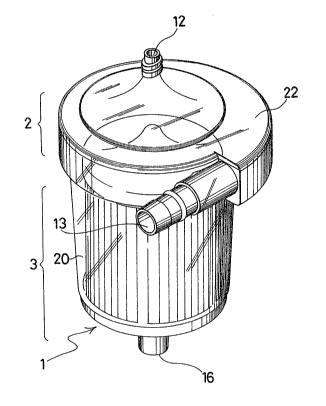
F~G. 1 is a perspective view of a blood filter
according to one embodiment of the present invention;
FIG. 2 is a side elevation of the blood filter of FIG.
1 ;
1 3 ~ 5707
EIG. 3 is a vertical cross section of the blood filter
taken along lines I-I in FIG~ 2,
FIG. 4 is a horizontal cross section of the blood
filter taken along lines II-II in FIG. ~, with the -filter
section omitted;
FIG. 5 illustrates partially in cross section the
filter member used in FIG. 1;
FIGS. 6 and 7 are cross sections similar to FIG. 4,
illustrating different embodiments of the present invention;
FIG. 8 is a schematic view showing an artificial
heart-lung circuit to which the blood filter of the present
invention is applied;
FIGS. 9 and 10 are schematic horizontal and vertical
cross-sectional views showing the interior configuration and
dimensions of the blood filter fabricated in Example;
FIGS. 11 and 12 are schematic horizontal and vertical
cross-sectional views showing the interior configuration and
dimensions of the blood filter fabricated in Comparative
Example;
FIG. 13 is a schematic view showing a test circuit
used to determine the performance of a blood fil-ter of the
invention and a comparative filter;
FIG. 14 is a vertical cross section of a prior art
blood filter; and
FIG. 15 is a horizontal cross section of the prior art
blood filter taken along lines III-III in FIG. 14.
DETAILED DESCRIPTION OF THE PREFERRED EMBODIMENTS
The invention will be described in further detail by
referring to several embodiments illustrated in the drawings.
The blood filter according to the present invention is
generally designated at 1, which comprises a bubble separat-
ing section 2 including a chamber 10 having a generally
circular cross section for allowing bubbles to separate from
blood, a vent 12 formed a-t an upper end of the chamber 10 for
1 3 1 5707
discharging air, an inlet conduit 13 horizontally extending
from the chamber 10 for introducing blood into the chamber,
the axis of the inlet conduit 13 extending substantially
parallel to a tangent to the generally circular chamber 10 at
the connection between the chamber and the inlet conduit, and
an inflow portion 15 merging the inlet conduit 13 to the
chamber 10; and a blood filter section 3 disposed below the
bubble separating section 2, including an outlet 16 disposed
at a lower end of the filter section Eor discharging blood
and a filter member 18 disposed between the inlet 13 and the
outlet 16; whereby the inflow portion 15 introduces incoming
blood from the inlet conduit 13 to flow as a substantial
laminar flow to a swirl flow of blood in the chamber 10 and
then merge with the swirl flow.
One embodiment of the blood filter of the present
invention is illustrated by referring to FIGS. 1 to 4
The blood filter 1 includes a bubble separating
section 2 and a blood filter section 3. The bubble
separat'ng section 2 includes an upper housing of a generally
circular cross section defining a chamber 10 therein for
allowing bubbles to separate from hlood. The upper housing
is a generally cylindrical housing over which a generally
conical lid 22 is fitted. A vent 12 is opened at an upper
end of the conical lid 22 in communication with the chamber
for discharging air. A tubular blood inlet conduit 13
horizontally extends from the side of the upper housing. A
blood inflow portion 15 merges the inlet conduit 13 to the
chamber 10. The blood filter section 3 includes a generally
cylindrical lower housing 20 which is concentrically disposed
below and contiguous to the upper housing. An outlet 16 is
opened at an axial lower end of the lower housing for
discharging blood therefrom. A filter member 18 is received
in the lower housing so as to axially extend between the
inlet and the outlet.
1 31 5707
More particularly, the blood filter 1 according to the
embodiment shown in FIGS. 1 to 4 includes an integral housing
20 having an upper opening, and a generally conical lid 22
fitted over the housing to close the opening. The integral
housing may be considered as compxising upper and lower
housings. The upper housing defines the bubble separating
chamber 10 therein and has the inflow portion 15 attached
thereto. The lower housing constitutes the filter section 3
with the filter member 18 received therein. The upper
housing and the lid 22 define the bubble separating chamber
10. That is, the lid 22 defines an upper portion of the
bubble separating chamber 10. The vent 12 is formed at the
top of the lid 22 to communicate the chamber 10 to the
exterior. The entire housing components including the lid,
upper and lower housings may be formed o~ any desired
synthetic resins includ.ing polycarbonate, polypropylene,
polyethylene, styrene-butadiene (SB) resin, and methylene-
butadiene-styrene (MBS) resin. These components are
preferably transparent as shown in FIGS. 1 and 2 because easy
observation of the contents in the housing is desirable. The
upper housing defining the chamber 10 is connected to the
lower housing through a step 20a in the illustrated
embodiment. It is also possible that the bubble separating
section 2 and the blood filter section 3 may have
substantially the same diameter so that they are smoothly
connected to each other. A tapered connection is also
possible~ It is to be noted that the blood filter section is
omitted in FIG. 4.
The housing 20 at its upper side wall is provided with
the blood inlet 13. The inlet 13 has a cylindrical blood
introducing conduit 14 which is provided outside the bubble
separating chamber 10 and extends substantially parallel to a
tangent to a cross sectional circle of the bubble separating
chamber 10. The blood inflow porti.on 15 is connected to the
blood introducing conduit 14 at a leading end and to the
1 31 5707
chamber 10 at a trailing end. The inflow portion 15 has an
outer wall 15a whose radial distance from the chamber 10
gradually decreases from the leading end to the trailing end.
The inflow portion 15 has the maximum cross sectional area at
the connection to the blood introducing conduit 14 and then
gradually reduces its cross sectional area or con-tracts until
the inflow portion 15 merges into the upper housing 1Ob or
chamber 10. The inflow portion 15 encloses a portion of the
outer circumference of the bubble separating chamber.
More particularly, the inflow portion 15 has the outer
wall 15a on a circle having a radius r1 about a center offset
the center of the upper housing as shown in FIG. 4. An
inflow of blood is smoothly introduced into the bubble
separating chamber 10 over an angle of 90 to 360,
preferably 135 to 225. The inflow portion 15 is a merging
portion where an inflow of blood incoming from the inlet 13
merges with a swirl flow of blood along the inside wall 1Ob
of the upper housing. The axis 13a of the inlet conduit 14
extends substantially parallel to a tangent 10c to the
circular cross section of the bubble separating chamber 10.
Thus in the inflow portion 15, an inflow of blood incoming
from the inlet 13 merges with a blood swirl flow in the
chamber such that their streamlines are substantially in
parallel to each other, and thus they flow as a substantial
laminar flow. The vertical cross section of the inlet 13,
the introducing conduit 14 and the inflow portion 15 may be
circular or rectangular.
The vent 12 is in communication with the top end of
the bubble separating chamber 10. The vent 12 may be
provided with valve means such as a three-way cock (not
shown). The vent 12 is located at the top of the conical lid
22 or aligned with the axis of the bubble separating chamber
10. The inflow of blood incoming from the inlet 13 to the
chamber 10 in a tangential direction thereof a-t a certain
flow velocity forms a swirl flow in the chamber 10 where
l3ls7n7
bubbles centrifugally se~regate from the blood, move toward
the axial center of the chamber 10, and collect in an upper
portion of the chamber 10. Then bubbles may be readily
discharged by releasing the vent 120
The lid 22 is fluid-tightly secured to the upper
housing 20 by adhesive bonding, ultrasonic sealing or RF
welding.
The filter member 18 is received in the lower housing
20 such that a bubble-containing inflow of blood incoming
from the inlet 13 may not directly impinge against the filter
member 18.
The filter member 18 is prepared as shown in FIG. 5,
by sandwiching a mesh screen 30 with an opening of abou-t 20-
50 ~m at its opposed major surfaces between plastic nets 31
and 31, folding the sandwich in pleats, rounding the pleated
sandwich and mating the ends thereof to form a generally
cylindrical filter member. The mesh screen 30 is generally
formed of a hydrophobic synthetic resin such as poly-
propylene, polyethylene, and polyester. The net 31 is also
formed of a similar resin such as polypropylene, poly-
ethylene, and polyester. Opposed end portions of the
cylindrical filter member 18 are bonded to form seals 24a and
24b (see FIG. 3) by casting any desired potting compounds
including polyolefins such as polypropylene and polyethylene,
ethylene-vinyl acetate (EVA1, polyurethane, styrene-
butadiene-styrene (SBS) and silicone rubber and other
elastomers. As shown in FIG. 3, the cylindrical filter
member 18 is axially received in the housing 20 with one seal
24a at the top and the other seal 24b in close contact with
the housing bottom. This construction prevents blood from
bypassing to -the outlet 16 without passing through the filter
member 18. A generally tubular holder 26 having a closed
bottom is inserted in a central bore of the filter member 18
to maintain the shape of the filter member. A generally
conical closure member 28 is mounted on the top seal 24a of
l3ls7n7
the filter member 18. The filter member 18 removes foreign
substances having a relatively lar~e mass r for e~ample,
thrombi from a blood flow.
Other embodiments of the blood filter of the present
invention are illustrated in FIGS. 6 and 7.
FIGS. 6 and 7 show a cross section of the blood filter
taken along an upper horizontal plane similar to FIG. 4,
schematically illustrating the blood filter section
constituting an internal structure of the filter. The
embodiment of FIG. 6 is different from that of FIGS. 1-5 in
the design of the blood inflow portion 15. The side wall 15a
of the inflow portion 15 encloses approximately less than a
half of the circumference of the bubble separating chamber 10
and has a fixed radius r1 in FIG. 4. In the embodiment of
FIG. 6, the side wall 15a of the inflow portion 15 encloses
approximately the entire circumference of the bubble
separating chamber 10. The side wall 15a consists of two
semi-circular segments of different curvatures, a first
segment of 180 having a firs-t radius r1 and a second segment
of 180 having a second radius r2, the first radius being
larger than the second radius (r1 > r2). Thus, the inflow
portion 15 in this embodiment is relatively long in a
circumferential direction. That is, the merge line between
the inflow portion 15 and the chamber 10 is long. A blood
inflow from the inlet 13 will more gradually merge with a
swirl flow of blood in the chamber 10.
The embodiment of FIG. 7 is similar to that of FIG. 6,
but different from the latter in that the side wall 15a of
the inflow portion 15 consists of four quarter-circular
segments of different curvatures, that is, first, second,
third and ~ourth segments of each 90 having different radii
rl, r2, r3, and r4 (r1 > r2 > r3 > r4). The distance between
the side wall 15a and the contour of the bubble separating
. .. ..
1 31 5701
1 0
chamber 10 gradually decreases from the leading end to the
trailing endO
The remaining construction and operation of the
embodiments shown in FIGS. 6 and 7 are the same as in FIGS. 1
to 5.
The blood filter of the present invention is described
only in conjunction with its removal o~ bubbles from blood.
The invention is not limited to blood, and may be applied to
any liquid medicaments.
1 0
The operation of the blood filter o~ the present
invention will be illustrated in connection with the embodi
ment shown in FIGS. 1 to 4 while also referring to FIG. 8.
The blood filter 1 is used in an artificial heat-lung
circuit 50 as shown in FIG. 8, for e~cample, for the purpose
oE removing bubbles and foreign substances from hlood. The
circuit 50 includes a patient 41, a blood reservoir 43, a
pump 45, a heat exchanger/oxygenator 48, and the blood filter
1 connected in this order. More particularly, the vein of
the patient 41 is connected to the reservoir 43 via a line
42. The reservoir 43 is connected to the pump 45 via a line
44, to the oxygenator 48 via a line 46, and then to t:he inlet
of the blood filter 1 via line 49. The outlet of the blood
filter 1 is connected to the aorta of the patient 41 via a
line 51. E~lood in the reservoir 43 is pumped to the inlet 13
of the filter 1 via the communicating lines by the pump 45.
Since the pump 45 feeds blood under pressure, a blood flow at
a certain flow speed reaches the inflow portion 15 through
the inlet 13 and the introducing conduit 14 (see FIGS. 3 and
4). Since the inflow portion 15 is designed such that its
side wall 15a gradually approaches the bubble separating
chamber 10, the blood incoming from the inlet 13 passes the
inflow portion 15 as a laminar flow with a blood swirl flow
in the chamber 10 and then merges with the latter. The blood
incoming -Erom the inlet 13 does not directly impinge against
1315707
1 1
the existing flow of blood swirling in the chamber 10 and the
filter section 3. The newly introduced blood passes along
the side wall 15a of the housing 20 defining the inflow
portion 15 while being converted into a swirl flow and then
merges with the existing flow of blood swirling in the
chamber 10 and the filter section 3. Therefore, the newly
introduced blood causes no substantial disturbance to the
existing swirl flow in the chamber 10. Since the blood which
has just been introduced from the inlet does not substantial-
ly interfere with the existing swirl flow in the filtervessel, few bubbles in the newly introduced blood will
migrate to the filter member 18. While blood flows in the
chamber 10 as a swirl flow, bubbles of a small mass in the
blood migrate toward the center of swirl due to a centrifugal
force of swirl and at the same time, move upward due to
buoyancy acting in blood, eventually collecting in a central
upper portion of the chamber 10. The thus collected bubbles
or air is discharged outside through the vent 12. The blood
entering the filter section 3 is filtered of foreign
substances. The filtered blood is fed back to the aorta of
the patient 41 from the outlet 16 of the blood filter 1
through the line 51.
An example of the blood filter of the present
invention is given below by way of illustration and not by
way of limitation.
Example
A housing and a lid were used having an interior
configuration and dimensions as shown in FIGS. 9 and 10. The
housing defined an interior volume of about 200 ml when the
lid was mounted thereon. A filter member was a generally
cylindrical mesh having an opening of 40 ~, a pitch of 12 mm,
an effective height of 68 mm, and pleat number 40
(manufactured by Izumi K.K., trade name T-350) which was
prepared by sandwiching a polyester mesh between a pair of
.. . .
1 31 57 07
polyester nets, and pleating and rounding the laminate as
shown in FIG. 5, The filter member had an outer diameter of
50 mm as shown in FIG. 9 and an e~fective sur~ace area of
about 650 cm2. The filter member at upper and lower ends was
bonded with a polyurethane potting compound. A tubular
holder in the form of a tapered tube having a closed bottom
and an annular flange around an upper opening thereof was
inserted into a central bore of the filter member from above.
A conical closure member was secured to the upper end of the
filter member so as to cover the holder. The conical closure
member had a diameter as shown in FIG. 9. The lower end of
the filter member was secured to the bottom inside of the
housing with a polyurethane adhesive. The lid was secured to
the housing, completing the blood Eilter of the design shown
in FIGS. 1 to 5.
Comparative Example
A housing and a lid were used havinq an interior
configuration and dimensions as shown in FIGS. 11 and 12.
The housing defined an interior volume of about 200 ml when
the lid was mounted thereon. A filter member was the same as
used in Example. The filter member had an outer diameter of
50 mm as shown in FIG. 12 and an effective surface area of
about 650 cm . The filter member at upper and lower ends was
bonded with a polyurethane potting compound. The filter
assembly was completed by the same procedure as in Example by
inserting a tubular holder into a central bore of the filter
member and securing a conical closure member to the upper end
of the filter member so as to cover the holder. The lower
end of the filter member was secured to the bottom inside of
the housing with a polyurethane adhesive. The lid was
secured to the housing, completing a comparative blood
filter.
1 3 1 5707
Experiment
An experiment was done with the blood filters
fabricated in Example and Comparative Example.
The experiment was carried out by incorporating the
blood filter in a blood circui-t as shown in FIG. 13.
Briefly stated, the circuit includes a blood reservoir
63, a pump 6~, parallel connected filters A and B, and a
ultrasonic bubble detector 61. The reservoir 63 is connected
to a suction port of the pump 65 via a line 64. A discharge
port of the pump 65 is connected to inlets of the parallel
filters A and B via a branched line 66. Outlets of the
filters A and B are connected to the detector 61 via a
branched line 60. The detector 61 is connected to the
reservoir 63 via a line 60 and a clamp 62. Vents of the
filters are connected to the reservoir 63 ~ia purge lines 73.
The line 64 is tapped with an air injector 74 for blowing air
into blood. The lines 66 and 60 are provided with clamps 75
and 76 50 as to switch a blood line between filters A and B.
It is to be noted that A designates the blood filter oE
Example and B is the blood filter of Comparative Example.
In the experiment, bovine blood (Ht 35%) was used.
Both the blood filters of Example and Comparative Example
were designed such that they filtrated bovine blood under an
average pressure of 200+20 mmHg at the upstream side of the
filter. The pump was designed to pump blood at a flow rate
of 4 liter/min. Air was blown into blood at a rate of 20
ml/min. by means of the air injector 74. Bubbles in blood
were measured using the ultrasonic bubble detector (Hps-1000,
manufactured by Microemboli Detection System Extracorporeal
Technologies, Inc., Indianapolis, USA). The priming
quantities required for the blood filters of Example and
Comparative Example were substantially equal.
Bubbles were counted in the blood flows which had
passed the blood filters of Example and Comparative Example.
The bubble count measurement was made 600 times per 30
1 31 5707
14
seconds. The count is a value of integration of maximum
bubble diameters sensed upon ultrasonic pulse exposure (an
average of five measurements). Since no accurate count could
be obtained as to bubbles having a small diameter of less
5 than 20 ~m, the corresponding data was omitted. The results
are shown in Table 1.
Table 1
Bubble size Filter A Filter B
(~m) (count) (count)
_
20-25 104 161
25-30 36 69
30-35 5 22
35-40 1 4
40-45 0
It is apparent that the structure of the present
invention is effective to assist the filter in removing
bubbles from blood.
As is apparent from the above teaching, the blood
filter of the present invention is designed as comprising a
bubble separating section including a chamber having a
generally circular cross section for allowing bubbles to
separate from blood, a vent formed at an upper end of the
chamber for discharging air, an inlet conduit horizontally
extending from the chamber for introducing blood into the
chamber, the axis of said inlet conduit extending
substantially parallel to a tangent to the generally circular
chamber at the connection between the chamber and the inlet
conduit, and an inflow portion contiguous to the inlet
conduit; and a blood filter section disposed below the bubble
separating section, including an ou-tlet disposed at a lower
end of the filter section for discharging blood and a filter
1 31 5707
, 15
member disposed between the inlet and the ou-tlet; whereby the
inflow portion leads blood incoming from the inlet conduit so
as to flow as a substantial laminar flow to a swirl flow of
blood in the chamber and then merge with the swirl flow. At
the interface between the inflow portion and the chamber, the
blood newly introduced through the inlet conduit does not
directly strike against the swirl flow of blood which has
been whirling in the chamber. The newly introduced blood
first passes as a substantial laminar flow to the existing
swirl flow at the leading end of the inflow portion, then
gradually converts into a swirl flow as it advances along the
side wall de~ining the inflow portion, and finally merges to
the existing swirl flow in the chamber until it reaches the
trailing end of the inflow portion. The blood inflow gives
rise to no disturbance to the existing swirl flow of blood in
the chamber. Since the blood which has just been introduced
from the inlet conduit does not substantially interfere with
the existing flow o~ blood in the chamber, most bubbles in
the newly introduced blood are readily entrained to the swirl
flow while few bubbles reach the filter member. Then bubbles
having a small mass will migrate toward the center of swirl
due to a centrifugal force of swirl while emerging upward due
to buoyancy, thus collecting at an upper portion of the
chamber. The thus collected bubbles are readily discharged
exterior of the housing through the vent.
The blood filter of the invention has a high
debubbling capacity irrespective of a simple structure and is
remarkably improved over a prior art blood filter using a
continuous foam for debubbling with respect to platelet
adherence, cell damage, pressure loss and debubbling upon
priming operation.
Obviously modifications and variations of the presen-t
invention are possible in the light of the above teachings.
It is therefore to be understood -that within the scope of the
1 31 5707
16
appended claims the invention may be practiced otherwise than
as specifically described.