Note: Descriptions are shown in the official language in which they were submitted.
" 1 ~ 1 6~22
The present invention relates to a system for in
vivo treatment of tumorous tissues on body surfaces.
The medical literature dealing with vari QUS
treatments for cancer is very expensive and the citation
of even a small portion thereof would certainly make the
present specifiGation prolix. In spite of worldwide
research and numerolls significant achievements, many
problems remain to he solved and any small inGrement of
improvement is significant.
It has long been known in the art that the
irradiation of t~morous tissues by speGific rays such as
X-rays or isotopes has a growth inhibiting effect. Such
treatments were based on the selective destructive effects
of these rays for tumorous and healthy cells. A different
branch of cancer research deals with chemotherapy.
Recently much effort has been conGentrated on
investigation of immunotherapy with tumor-infiltrating
mononuclear cells, Of the pertinent literature a few
papers will be cited. The first. i8 the work of Steven A.
Rosenberg, Paul ~piess and Rene Lafrenier: "A new
Approach to the Adoptive Immunotherapy of Cancer with
Tumor-Infiltrating Lymphocytes" ~Science, Vol. 233, 19
September l9~Ç, pp. 1318-1321). The second reference is
Richard L. Kradin and James T. Kurnick: Adoptive
2S Immunotherapy of Cancer with Activated Lymphocytes and
Interleukin-2 published in "The Year in Immunopathology,
Pathol. Immunpathol. Res. pp. 1~3-202 (1~6)". Rosenberg
et al have reported that the adoptive transfer of tumor-
infiltrating lymphocytes (TIL) expanded in interleukin-2
(IL-2) has proved to be su~stantially more effective to
mice bearing micrometastases from various types of tumors
than are lymphokine-activated killer (LAK) cells. The
combination of TIL and cyclophosphamide was further
potentiated by the simultaneous administration of IL-2.
~5 Kradin et al have disclosed that a measure of therapeutic
success has been achieved by the administration of
interleukin-2 (IL-2) and IL-2 activated lymphocytes in
mice with metastatic malignancies. The April ~ 7
'~
:
.
2 1 31 622?
issue of The New England ~ournal of MediGine includes
reports by two different groups of investigators
concerning their experienGe with adoptive immunotherapy
for human cancer.
~ These references demonstrate that there exists a
certain degree of c.orrelation between the responses of
tumor cells on various treatments in mice and in humans.
In a quite different field of art, ~.S. patent
4,686,~86 isffued to Fenyo et al, discloses a method for
the stimulation of biological processes relating to
cellular activity, particularly for promoting the healing
of wounds, ulcers and epithelial injuries whiGh was baffed
on the recognition that the polarization property of laser
light was responsible for the well-demonstrated wound-
healing effect of laser light, thus the expensive and
bulky laser could ~e replaced by a light source emitting
incoherent polarized light.
The polarized lamp has received a fairly
moderate acceptance and its medical use has been rather
limited. A number of patent applications have been
directed to particular designs of polarized light sourceff
for biostimulation, including P~T publication W0-A-8 403
049 and published European patent application 84850~5.9.
An object of the invention is to provide a new
system for in vivo treatment of tumorous tissues on body
surfaces which can enhance the available arsenal of means
and methods of overcoming or moderating this disease.
The essence of the invention lies in recognition
of the fact that polarized light (be it incoherent or
laser light) can positively influence the tumor-host
relationship leading to suppression or rejection of
tumors. The exact mechanism of this effect remains to be
elucidated.
According to the invention a system is providçd
for in vivo treatment of tumorous tissues on a body
surfaçe, which comprises means for emitting ~olarized
light by which a major ~ody surface of a subject including
the portion bearing tumorol1s tissues iff irradiated by
- 1 ~t 62~
polarized light of predetermined intensity ~etween a~out
20 and 150 mW~cm2, whiGh inclu~es wavelength components
exceeding 300 nm and substantially exGludes ultraviolet
components. The term "body surface" is intended to
include are~s in body cavities to which light can be
administered as well as under-surface tissues within the
range of penetration of polarized rays.
The term "major body surface" may inGlude the
whole free surface of the body of a subject when
irradiated from one direçtion, or a dominant portion
thereof. A biologiGally dominant portion of the skin
surface can be interpreted as a one which, when being
irradiated, substantially stimulates protective cells in
the circulating blood. The light has certain depth of
penetration, and thus affeçts circulating blood. If a
larger area is irradiated, then the number of stimulated
blood cells will be high and the blood stream ensures that
a major portion of the cirçulating blood beçomes exposed
to such effects during the time of treatment. In case of
human sllb~ects an area of at least 1000 çm2 can be
considered as biologically dominant.
The highest intensity of the irradiation is
limited only by the sensitivity of tissues against heat
load concomitant with the irradiation.
In a preferred embodiment of the invention, the
polarized light of the source includes polarized infrared
components. With such components the depth of penetration
will be higher than in the case of using visihle
components only. A further advantage of using infrared
3~ components lies in the incrçased efficiency of utilizing
the available light output of generally available light
sources.
The irradiating light source preferably consists
of parallel rays, is produced by a metal halogen bulb and
does not comprise spectral components under about 400 nm.
In a further preferred embodiment a plurality of
sources of polarized light can be arranged in a spatial
4 1 31 6222
Gonfiguration that corresponds substantially to a major
~ody surface.
These sources Gan be spaced in such a way that
the suhject can be placed between them to irradiate
.~ opposite body surfaces, e.g. opposite sides of the ~ody.
In such embodiments at least a group of light
sources or two opposing groups thereof are arranged in
side-by-side relationship beside each other. Ry this
arrangement sources with smaller light cro~s-section can
be used to irradiate the required large areas.
It is preferable for the time of the daily
treatment to ~e between about 5 and 30 minutes. These
data are, however, largely dependent on the individual and
on several other factors.
Embodiments of the invention will now be
described, by way of example, with reference to the
accompanying drawings, in which:
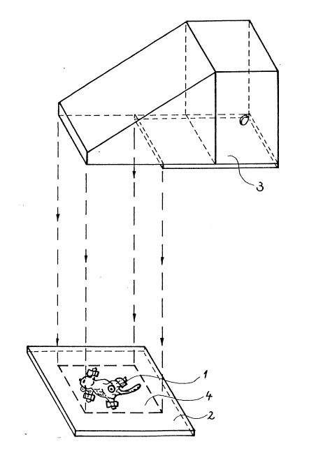
Figure 1 shows a system for the irradiation of
mice with polarized light;
Figure 2 shows graphically the increase of tumor
area versus time for mice of a control group;
Figures 3 to 8 are curves similar to those of
Figure 2 relating respectively to six treated groups;
Figure ~ shows graphically the average of tumor
growth curves;
Figure 10 shows diagrammatically the average
tumor surface for the seven groups on the 2~th day of
treatment; and
Figure 11 shows diagrammatically the results of
~0 toxicity tests.
All experiments were preformed with inhred
BALB/c mice of our own breeding colony originally derived
from the Sloan-Kettering Memorial Institute (New York,
USA~. The tumor used was the methyl-cholanthren induced
.3.~ fi~rosarcoma (BALB/c Meth A) originally derived from ~r.
Old's Laboratory (Sloan-Kettering Memorial Institute).
The tumor was maintained in its ascites form
through serial passage in the peritoneal cavity of RALR/c
1 31 62~2
mice. The Gells were harvested, washed three times in
Hanks' solutiQn, examined for vitality and inoclllated
subGutaneously (s.c.) into the abdomen ~f syngeneic mice.
From prçvious experiments it is well known that the
injection of ~X106 vital cell~ represents 100% take of the
tumor. Therefore this amount of cells was administered in
all cases. For the experiments only male mice were used
with body weight of more than 25 g and of equal age and
breed.
In the experiment~s 7 groups were formed, each
including four or five mice. These groups were first
treated 48, 72 and ~ hours, respectively, after
transplantation and subsequently ea~h day until their
death by linearly polarized light. A group of four mice
served as tumor bearing untreated cQntrQl. The treatment
consisted of irradiation by polariæed light of about 5Q
mW/cm2 power density using a lamp sold under the trade
mark EV~LITE by Rildsystem AB, of Malmo, .Sweden, designed
according to p1~blished European patent application
fl4850395.9. Figure 1 shows a general view of the
irradiation arrangement. Mouse 1 under treatment was kept
on its back and was temporarily fixed on a board 2. Light
source 3 emitted parallel rays of polarized light covering
a rectangular cross-sectiQnal area of 200 x 3Q0 mm
indicated by reference numeral 4. The spectra~
distribution of the irradiating light was in
correspondence with that of a usual metal halogen bulb,
however, components under a wave len~th of about 400 nm
were effectively suppressed. The distance of the treated
~0 surface of the mouse 1 from the exit opening of the light
source .~ was about 18-20 cm. The cross-sectional area of
the irradiating light was larger than the whole surface of
the mouse 1, hence the irradiation was not limited to the
tumorous area.
~5 Each irradiation was performed under neuroleptic
anaesthesia (Vetran~uil 0.01% 0.1 ml intramusGular inj.).
The duration of the daily irradiation was 6, 10, 15 and ~0
minutes in the respective group.s. Each day the mice ~ere
.
6 l 3l 6 222
irradiated at about the same time. Ta~le 1 summarizes the
data of irradiation for groups 1 to fi.
Table 1
Number Group Time of the first naily time
size treatment after tumor of irradiation
in~ection
1 4 4~ h 30 min
2 4 4~ h 15 min
3 5 48 h 10 min
4 5 4~ h 5 min
4 ~2 h 30 min
6 5 96 h 30 min
The growth of the tumor grafts wçre monitored
twice every week by measuring the size thereof by means of
calipers. The tumor size is expressed in mm2 units
obtained by the product of the largest two diameters of
the tumor. Figures 3 to 8 illustrate the tumor growth as
a function of time for the six groups. A similar diagram
for the untreated control group is shown in Figure 2.
On the basis of the measured data, statistical
work was performed using the Mann-Whitney test on the 23rd
day and the Wilcoxon test on the 29th day. The average of
the tumor growth curves for the treated and for the
control groups is shown in Figure 9, while Figure 10
illustrates the average tumor surface on the 29th day for
the respeGtive groups including the control group.
The tumor growth curves demonstrate without
exception that the treatment with polarized light
effectively inhibited tumor growth in each group. On the
23rd day the average surface area of the treated groups
was only 31% of the avorage of the control group, while a
similar ratio at the end of the 29th day was only 23%.
Figure 9 shows that the growth rate of the treated animals
is practically constant from the 6th day to the end of the
29th day, while this rate in the control group was uneven,
with the steepness increasing with time.
.~.
,
7 1 31 6~22
The fact is reflected also from Figure 10, in
which the group averages can be seen separately. This
figure shows that there is no direct relationship between
the irradiation time and the extent of response. The
timing of the first treatment probably has more influence
at least on the initial tumor growth. The survival of the
animals was not investigated in this sort of experiment.
To exclude the possibility that a non-specific
toxic effect of the polarized light Gould be responsible
for the differences in the tumor growth, a separate
toxiGity test was carried out. In this test series, 10
female mice were irradiated by polarized light after
weaning for twelve hours per day. Another group of 10
similar mice was irradiated with non-polarized (normal)
light with identical intensity, also for twelve hours a
day. The increase of weight was measured over a longer
period of time. The results of this test, i.e. average
weight of the respective groups versus time, are
illustrated in Figure 11. This figures shows that apart
from a slower weight increase in the first two weeks, the
weight gains after the first month became very close to
each other and by the end of the sixth week the average
weight of both groups became equal. This demonstrates
that the treatment with polarized light does not have a
toxic effect on the experimentary mice, and the tumor
growth inhibiting effects demonstrated by the tests are
due to specific effects of polarized light on the tumor-
host system.
The experience obtained with polarized light
treatment on mice during other series of tests than
demonstrated here suggest that the effects of polarized
light do not last longer than about 24 hours, and this
explains the daily irradiation rate has been chosen.
The essence of the test-series described
hereinabove is the fact that polarized light with specific
intensity can positively influence the tumor-host
relationship leading to suppression or rejection of
1 31 62,,
otherwise untreatable tumors. The exaGt mechanism of this
effect remains to ~e eluGidated.
There are, however, certain facts and phenomena
which can assist in understanding the way in which
polarized light can accomplish its beneficial effects. It
is known from the literature ~Dvorak, HF; Senger, DR;
Dvorak, AM: Fibrin as a component of the tumor stroma;
origins and biolog.ic significance, published in Cancer
Metastasis Review 1983, 1 pp. 41-73~ that the tu1nor
structure is composed of malignant Gells surrounded ~y
Stroma. The latter regulate the access of inflammatory
cells to tumors. In many transplanta~le tumors,
lymphocytes are confined largely to the tumor-host
interface and do not penetrate into mature tumor stroma or
provisional matrix to any important extent (see also
Dvorak, HF; Dvorak, AM: Immunhystochemical
characterization of infl~mmatory cells that infiltrate
tumors; In: Haskil S. ed: Tumor immunity in prognosis:
The role of mononuclear cell infiltration, Vol. 3 New
York, Marcel Dekker 1~82, pp. 297-307).
According to recent investigations, a well-
defined permea~ility factor is produced by the tumor cells
which renders local blood vessels permeable for protracted
periods (See Senger, Dr; Galli, SJ; Dvorak, AM; Peruzzi,
CA; Harvey, VS; Dvorak, HF: Tumor cells secrete a
vascular permeability factor that promotes accu~ulation of
ascites fluid, Science, 1983; 219: ~33-6). The
discrepancy between rapidly growing tumor cells and
imperfect metaholic supply leads to necrosis which
phenomenon is often referred to as spontaneous
disappearance of tumors (See Folkman J: Tumor
angiogenesis, Adv. Cancer Res.: 1~35; 43 pp. 17~-203).
One of our interestin~ o~servations during the
experiments was that central necrosis occurred earlier and
to a ~reater extent after treatment using the system of
the invention than on the animals without polarized light
therapy. Although such observatinns require mnre
investigation, this situation can mature to a further
13162'2
verification that polarized light stimulates the
immunological defen~e system.
The tumor used for the experiments was a
chemiçally induced type. It is known from our preViQus
experiments (See Rorberg, H; Abdallah, A; ~chwulera, ~;
.Sonneborn, H: Inhibition of tumor growth in a mouse
fibrosarcoma after interleukin 2 applicatl~n, Immun~iol.
1~2. 1~8fi, pp. 383-390 and the references includecl
therein) that the growth of chemically induced tumors can
ke inhibited by mean~ of non-specific immunostimulants, by
lymphocytes from immunised donors and hy soluhle products
of activated lymphocyte~. The fact that polarized light
proved to be beneficial for decreasing the growth rate of
a chemically induced tumor, can also be regarded as
further support for the hypothesis that the mechanism by
means of which polarized light can ~e effective is the
general stimulation of the immune system.
In view of the present invention and the above
outlined hypothesis, the experiments obtained with wound
healing permit a new interpretation. In such test the
~ompositions of the wound 6ecretions before and after
treatment with polarized light were examined and compared
to each other. The treatment resulted in a signifioant
increase in immunoglobulins and other proteins. Also the
cellular composition showed a marked difference: among
neutrophil, granulocytes, lymphocytes and monocytes
appeared and demonstrated activity within 'the wound
secretion. These were caused by changes in vascular
permeability and/or subsequent chemotaGtic effects which
were otherwise lacking. Thus, the particular references
dealing with the adoptive immunotherapy of cancer cited
hereinabove, point out the fact that an increase in
activity of lymphocytes at the close proximity of tumor
cells has beneficious effects. The above demonstrated
increase in activity of the immune system in response to
irradiation with polarized light can cause similar
effests. Thus it can be expeoted that irradiation with
polarized light can be an alternative (or complçment) to
.~.
1 31 62~'~
lQ
the effect of immunomodulatQrs and cytokines as for
instance of IL-2 and,/or IL-2 activated lymphocytes.
It is considered to be significant that during
the experiments not only the tumor area, but the whQle
body surface of the mice facing the source of polarized
light was irradiated. ~ince polarized light with infrared
c~mponents has a certain depth of penetration in tissues,
it can be expected that a treatment with polarized light
on human applicatiQns could be effe~tive if irradiation is
not limited physically to the tumor areas.
