Note: Descriptions are shown in the official language in which they were submitted.
`- 1317180
This invention relates to a surgical stapling
apparatus.
Heretofore, various types of surgical stapling
devices have been known wherein a stapling function takes
place at a location which is relatively remote from the
location at which the stapling device is held and actuated
by an operator. For example, linear closure surgical
stapler devices are described in U.S. Patent 3,494,533 and
circular anastomosis surgical stapler devices are described
in U.S. Patents 4,304,236; 4,351,466; 4,473,077 and
4,488,523 as well as U.S. Design Patents 273,041 and
271,944~ Typically, the stapling devices described in
these patents operate by placing tissue to be stapled in a
clamped manner between an anvil assembly and a fastener
holding assembly, both of which are located at the distal
end of the instrument. The clamped tissue is stapled by
driving one or more fasteners from the holding assembly so
that the ends of the fasteners pass through the tissue and
are formed properly by contact with the anvil assembly.
The forces required to operate the instrument are applied
by the operator of the instrument to one or more actuator
elements located at or near the proximal end of the
instrument. The distal and proximal portions of the
instrument are joined by a longitudinal connecting shaft
structure along which the actuating forces and motions are
transmitted to the distal operation elements. This type of
construction, including relatively widely spaced distal and
proximal portions, may be employed for any of se.veral
reasons, such as the relative inaccessibility of the tissue
to be stapled, the need for good visibility of the tissue
during stapling, and the like. These known types of
surgical stapler devices generally use a plurality of small
and discrete fasteners re~uiring precise registration with
the anvil assembly to ensure that proper fastener formation
occurs during the stapling operation.
~9~
13171SO
Accordingly, it is an object of the invention to
provide a surgical stapling apparatus for applying a
unitary surgical fastener having a multiplicity of
fasteniny points.
It is another object o the invention to
eliminate the need for a plurality of precisely registered
small and discrete fasteners in anastomosis stapling
devices.
It is another object of the invention to provide
a surgical stapler which can be easily and quickly
installed in tissue by a surgical stapling apparatus.
It is another object of the invention to provide
a surgical stapling apparatus with an anvil head capable of
functioning with a unitary surgical fastener.
It is another object of the invention to
eliminate the need for a high degree of rotational accuracy
in the registration of a fastener holder relative to an
anvil assembly in an anastomosis stapling device.
Accordingly, the present invention provides an
anvil assembly for the mounting of a surgical fastener.
The anvil assembly includes a plurality of radiating
fingers which extend angularly of a longitudinal axis, an
annular cutting block which is removably mounted on the
fingers for engagement with an annular knife blade and
means for biasing the fingers radially inwardly of the
block in order to permit movement of the fingers radially
inwardly in response to removal of the cutting block from
the fingers. In one embodiment, the fingers are integral
with a central hub and are resiliently deformable while the
means for biasing the fingers inwardly constitutes an anvil
head having an internal wall receiving the anvil with the
fingers abutting the wall and a retainer which is movably
mounted within the anvil head and anvil in order to abut
the hub and tension the resilient fingers radially
inwardly. In another embodiment, the anvil head has an
annular recess while each finger is separately mounted
.~
1 3 `1 7 1 ~ ()
within the recess. In this embodiment, means for biasing
the fingers includes an axially movable retainer ring
concentrically within the fingers and mounted on the anvil
head and a circular spring encompassing the fingers within
the recess of the anvil head in order to bias the ~ingers
onto the retainer ring. In addition, each finger is
provided with a recess so as to receive the retainer ring
after axial movement of the ring in order to permit inward
radial movement of the fingers under the action of the
circular spring.
A surgical fastener to be mounted upon said anvil
assembly may suitably be comprised of: 1) an annular
stapling part having a plurality of axially extending
circumferentially spaced prongs each of which has a sharp
tip for piercing tissue; 2) an annular retaining part with
an annular gap for receiving the prongs of the stapling
part; and 3) catch means for holding the prongs in the
retaining part in order to clamp pierced tissue
therebetween. For example, the catch means may include a
radially extending barb on at least one of the prongs and
a retaining ring on the retaininy part for butting of each
barb thereon.
The retaining ring may also include a cylindrical
guide wall about the retaining ring in order to define an
annular gap to receive the prongs of the stapling part.
Suitable means are also provided for securing the guide
wall to the retaining ring.
The invention also provides a surgical stapling
apparatus which is comprised of a central shaft, an anvil
assembly mounted on the distal end of the shaft and a
surgical fastener holding assembly mounted on the shaft for
relative movement with the anvil assembly to releaseably
retain two ends of tubular tissue therebetween. In this
regard, the anvil assembly includes a plurality of
radiating fingers extending angularly outwardly of the
shaft, an annular cutting block removably mounted on the
- 13171~0
fingers and means for biasing the fingers radially inwardly
of the block. The holding assembly încludes an annular
knife blade coaxially opposite the cutting block for
severing tissue disposed therebetween. This blade also has
an edge for penetrating and holding the block thereon
whereby upon movement of the holding assembly and the anvil
assembly from each other, the block is removed ~rom -the
fingers to permi~ the fingers to move radially inwardly for
passage through a stapled seam between the two ends of the
tissue.
The aforesaid stapling apparatus permits the user
to staple the tubular ends of a pair of vessels together by
following the steps of clamping the tubular ends of the
vessels between an anvil assembly and a surgical fastener
holding assembly, driving an annular stapling part through
the clamped ends of the tissue into an annular retaining
part removably supported on the anvil assembly and severing
the clamped ends on a circular cutting line disposed
radially within the stapling part. Subsequently, the anvil
assembly may be moved away from the stapled ends of the
tissue to withdraw the anvil assembly from the annular
retaining part, thereafter, collapsing the anvil assembly
radially inwardly of the retaining part and the cutting
line and then withdrawing the stapling
, ~
1 3 1 7 ~ ~0
1 apparatus from the stapled-to~ether vessels~
These and other ob~ects and advantages of the
3 invention will become more apparent fxom the following
1~ detailed description taken in conjunction with the accompanving
drawings wherein:
6 Fig. 1 illustrates a perspective view of a surgical
7 stapling appara~us constructed in accorflance with the invention
~3 in place within an intes~ine;
g Fig. 2 illustrates a ~erspective view of the distal
1~ end of the stapling apparatus prior to fastening;
11 Fig. 3 illustrates a view similar to Fig. 2 of the
12 distal end of the stanling apparatus during a staplinq
13 operation;
14 Fig. 4 illustrates a part sectional side view of the
~pparatus of Fig~ l;
lZ Fig. 5 illustrates an exploded view of the distal
~7 end of the apparatus of Fig. l;
18 Pig. 6 illustrates an exploded view of a surgical
19 ~astener, anvil assembly and holding assembly in accoxdance with
the invention;
21 Fig. 7 illustrates a front view of an annular retaining
22 part in accordance with the invention;
23 Fig. 8 illustrates a front view of an annular stapling
24 part in accordance with the invention;
Fig. 9 illustrates an exploded view of the retaining
26 part and stapling part in accordance with the invention:
27 Fig. lG illustrates side views of the retaining part
28 and stapling part of Fig. 9;
29 Fig. 11 illustrates a view of the distal end of the
3 surgical stapling apparatus prior to stapling;
_5 _
1 ~ 1 7 1 ~0
~ Fig. 12 illustrates a cross sectional view of the
2 stapling components of the apparatus during an initial phase
~f stapling;
Fig. 13 illus~ra-tes a view similar to FigO 12 of the
~, stapling components after stapling;
Fig. 14 illustrates a view similar to Figs~ 12 and 13
7 during withdrawal of the anvil assemblv;
rO Fig. 15 illustrates an exploded view of the distal end
g of a surgical apparatus employing a modlfied anvil assembly in
accordance with the assembly~
1~ Fig. 16 illustrates a cross sectional view of the
12 distal end of the surgical apparatu- employing the anvil
13 assembly of Fig. 15 during a stapling operation;
14 Fig. 17 illustrates a view similar to Fig. 16 of the
apparatus after stapling; and
1~ Fig. 18 illustrates a view similar to Figs. 16 and 17
17 after collapsing of the anvil assembly in accordance with the
18 invention.
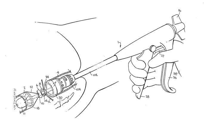
19 Referring to Figs. 1 and 4, the surgical stapling
~o apparatus 1 is used, for example for ~he stapling of two
~1 ends 2, 4 of an in-testine wherein a section of the intestine
~ has been surgically removed between cut ends 6, 8. As
23 indicated in Fig. 1, the cut ends 6, 8 of the intestine are
24 generally tied with suture material 10, 12 with conventional
purse-string suturing being used.
26 The staplin~ apparatus 1 includes a shaft 14 and a
27 hand screw 16 which is articulated to the shaft 14 in order to
28 move the shaft 14. As indicated in Fig. 4, the apparatus 1
29 includes an anvil assembly 17 which is mounted on a distal end
of the shaft 14 and includes an anvil 18 which faces a surgical
--6--
1 ~1 7 1 ~0
1 fastener holding assembly 20 which is also mounted on the
2 shaft 14 fo~ ~elative movement with the anvil assemblv 17 to
3 maintain the two ends of the intestine areas 22, 2~ therebetween.
4 Referring to Fig. 4, the shaft 14 is provided with a
5 sc~ewthread 30 at the proximal end which mates in an internally
6 threaded sleeve extensio~ 32 of the hand screw 16~ The sleeve
7 extension 32 is secured to the hand screw 16 so that both turn
8 together and thus the rotation of the hand screw 16 causes
9 longitudinal movement of the shaft 14. By tightening the hand
screw 16, the anvil assembly can be ~oved towards the holding
11 assembly 20 so that the tissue can be clam~ed therebetween
12 with proper spacing between the anvil assembly 17 and the
~3 holding assembly 20. Calibration means (not shown) may be
14 provided to ensure proper spacing, for example as described in
~.S. Vatent 4,473,077
1~ The apparatus 1 is also provided with a handle 36 and
17 a trigger 28 which is pivotally mounted on a pivot pin 40 secured
18 in the housing of the apparatus 1. A safetv latch 27 is also
19 pivotally mounted on a pivot pin 34 on the handle 36 in order
to prevent pivoting of the trigger 28. In addition, the
21 trigger 28 is articulated in known manner, for example via a
~2 pusher 42 to a slider 44 disDosed about -the shaft 14. This
23 slider 44 abuts a compression sprin~ 46 in order to applv a
24 biasing force on a tube 48 concentric of the sleeve 14 in order
to move the tube 48 distally upon actuation o the trigger 28.
2b The tube 48, in turn, cooperates with an actuator 50 in order to
~7 perform a stapling operation.
28 Referring to Figs. 4 and 5, the shaft 14 has a threaded
29 distal end on which the anvil assembly 17 is mounted in threaded
manner. In addition, a bayonet mount 104 is secured, in known
1 31 7 ~ ~0
l manner on the tube 48. In this regard, ~he holding assembly 20
2 includes a sleeve at the proximal end which carries a pin or
3 fitting into the ba~onet connection of the mount 104.
4 As indicated in Fi~. 4, a sheath 102 is provided over the
central part of the instrument and has a tubular portion 53
Fitting within the bayonet mount 104 (see F.ig. 12).
v~ Referring to Figs. 2 ~nd 3, when the ~pparatus is
initially put in place, the cut ends 6, 8 of the body tissue are
9 drawn in about the shaft 14 with the anvil assembly l~ .in a
spaced condition relative to the holding assembly 20.
ll During stapling, the anvil assembly 17 is drawn against the
12 holding assembly 20 so as to clamp the ends of the tissue between
13 the anvil assembly 17 and the holding assembly 20 (see Fig. 3).
l4 Referring to Figs. 6 and 12, the actuator means includes
an actuator 50 which is abutted against the distal end of the tube
16 48 and is disposed within the holding assemblv 20. In this
17 respect, the holding assembly is ~rovided with a pad 51 which
l8 ~rictionally retains the actuator 5~ in place until the actuator
~g .50 is driven clear of the pad 51 by the tube 48. The actuator
50 carries an annular push ring 54 as well as an annular
21 blade or scalpel 52 which is retained between the actuator 50
22 and push ring 54. In addition, a plastic spacer ring 56 is
23 concentrically disposed between the push ring 54 and the
24 annular scalpel 52. As indicated in Fig. 6, the push ring 54
is provided with a plurality of circumferentially disposed
26 slots at the distal end.
27 An annular stapling part 58 is mounted about the
28 spacer ring 56 and against the push ring 54 (see Fig. 12) and
29 has a plurality of axially extending circumferentially spaced
prongs 60.
1 3 I 7 1 8~)
1 The anvil 18 is made of resilient material and has a
2 plurality of radiating fingers 80 extending angularly outwardly
3 from a central hub 82 concentrically disposed about the
longitudinal axis of the shaft 14. In this regard, the anvil 18
~ is made of one piece with several fingers 80. As indicated
o in Fig. 6, the anvil 18 has a somewhat frustum-like shape with
7 the cylindrical hub 82 at the distal end. In addition, each
8 finger 80 tapers in bo~h thickness and width from the free end to
g the hub 82. In addition, the free end of each finger 80 has an
extension 78 which forms a basal ridge as well as an annular
11 reaction surface 77 within the extension 78.
1~ An annular cutting block 76 is removably mounted within
13 the outer ends of the fingers 80. That is, the cutting block
14 76 abuts against the reaction surfaces 77 of the fingers 80
within the extension 78. As indicated in Fig. 12, the cutting
lS block 76 is aligned with the annular scalpel 52 and is made of a
17 material so as to be penetrated by the cutting edge of the
18 scalpel 52. The cutting block 76 is also shaped so as to be
19 fitted into and about the extensions 78 in a slide fit manner.
An annular retaining part 62 is also mounted at the
21 free ends of the finyers 80 of the anvil 18. As indicated,
22 the retaining part 62 includes an inner cylindrical guide wall 64
23 and an outer retaining ring 66 which are concentrically disposed
24 relative to each other to define an annular ga~ for receiving
the prongs 60. Suitable means in the form of posts 68 (Fig. 8)
26 are provided to secure the retainin~ rings 66 to the cylindrical~
27 wall 64. An annular flange 67 is also provided between the
28 post 68 and the guide wall 64 ~Fig. 7).
29 Referring to Figs. 9 and 10, wherein like reference
characters indicate like parts as above, each prong 60 of the
~ g _
13171~0
1 s~apling part 58 has a sharp tip for piercing tissue while the
2 retaining par~ 62 is positioned to receive the pr~ngs 60. Catch
3 means are also provided for holding the prongs 60 in the
~ retaining part 62 in order i~o clamp the pierced ti,ssue there-
between. As illustrated, ~he catch means includes a radially
6 extending barb 69 on each prong 60 with a proximally facing
7 surface 70 which can be engaged against the retaining ring 66
~ of the retaininy part 62. As indicated, each barb 69 extends
9 radially outwardly of a prong 60 so that the proximally facing
surface 70 can be engaged against the retaining ring 66.
11 The annular stapling part 58 and annular retaining part
12 62 form a surgical fastening 98 which is of relatively simple
13 construction. Both parts 58 62 can be rotated relative to the
14 other and need n~t be precisely registered in order to provide
for stapling.
16 When the two parts 58, 62 are brought together, the
17 prongs 60 pierce the tissue and then enter into the gap between
18 the guide walls 64 and retaining ring 56. At this time, the
19 guide walls 64 and rings 66 temporarily deform due to the
wedging action of the barbs 69. ~fter the barbs 69 clear the
21 retaining ring 66, the ring 66 and wall 62 snap back into their
22 normal relationship in which the surfaces 70 of the barbs 69
23 engage against the retaining ring 66 thus securing the parts
24 58, 62 together while also clamping the two ends of tissue
together. Of note, the flange 67 protects uninvolved tissue
26 from ~he sharp ends of the prongs 60 ~see Fig. 13).
27 j Referrins to Figs. 6 and 12, the anvil assembly 17
28 also includes an anvil head 81 having an internal conical wall
29 which receives the anvil with the fingers 80 abutting against
the wall. In addition, the anvil head 81 has a central shaft 86
_ 10 --
1 3 1 7 1 ~0
1 about which the hub ~2 is mounted via an ~xial opening 84. The
~2 shaft 86 also has an external screw thread 88 on which a huh
3 retainer 92 is ~eaded via internal scrPw threads 94. The
4 hub retainer 92 is thus able to free~heel in relation to the
anvil head 81, that is, the hub retainer 92 can be threaded into
abuttment with the hub 82 with a greater or lesser degree o~
7 force. ln this way, the anvil head 81 and retainer 92 cooperate
8 to form a means for biasin~ the fingers 80 o~ the anvil 18
9 radially inwardly of the cuttin~ block 76 to permit movement of
the fingers 80 radially inwardly in response to removal of the
11 cutting block 76 from the fingers 80. As indicated in Fig. 12,
12 cuttin~ block 76 holds the fingers ~0 in a tensioned state. In
13 addition, the fingers 80 are suitably shaped so as to hold the
14 retaining part 62 in a snap fit relation (see Fig. 12). When
~5 the cutting block 76 and retaining part 62 are in place,
16 dimensional stability is imparted to the resilient fingers 80.
17 As indicated in Fi~. 12, while the anvil 18 is made of
18 a plastic, the anvil head 81 and retainer ring 92 are made of a
19 metal, such as aluminum. Further, the anvil head 81 is provided
with a threaded bore so as to be threaded onto the distal end of
21 the central shaft 14.
22 Referrlng to Fig. 11, in use, in order to staple the
23 tubular ends of the tissue together, the stapling apparatus
24 is inserted in a conventional manner. Thereafter, the ends
of the tissue 2, 4 are pulled to~ether as indicated in Fig. 11
26 about the central shaft 14 so as to dispose two areas 22,
27 24 between the anvil assembly 17 and the fastener holdin~
28 assembly 20. Thereafter, the shaft 14 is moved proximally via
29 the hand screw 16 so as to move the anvil assembly 17 into a
clamped position with the holding assembly 20. In this position,
13171~0
1 the areas 22, 24 of the tissue 2, ~ are clamped between
2 the anvil 18 and the holding assembly 20. Next, triggering
;~ oE the in~trument via the trigger 28 (see Fig. 1) causes the tube
l~ 48 to be moved distally. This in turn moves the ac~uator 50
distally. As a result, the annular scalpel 52 severs the
S clamped ends of the tissue on a circular cutting line 99
7 while penetrating intc the cutting block 76O At the same
r~ time, the push ring 54 p~lshes the prong 60 oE the stapling par~
9 58 through the clamped ends of the tissue into the annular
retaining part 62 with the barb 69 engaqing behind the retaining
11 ring 66 as indicated in Fig. 13.
12 Next, the anvil assembly 17 is moved away from the
13 holding assembly 20 by turning of the handscrew 16 [see Fig. 1~.
14 During this time, the annular cutting block 76 which has been
imbedded by the annular scalpel 52 remains in place on the
16 scalpel 52 as indicated in Fig. 14. At the same time, the
17 annular stapling part 58 remains engaged in the retaining
18 part 62 so as to staple the tissue ends together in a seam as
19 indicated in Fig. 14. In addition, since the cutting block
76 has been withdrawn from the fingers ~0 of the anvil 18,
21 these fingers 80 collapse radially inwardly as also indicated
22 in Fig. 14. The degree of collapse of the fingers ~0 is such
23 that the fingers 80 fall inside of the cutting line 99 defined
24 by the seamed tissue. Thus, the staPling apparatus 1 may then
be removed from within the stapled-together ends. Of note,
26 when the trigger 3B (see Fig. A) is released, the compression
27 spring 46 biases the slider 44 to return to a proximal position
28 which, in turn, pulls back the shaft 48 into a position as
29 shown in Fig. 14. The actuator 50 remains within the holding
3 assembly 20, for example, as indicated by means of a detent
- 12 -
13171~0
1 and a holding ring of the holding assembly 20.
2 The stapling ~art 58 and retaining part 62
~ can be made of any suitable mat~rials, such as nylon,
4 polycarbonate or other material. If a non-permanent fastener
5 is to be used, these parts may be made of a tissue
~ absorbable polymer.
7 Referring to Figs. 15 and 18, wherein like reference
8 characters indicate like points as above, the stapling apparatus
9 may be provided with a modified anvil 118 for the stapling
of the surgical fastener parts 58, 62. In this respect, the
11 anvil assembly has an anvil head 188 which is threaded onto a
12 threaded distal end of the shaft 14 and which includes an
13 elongated sleeve 182 with an annular recess defined by the
14 sleeve 182 and the outer peripherv of the anvil head 188.
As indicated in Fig. 16, the sleeve 182 may be abutted against
16 a shouldered portion of the shaft 14. In addition, a plurality
17 of individual fin~ers 180 are circumferentially disposed with
18 one end 181 within the recess of the anvil head 188. As shown in
19 Fig. 16, the distal end 181 of each finger 180 rests on a sloped
surface on the sleeve 182 while a proximal end rests by way
~1 of plane suraces 184 on the outer surface of an axially movable
22 retainer ring 185 which is mounted on the sleeve 182. In
23 addition, a split circular spring 183 encompasses the fingers
24 180 and is disposed within a groove 184A in each finger so as
to bias the fingers 180 onto the retainer ring 185. Each
26 finger 180 is also provided internally with an intermediately
27 disposed recess 187 whichis spacedaxially from the ring 185 and
28 which is sized to receive the ring 185 upon axial movement of
29 the ring 185 thereinto to permit inward radial movement of
the finger 1~0 under the bias of the sPring 183.
~3l7lao
1 The fingers 180 may be made of plastic, as indicated,
or may be made of anodized aluminum or other metals.
3 The fingers 180 of the anvil 118 are mounted within the
anvil head 188 so as to be biased outwardly by the retaining
~ ring 185 against the interior surface of the anvil head 188.
6 The spring 183 which is in the form o a split ring serves to
7 bias the fingers 180 against the retaining ring 185. In this
8 respect, the fingers 180 tend to pivot about the ends 181
g (see Fig. 17) within the anvil head 188.
As described above, the anvil 118 carries an annular
11 cutting block 176 and an annular retaining part 62 of the
12 fastener at the proximal end.
13 Referring to Fig. 16, the actuator of 150 in addition
14 to carrying an annular scalpel 152 and the fastener holding
~5 assembly 20 also abuts a pusher 186 in the form of a sleeve at
16 a distal end. As indicated, the sleeve 186 has a recessed
17 distal end to slide over the sleeve 182 of the anvil head 188 in
18 order to abut against the retainer ring 185 when the actuator
19 lS0 is moved distally.
Referring to Fig. 16, 17 and 18, the operation of the
21 stapling apparatus is similar to that as described above. In
22 this respect, after the anvil assemblY and anvil head 118 have
23 been drawn towards the fastener holding assembly 20 to clamp the
24 tissue areas 22, 24 therebetween, the triyger (see Fig. 1) is
25 actuated to push the tube 48 and actuator 150 distally. At this
26 time, the pusher 186 slides over the sleeve 18~ of the anvil
27 head 188 while th~ scalpel 152 severs the tissue and penetrates
28 into the cutting bl~ck 176 an~ ~he prongs 60 of the stapliny
29 part 58 pierce the tissue and become retained in the retaining
part 62 as indicated in Fig. 17. The motion of the actuator 150
- 14 -
~3171~0
is such that the pusher 1~6 moves the retainer ring 185
into alignment with the recess 187 of the fingers 180.
Thus, the fingers 180 move under the bias of the spring 183
radially inwardly by pivoting about the ends 1810
Thereafter, the apparatus is manipulated as
described above to displace the anvil 118 from the holding
assembly 120 in order to insure displacement of the anvil
118 from the cutting block 176. At this time, the
apparatus can be withdrawn from within the seamed tissue as
indicated in Fiy. 18. In this respect, the cutting block
remains on the scalpel 152 while the fingers 180 are
collapsed about the retaining ring 185. At the same time,
the sleeve 186 has been moved proximally a slight distance
away from the retaining ring 185. In this condition, the
outside diameter of the fingers 180 has been reduced so as
to pass through the stapled-together tissues.
Of note, the anvil head 188 is sized to be
smaller than the fastener 98 and the sleeve 182 is
contoured so that the anvil fingers 180 are retained in a
snap fit relationship.
The stapling apparatus of the present invention
may employ a surgical fastener of two-part construction
which can be readily manipulated and oriented in place for
stapling of body tissue in an anastomosis procedure.
Because the fastener paxts are annular with the prongs of
one part fitting into an annular groove of the other part,
precise registration of the prongs relative to the annular
gap is not required.
Furthermore, the invention provides an anvil
assembly of relatively simple construction for holding
fastener parts in place for stapling purposes while also
permitting collapse of the anvil assembly for withdrawal
after use. In this respect, the anvil assembly is reduced
in outer contour so as to avoid any injury to the seam
which has been formed by the surgical fastener.
\: '
13171~3()
The invention also provides a surgical stapling
apparatus which is relatively simple to use. Further, the
apparatus can be readily actuated in a single motion to
effect stapling and thereafter, with little effort, be
withdrawn from a vessel.
The stapling apparatus of the present invention
may be used to effect the fastening together the ends of
two tubular body organs in a relative minimum of time and
with relatively minimal effort with respect to previously
known techniques.