Note: Descriptions are shown in the official language in which they were submitted.
~ 3 ~
INTRAOCULAR LENS WITH FOLDABLE SIDES
Field of the Invention
The present invention is directed to an intraocular
lens of a type having foldable sides so that the width of the
lens during insertion in the eye may be made smaller than the
width when implanted, thereby requiring a smaller incision in
the cornea than is now the case for implantation.
Background of the Invention
. .
Many different types of synthetic intraocular lens
structures have been developed to replace the natural lens of
the human eye after lens removal during cataract surgery. In
such operations, an opening or incision is made in the cornea
and in the anterior surface of the capsular bag, commonly in
the area adjacent to the pupillary aperture. The damaged
lens tissue is removed by means of a vacuum tool resulting in
total loss of vision to the affected patient. In order to
restore normal or correctable vision, a variety of lens
structures have been developed which are designed to be
affixed in the intraocular space of the eye. Such structures
commonly comprise a centrally positioned lens and a plurality
of appendages attached to the lens which function to position
`, and secure the lens in front of or just behind the pupil.
The artificial lens is formed from an optically
clear substance and shaped so as to focuse impinging light
onto the retina of the eye. Such lens are commonly optically
formed to be plano-convex, convex-plano or bi-convex. The
appendages attached to the lens typically comprise ~lexible
legs of resilient plastic or metal fibers which are designed
to make contact with appropriate structure in the interior of
the eye.
One commonly employed type of intraocular lens
structure is designed to position the lens in the anterior
. : : ,
; :
: ~ : ~: . ' : '
. :' , : : ' : . :
~1 3 ~
--2--
chamber of the eye just in front of the pupil. A structure
of this type is disclosed, for example, by Kelman (IJ. S.
Patent 4,451,938). Another commonly employed type of
intraocular lens structure is designed to position the lens
in the posterior chamber of the eye just in back of the
pupil. Devices of this type are disclosed by Faulkner (U. S.
Patent 4,366,582) and Shearing (U. S. Patent 4,159,546).
Streck (U. S. Patent 4,361,913) discloses a lens which is
indicated for possible use in either the anterior or
posterior chambers.
Each of the structures mentioned above, except that
of Kelman, is comprised of a single element lens with a
plurality of haptics or position-fixation members attached
to the lens. The lenses ordinarily have a circular peri-
meter~ Thus~ the incision in the cornea of the eye must beat least as long as the diameter of the lens. It is clear
that the longer the incision, the greater will be the trauma
to the eye and the longer will be the recovery time.
Furthermore, since cataract surgery is usually performed on
older patients, the general health of the patient may make i-t
exceedingly important to keep the incision as short as
possible. With this in mind, Kelman discloses in U. S.
Patent 4,451,938 an intraocular lens which is separable into
two body portions. Each body portion is inserted separately
through the cornea and the lens is then reassembled inside
the eye during implacement. Such lens structure certainly
leads to the necessity for a shorter incision in the cornea
than would otherwise be the case. The Kelman device,
however, leads to delicate manipulation of the parts within
the eye in order to reassemble the intraocular lens.
Furthermore, there is a possibility that the mating line of
the two halves of the lens will cause distortion and other
vision problems in the center of the field of view of the
patient. The present invention addresses the problem of
keeping the incision as short as possible in another way.
, .
':
7 ~ ~.
-3
Summary of the Invention
The present invention is directed to an intraocular
lens having a lens body with a primary portion and a secon-
dary portion, wherein the secondary portion may be Eolded
with respect to the pr:imary portion. In this way, the lens
body may be manipulated from an implanted or operational con-
figuration to a smaller insertion configuration so that the
lens may be inserted through a smaller cut in the cornea than
would otherwise be possible. A plurality of position-
fixation members are attached to the lens body for holdingthe lens body in place relative to the eye.
In a preferred embodiment, the lens body has a pair
of side secondary portions which are formed by separations
between the primary portion and the secondary portions along
chords of the preferable circular perimeter of the lens body.
The chords are generally parallel to one another so that each
secondary portion is on an opposite side of the main portion
of the lens body. A connect member forms the attaching ele-
ment between each secondary portion and the primary portion
of the lens. The connect member has an outer edge which
forms a part of the perimeter or circumference of the lens
body. There is a slot between the connect member and the
primary portion of the lens. The slot allows the connect
member to bend inwardly so that the secondary portion may be
moved on top of or beneath the primary portion and within an
envelope formed by the perimeter of the primary portion.
Just as there are opposite secondary portions, there are also
opposite connect members. In addition, a pair of haptics or
position-fixation members are attached to the primary portion
of the lens in the region where the connect members attach to
the primary portion. Opposing haptics hold the lens in place
when implanted in either the anterior or posterior chambers
of the eye.
In an alternate embodiment, the connect member
::
~ :,
, O~
`~
:,
.~.. ". ... - , , ,
: ~ :
7 ~3 ~
--4--
includes a portion parallel to the axis of the lens and a
portion perpendicular to the axis of the lens such that the
secondary portions of the lens body are held beneath or out-
wardly on one side from the primary portion. With this embo-
diment, the secondary portions are easily compressed behindor, in other words, along the one side of the primary por-
tion. Since the secondary portions are not present to per-
form a refractive function, but rather to perform a shading
function, displacing the secondary portions axially to one
side from the primary portion does not detract from their
intended function.
The present intraocular lens is particularly advan~
tageous since the secondary portions may be folded inside the
envelope of the perimeter of the primary portion of the lens
so that during insertion, the lens may be snaked through a
shorter cut in the cornea than would be possible if the lens
were in its operational configuration represented by a cir-
cular perimeter.
The present invention is also advantageous in that
the smaller insertion configuration is obtained with a lens
which keeps the central portion of the lens always intact.
Furthermore, the secondary portions are not missing, but are
simply foldable between insertion and operational con-
figurations so that when implanted, the side portions are
present to shade the retina from direct rays of light at the
edges of the pupillary opening. Although such rays of light
are not needed for focusing the images observed by the
patient, unless such rays are shaded, they irritate and are
otherwise troublesome. Thus, the present invention provides
for a way to insert a lens through a cut smaller than the
diameter of a circular lens, but does so without giving up
the benefits of a circular device.
It is further advantageous that the connect members
between the secondary portions and the primary portion of the
;
~ ",
'^'' .
" \
_5_ ~3~
lens body are resilient so that when the secondary portions
are released after insertion, they automatically assume a
proper operational position.
The present invention is still further important
because in spite of movable side portions and connect members
along the outer perimeter of the primary portion of the lens,
a region is still available for attachment of a pair of hap-
tics on opposite sides of the lens. Furthermore, the haptics
are designed to be yieldable and flexible and provide more
than point contact on each side of the eye.
These several advantages and objects obtained by
this invention are explained further hereinafter and, con-
sequently, may be better understood by reference to the
following drawings and descriptive matter wherein a preferred
embodiment of the invention is illustrated and described in
detail.
Brief Description of the Drawings
FIGURE 1 is a top, plan view of an intraocular lens
in accordance with the present invention;
FIGURE 2 is an end, elevational view of the lens of
FIGURE l;
FIGURE 3 is a cross-sectional view, taken along
line 3-3 of FIGURE l;
FIGURE 4 is a top view of the lens of FIGURE 1 when
the secondary portions are folded;
FIGURE 5 is a side elevational view of the folded
lens showing a tweezers in phantom lines holding the lens in
the folded configuration;
FIGURE ~ shows a dotted line and solid line top
view of the folded lens as held by a tweezers illustrating
two different positions during insertion and also shows in
dotted line representative, implantation configuration rela-
tive to the eye structure;
~ FIGURE 7 is a side elevational view of the lens,
: :
:
.
.
~3~ ~7~
--6--
relative to a cross-sectional view of an eye, with the lens
implanted in the posterior chamber;
FIGURE 8 is a top, plan view of an alternate embo-
diment of an intraocular lens in accordance with the present
invention;
FIGURE 9 is a side view of the lens of FIGURE 8;
FIGURE 10 is a top, plan view of the alternate
embodiment showing the secondary portions folded toward one
another; and
FIGURE 11 is a cross-sectional view taken along
line 11-11 of FIGURE 8.
Detailed Description of the
Preferred Embodiment
Referring now to the drawings wherein like
reference numerals designate identical or corresponding parts
throughout the several views, and more particularly to FIGURE
7, an intraocular lens in accordance with the present inven-
tion is designated generally by the numeral 10. Lens 10 is
shown after implantation in a representative eye 12.
FIGURES 6 and 7 show simplified illustrations of
the eye wherein portions not believed to be necessary to an
understanding of the invention have been omitted for the sake
of clarity. As shown in FIGURE 7, the eyeball includes a
cornea 14 having a scleral spur 16 near the base of the cor-
nea. The diaphragm of iris 18 extends outwardly from the
sides of the eyeball to define a pupillary or irial opening
20. The scleral spur 16 is spaced from the iris near the
base of the cornea to define a groove 22. The natural lens
has been removad with only the rear portion of the capsular
bag 24 remaining. An aqueous zone between the cornea 14 and
the capsular bag 24 is subdivided by the iris 18 into an
anterior chamber 26 and a posterior chamber 28. Artificial
lens 10 is shown installed in posterior chamber 28. It is
understood that lens 10 may be implanted in either the
anterior or posterior of chambers 26 or 28.
''
., -
.: . . ..
.
.. . .
--7--
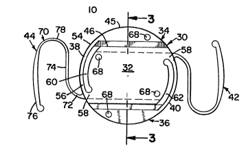
As shown in FIGURES 1-3, intraocular lens 10 in
accordance with the preferred embcdiment of the present
invention includes lens body 30 having a primary portion 32
and a pair of secondary portions 34 and 36. Lens 10 can be
formed from any suitable material which is compatible with
the environment of the eyeball, such as a non-toxic plastict
for example, polymethylmethacrylate. Secondary portions 34
and 36 are connected to primary portion 32 by a pair of con-
nect members 38 and 40. Haptics or position-fixation members
42 and 44 extend from primary portion 32 outwardly for
yieldingly pressing against the side of the eye at either the
soft tissue at the edge of the capsular bag behind the iris
in the posterior chamber or in groove 22 in the anterior
chamber.
Lens body 30 is preferably shaped to have a cir-
cular perimeter when connect members 38 and 40 are relaxed.
As shown in FIGURE 4, the insertion width of lens body 30
with connect members 38 and 40 bent is significantly smaller.
Primary portion 32 forms the central refractive part of lens
body 30. Secondary portions 34 and 36 form opposite sides of
lens body 30 and are separated from primary portion 32 along
substantially parallel chord lines. Thus, each of secondary
portions 34 and 36 preferably have a portion of the circular
circumference of lens body 30 as one edge 45 and a chord line
as the other edge 46 as shown with respect to secondary por-
tion 34 in FIG~RE 1. Although secondary portions 34 and 36
; may mate with the curved or planar top and bottom surfaces of
primary portion 32, it is only necessary that the top and
bottom surfaces 48 and 50 of secondary portions 34 and 36 be
planar. This is the case since the greatest percentage of
light rays which are focused on the retina pass through pri-
mary portion 32. Although some light passes through secon-
dary portions 34 and 36, which may be opaque, such light need
not be focused to provide adequate vision. It has been
,,
~ i
.
- ': : '
.~3~7~
--8--
found, however, that secondary portions 34 and 36, which may
be opaque, should not be eliminated in an attempt to obtain a
smaller lens body for insertion through the cornea since they
serve the valuable purpose of shading the retina from side
rays of light, which side rays otherwise irritate the retina.
Connect members 38 and 40 extend along -the edge of
primary portion 32 to secondary portions 34 and 36. As shown
for connect member 38, for example, one end 54 of connect
member 38 is attached preferably integrally at one end of
secondary portion 34. The other end 56 is attached pre-
ferably integrally with primary portion 32. End 56 is
attached to primary portion 32 at a region 58 which is a part
of primary portion 32 and adjacent to the other secondary
portion 36. The other connect member 40 is connected in a
similar fashion and attached to primary portion 32 in a simi-
lar region at the other end of a diameter across lens body
30. Slots 60 and 62 separate connect members 38 and 40 from
primary portion 32. Slots 60 and 62 provide space within
which connect members 38 and 40 may bend as secondary por-
tions 34 and 36 are folded under primary portion 32 to formthe second perimeter for insertion as shown in FIGURE 4.
Since connect members 38 and 40 are preferably integral both
with primary portion 32 and secondary portion 34 or 36, con-
nect member 38, for example, has the thickness of primary
portion 32 at end 56 while decreasing gradually in thickness
to the thickness of secondary portion 34 at end 54 as shown
in FIGURE 2. Also, note that the outer edges of connect mem-
bers 38 and 40 are a part of the circumferential perimeter of
lens body 30. :~
Although not shown, secondary portions 34 and 36
may be separated from primary portion 32 by an axially
~ aligned slot. Preferably, however, the separation is made in
:~ a way which creates as small a space as possible between
~ secondary portions 34 and 36 and primary portion 32. As
.
' '.~
: '.' :
: '
'
. , :
~ 3 ~
g
shown in FIG~RE 3, grooves 64 may be formed in back 52 of
primary port;on 32 near edges 46. Grooves 64 have a depth
approximately one-half the thickness of secondary portions 34
and 36. Grooves 66 are formed adjacent to edges 46 in top
surfaces 48 of secondary portions 34 and 36. Grooves 66 also
have a depth of approximately one-half the thickness of
secondary portions 34 and 36. In any case, grooves 64 and 66
have depths just sufficient so that preferably a corner of
one of grooves 64 intersects a corner of one of grooves 66.
With a wall of one of each of grooves 64 and 66 in common,
the intersection corner provides a separation space between a
secondary portion 34 or 36 and primary portion 32 wherein the
separation space has as small a thickness as possible.
A plurality of openings 68 are formed in secondary
portions 34 and 36 and primary portion 32 so that an
appropriate instrument may engage one or more of the openings
for the purpose of positioning lens 10 during implantation.
Preferably, an opening 58 is formed near the end of each of
secondary portions 34 and 36 and in a pair of opposite cor-
ners of primary portion 32 at opposite ends of edges 46 fromopenings 68 in secondary portions 34 and 36.
Haptics or position-fixation members 40 and 42 are
preferably integral with primary portion 32 and attached at
regions 58. Haptics 40 and 42 are identical. Haptic 40, for
example, has a U-shaped portion 70 with a connecting portion
72 extending from one leg 74 of U-portion 70. The other leg
76 is preferably formed to fit along an imaginary surface
substantially concentric to the circular perimeter of lens
body 30. Such shape allows for bending of haptic ~0 not only
in the region of connect portion 72 but also along other por-
tions of U-shape 70, and especially near base 78. Haptic 40
serves to provide more than a point contact against the side
tissue of eye 12. In this way, intraocular lens 10 need have
only a pair of haptics, rather than a larger number.
::
:
~,
~1 3~7~
--10--
Furthermore, haptics 40 and 42 are flexible and yieldable
and, consequently, provide an appropriate tight fit. Haptics
and 42 are formed to extend sidewardly and slightly
beneath back side 52 of primary portion 32, as shown in
FIGURE 3.
As indicated hereinbefore, the present invention
provides for a preferred circular first perimeter which is
the desired shape after implantation of lens 10. In addi-
tion, the present invention provides for a reduced, second
perimeter for insertion so that a smaller cut may be made in
the cornea. The reduced second perimeter is shown in FIGURE
4, wherein connect members 38 and 40 are bent so that secon-
dary portions 34 and 36 fold behind primary portion 32 and
within the envelope of the perimeter of primary portion 32.
FIGURE 5 shows a tweezer 43 or other tool in phantom lin~s
holding the secondary portions 34 and 36 in the folded con-
figuration. FIGURE 6 shows one of the haptics being snaked
through the cut in cornea 14 while the solid lines in FIGURE
6 show lens 10 at a position where lens body 30 is ready to
be inserted through the cut. Once inside the cornea, the
tweezer or pinching tool may be released and secondary por-
tions 34 and 36 automatically spring back to the operational
configuration wherein connect members 38 and 40 are relaxed
and lens body 30 again assumes a circular perimeter. FIGURE
6 includes a further phantom illustration showing lens 10
relative to eye 14 after implantation.
Thus, in use, secondary portions 34 and 36 are
folded with respect to primary portion 32 to form a folded
configuration having a smaller perimeter than the unfolded
configuration. The lens body is held in the folded con-
figuration, and the intraocular lens is inserted through the
incision in the cornea. The lens is positioned and secured
to the eye with the position fixation members so that the
lens body is in line with and substantially parallel with the
.,
-
.:
~ 3 ~
pupillary or irial opening of the eye. The folded secondary
portions 34 and 36 are then released for return to the
unEolded configuration. The release of the folded secondary
portions may also occur immediately after the lens has been
inserted through the incision. A tool engages one or more of
openings 68 so as to properly position lens lO.
In an alternate embodiment, as shown in FIGURES
8-11, the various elements which are similar to the earlier
described embodiment are identified by identical numbers,
only the numbers are primed. In this embodiment, lens lOI
includes a lens body 30' having a primary portion 32' and a
pair of secondary portions 34' and 36'. Secondary portions
34' and 36' are connected to primary portion 32' by a pair of
connect members 38' and 40'. Haptics 42' and 44' extend out-
wardly from lens body 30'.
Lens lO' is generally similar to lens 10. Lens 10'is different from lens 10 in that secondary portions 34' and
36', as well as haptics 42' and 44' are attached and held to
primary portion 32' at a different side elevation. As shown
in FIGURE 9, primary portion 32' includes first and second
opposite sides 80 and 82. Primary portion 32' has an axis
84. Side 82 of primary portion 32' is planar and is perpen-
dicular to axis 84. It is understood, however, that primary
portion 32' may be any type of lens, e.g., convex, concave,
etc. In any case, connect members 38' and 40' and haptics
42' and 4A' are held by a neck 86 (see FIGURE 11 ) at a level
spaced outwardly from a plane, in a direction along axis 84
opposite from first side 80, where the plane is perpendicular
to axis 84 and is nearer second side 82 than side 80 and
includes at least some of primary portion 32'. That is, in
FIGURE 9, the plane could be side 82 or at an elevational
level spaced somewhat upwardly from side 82. Secondary por-
tions 34' and 36' are held spaced from the plane at the same
elevational level with respect to primary portion 32' as con-
nect members 38' and 40'.
~ ~,
. ~
:
:
-:
:
11 3 ~
-12-
Neck 86 is connected to primary portion 32' at an
end 96. Ends 96 are an edge of primary portion 32' which is
not adjacent along most o its length to one of secondary
portions 34' ant 36'. In FIGURE 8, one of necks 86 is
located near a corner of one of ends 96 and one of edges gn
of primary portion 32'. The other neck 86 is located simi-
larly at a diagonally opposite corner. Necks 86 extend in an
axial direction sufficiently far so that when one of haptics
42' and 44' and one of connect members 38' and 40' connect to
and extend away from neck 86, they do so in the relationship
discussed hereinbefore.
Although necks 86 have been described with par-
ticularity and specifically located, it is understood that
they could restructure and locate differently, or that the
haptics and connect members be formed to angle axially in the
same direction away from the primary portion in the fashion
of haptics 42 and 44 as shown most clearly in FIGUR~S 2 and
3.
It is noted that since secondary portions 34' and
36' are present to provide a shading function, as opposed to
a refractive function, it does no-t matter whether the secon-
dary portions have an optically critical relationship with
respect to primary portion 32'. The important relationship
concerns minimizing light from passing between primary por-
tion 32' and secondary portions 34' and 36'. In this regard,in FIGURE 8, secondary portions 34' and 36' are shown in a
plan view as being slightly spaced from primary portion 32'.
It is preferable, however, that a separation space 88 not
occur between a first edge 90 of primary portion 32' and a
second edge 92 of secondary portion 34', for example, but
rather that space 88 would occur between a portion of a side
94 of secondary portion 34' and side 82 of primary portion
32'. It is understood, however, that space 88 may occur bet-
ween first and second regions where the first region includes
. : ' ~ ' ' '
-13- ~97~
an edge 90 of primary portion 32' and adjacent parts of oppo-
site sides 80 and 82 and where a second region includes an
edge 92 and opposite sides 94 of secondary portion 34'.
Although the embodiments described have generally
had a circular perimeter when viewed in plan view, it is
understood that other shapes and configurations are encom-
passed within the spirit of the present inventionD In par-
ticular, it i5 envisioned that both secondary portions could
be attached to connecting members which have a common attach-
ment to the primary portion at one of the ends o the primaryportion, an end being an edge of the primary portion not
adjacent to a secondary portion. This configuration would be
particularly applicable with respect to the concept of the
alternate embodiment wherein the secondary portions are held
at a different elevational level than the primary portion.
In any case, the details of the structure and func-
tion, including advantages, have been set forth with respect
to the preferred and an alternate embodiment of the present
invention. It is understood, however, that such details are
exemplary. Therefore, any changes made, especially in mat-
; ters of shape, size, and arrangement, to the full extent
extended by the general meaning of the terms in which the
appended claims are expressed, are within the principle of
the invention.
:
:- - .