Note: Descriptions are shown in the official language in which they were submitted.
` TRAP:010
O~JECTIVE LENS POSITIONING SYSTEM
FOR CONFOCAL TANDEM SCANNING
REFLECTED LIGHT MICROSCOPE
The present invention relates to light microscopes,
and more particularly, it relates to scanning the plane of
focus up and down through a specimen to optically section
the specimen.
Confocal scanning light microscopy involves use of an
objective lens to bring light to a focal point in an
object plane. Reflected light from the focal plane is
brought into focus on a viewing eyepiece. To image an
entire field, a mechanical scanning disk havlng light
transmissive areas is used.
The object to be imaged is placed on a scan table or
stage in the focal ~lane between the objective lenses.
Heretofore, this table has been moved in the X-Y plane by
electromechanical drivers and in the vertical Z direction
by mechanical and piezoelectric element drivers. Movement
of the specimen stage relative to the objective lens has
been used for optical sectioning of the specimen.
: ~
: :. , : :, :,
,. .
:. ': ,- ,
:: : ~ . :: , :,: .
-
-2- ~3~ 8
In the prior art, movement of an objective lens has been
used exclusively for op~ical alignment to ensure confocal
operation. OnlY the vertical movement of the stage has been used
to perform optical sectioning.
The present invention in one aspect provides a scanning
light microscope, comprising a specimen stage, a confocal
reflected light image scanning system including an objective lens
movahle relative to the specimsn stage along first and second
mutually perpendicular axes and a control system for positioning
the objective lens to a predetermined location within the plane
of the perpendicular axes/ the control system including first and
second piezoelectric elements to translate the obiective lens
relative to the stage and first and second eddy current sensors
to monitor the position of the objective lens.
A further aspect of the inverltion provides a method of
stereo image collection with a confocal tandem scanning reflected
light microscope having a specimen stage and an objective lens,
comprising the steps of moving the objective lens vertically and
laterally relative to the specimen stage under feedback position
control between first and second imaging positions to produce two
range images at the same vertical depth of the plane of focus
within a specimen.
The present invention allows an operator to scan the plane
of focus up and down through the specimen. A relatively thick
specimen can be optically sectioned to a depth that depends only
upon the working distance of the objective and the degree of
translucency of the specimen. The tedious and dlfficult task of
slicing the specimen into thin sections is avoided. Also avoided
is the complex, expensive scheme of a motor-controlled stage.
The present invention also provides for stereo images to be
collected using two axes of motion to produce the required
parallax without moving the specimen.
A written description setting forth the best mode presently
,~ ",
--3- ~3~ 8
known for carrying out the present invention and of the manner of
implementing and using it, is provided by the following detailed
description of a preferred embodiment which is illustrated in the
attached drawings wherein:
FIGURE 1 is an illustration of a confocal scanning light
microscope;
FIGURE 2 is a diagram of the interior of the head of ~he
microscope;
FIGURE 3 is a diagram of the objective lens positioner
viewed from the top of the head;
FIGURE 4 is a diagram of the lens positioner viewed from the
front of the head;
FIGURE 5 is a block diagram of the objective lens positioner
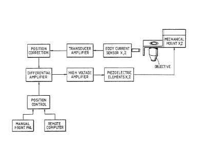
and control system; and
FIGURE 6 is a schematic diagram of the circuitry for
position control system. (FIGURE 6 is composed of FIGURES 6A and
6B).
Tandem scanning microscopy (TSM) involved confocal imaging.
Illumination enters through an illuminating aperture on a
scanning disk and is focused by objective lenses. Only
reflective light from the focal plane of the objective passes
through a viewin~ aperture. Light from above or below the focal
plane is not brought to focus on the viewing aperture and is
blocked by the disk. The viewing aperture is a conjugate
aperture on the observation side of the scanning disk. In
practical devices, there are thousands of apertures on the disk.
Objects in the focal plane of the objective are
illuminated by the point source and the liyht reflected
by the specimen is seen by a point detector.
In practice, the point source and point detector
are obtained by placing apertures between a conventional source
and detector and the objective lens. Confocal imaging is
achieved when the system is preciselY aligned via a system
. " .
,,
.
: : . : , ,
- 132~
of adjustable mirrors and a beam splitter so that rays
from the source aperture pass through the viewing
aperture. Rays that emerge from objects out of the focal
plane are not focused at ~he viewing aperture and are
blocked from reaching the detector. The result is a high
contrast image of a small portion of the specimen at the
focal plane. To see an entire field, a means is required
to scan either the specimen or the illumination and
detector. This is accomplished by scanning the source and
detector by means of a scanning disk having light
transmissive areas.
The diagram of Figure 1 illustrates a physical form
for a practical TSM instrument. The instrument 10 has a
stand 12 for placement on a planar surface such as a table
top. The stand 12 supports specimen platform or stage 14.
Vertical adjustment of platform 14 is provided by rotation
of knobs 16. Mounted atop stand 12 is the head 18 which
includes the optical components and scanning disk. Also
included is Epi-illuminator 20 which conveys light from
the lamphouse 22 into the head. The Epi-illuminator
contains several lenses, iris diaphragms and filter
holders in order to adjust the apparent brightness and
emission spectrum of the light source.
For further information as to the structure for
realizing a TSM, U.S. Patent No. 3l517,980 may be
referred to.
A top view through the interior of the head is shown
in 2. From this view, the components of the microscope
are in view. The optical light path through the device
begins at the input mirror 24~ Light from this mirror is
reflected by steering mirror 26 to the beam splitting
pellicle 28 The beam splitter directs light to the
objective mirror 30, and the objective lens 36. The
.. ., . ~
Y
~32~
objective lens 36 directs the reflected i~age to the
objective mirror 30 and onto the beam splitting pellicle
28. The image is reflected from the pellicle 28 and
eyepiece turning mirror 32 and is projected on the
scanning disk 34. Also in Figure 2, the mechanical
mounting of the control system for positioning the
objective lens is shown.
The confocal scanning system includes an objective
lens movable laterally and vertically to its optical axis.
Movement of the objective lens is by piezoelectric
elements. The Z-axis element is item 38 r and the X-axis
element is item 40. Movement of the objective lens in the
Z-axis direction is detected by eddy current sensor 42.
Similarly, movement of the objective lens in the X-axis
direction is detected by eddy current sensor 44.
Referring to Figure 3, the objective lens positioning
mechanism is further shown in a top view of the microscope
housing head. The objective lens mount and the X-axis and
Z-axis piezoelectric positioners are shown. Also depicted
are the position sensors. The piezoelectric positioners
are available from Burleigh Instruments, Inc. of Fishers,
New York, 14453. The preferred device is the model PZS-
50TN MICROSTAGE* The eddy current sensor is a two part~non-contacting transducer for proximity detection. One
part of the proximity transducer system is the proximity
probe and the other part of the system is the sensor
amplifier. Shown in Figure 3 is the proximity probe
portion. The preferred proximity probe is the model
82015-00-08-15-02 device available from Bentley Nevada.
The prefPrred sensor amplifier is the model 40892-03 Micro
Prox~which is also available from Bentley Nevada.
The eddy current sensors detect the movement and the
position of the objective lens mount. The proximity
* Trademarks
,
:
.
.
" ~ -
.. ' .-' ~ ' , .
- ~2~8
transducer sensors operate on the edd~ current principle.
The proximity transducer senses the distance between the
probe tip and the surface it is observing, which is shown
in Figure 3 as the sensor target. A radio frequency is
generated through the probe tip into t:he observed
material, setting up eddy currents. The loss of energy in
the return si~nal is detected, and an output signal is
generated.
The piezoelectric positioning devices are miniature
electromechanical devices. The active element is a piezo-
electric actuator that expands when voltage is applied.
The positioner has a unique position for each applied
voltage level.
Referring to ~'igure 4, a view from the front of the
microscope housing shows the objective lens mount and its
position relative to the Z-axis piezoelectric positioner,
and the Z-axis eddy current sensor. Similarly, the
piezoelectric positioner and eddy current sensor for the
X-axis direction of lens movement is shown.
Figure 5 illustrates in block diagram form the
objective lens positioner system in accordance with the
~5 present invention. The mechanical mounting of the
objective lens and an X-Z plane is indicated. Mechanical
input to move the objective lens is provided by
piezoelectric elements. The position of the objective
lens is detected by eddy current sensors. As indicated,
the output signal from each sensor transducer is amplified
and combined with a position correction input. The
composite signal is applied as an input to a respective
X-axis or Z-axis differential amplifier. The other input
to each differential amplifier is a position control
signal obtained from either a manual, local input, or from
a remote, automatic input. The manual input can be a
~ 3 2 ~
control on the front panel of the microscope. The remote
input can be a computer generated signal. The output of
the differential amplifier is applied to a high voltage
amplifier for driving the piezoelectric elements. It will
be appreciated that the system shown in Figure 5
illustrates a novel feedback system in the scanning
microscope art.
In Figure 6, a schematic diagram of the Z-axis
position control system for the lens positioner is
presented. The voltage of the eddy current sensor
amplifier output is applied via terminal 50 as an input to
amplifier stage 52. A reference voltage established on
potentiometer 54 provides an offset correction to
compensate for positioning error in the sensor. The
reference voltage is applied by amplifier 56 to the input
of gain stage 52. The offset reference voltage and the
eddy current sensor voltage are summed by amplifler 52.
Potentiometer 58 in the feedback network of amplifier 52
establishes the gain setting.
The output of amplifier 52 is applied to a unity gain
amplifier stage 60. The output of amplifier 60 is applied
to the inverting input of differential amplifier 62. The
non-invertlng input of differential amplifier 62 receives
an input from analog switch 64. This switch selects
either the Remote or the Local position setting input. If
the Local input is selected, the signal input to
differential amplifier 62 is from voltage follower 66.
3Q The input to staye 66 is from a potentiometer 68 coupled
to a reference voltage. If the Remote input is selected
by switch 64, the input to differential amplifier 62 is
obtained from a Remote source coupled to input terminal
70. Preferably, the Remote source would be a computer
generated signal. Either the Remote or the Local input
.
- ~ , , . .:
-: .
... . . .
:~ ~ 2 ~
provides the desired position-setting input to the
feedback control system.
The output of differential amplifier 62 provides an
output error signal for driving the lens positioning
mechanism to a proper location. The output signal is
applied to a high voltage amplifier stage 72. The output
voltage to the piezoelectric positioning element is
available at terminal 74. The voltage at terminal 74 is
established between ~150 volts and -15 volts based upon
the amount of current drive through transistor 76.
Current limiting protection for the piezoelectric elemient
is provided by transistor 78. When current flow through
resistor 80 develops a sufficient voltage to turn-on
transistor 78, base current is removed from transistor 82.
This in turn reduces the current flow through transistor
76. Amplifier stage 84 and t~ansistor 86 provide
buffering of the differential amplifier 62 output signal.
The circuitry for the X-axis position control is
identical to that of Figure 6.
The X-axis, Z-axis position control of the objective
lens provides a unique feature of recording extended focus
or range lmages by continuall~ changing the focus level
while recording one photographic image. Two range images
of the same vertical depth "slice" but with inclined axes
of focusing, achieved by a combination of a small lateral
movement (X) while focusing (Z), constitute a stereo pair.
The foregoing description of the invention has been
directed to a particular preferred embodiment for purposes
of explanation and illustration. It will be apparent,
however, to those skilled in this art that many
modifications and changes may be made without departing
from the essence of the invention. It is the
~32~8
applications' intention for the following claims to cover
such equivalent modifications and changes as fall within
the scope of the invention as defined by the following
claims.
- . . ,
:
::: ...
, . , :
