Note: Descriptions are shown in the official language in which they were submitted.
1 32 1 520
APPARATUS FOR
REDUCING A FRAC~URE
FIELD OF THE INVENTION
The present invention generally relates to
an apparatus for reducing the fracture of a bone and,
in particular, to a tool for reducing the fracture of
a femur.
BACKGROUND OF THE INVENTION
In the field of orthopedics, various
techniques are employed for holding together parts of
a fractured bone during the healing process. However,
prior to the fixation of the bone fragments, it is
first required that the fracture be reduced, that is,
the various bone fragments or pieces must be
repositioned in their proper relative arrangement
before the fractured bone can be fixed or stabilized
for healing. U.S. Patent No. 4,103,683 generally
refers to reduction of a fracture which is maintained
with suitable bone clamps.
Another reduction technique is illustrated
and described in U.S. Patent No. 4,127,119 which
includes upper and lower pin holder assemblies that
have a ring-like configuration and can be positioned
about the limb to be reduced. Bone penetrating pins
are secured to the appropriate pin holders in these
assemblies. A solely external fracture reduction
system is described in U.S. Patent No. 3,850,166.
However the apparatus of this '166 patent is intended
for use only for lower limb fractures. Moreover, it
is not suitable for complicated fractures which result
in disorientation of the bone fragments.
I
X ~
321 520
Still yet another fracture reduction apparatus is
described in U.S. Patent No. 4,628,922 which illustrates
single-sided fixation of a bone fracture which requires
fixation pins to be inserted through the bone fragments.
Although this device is said to be able to reduce the
fracture, it involves a relatively complicated procedure in
that movement of one component will affect the orientation
of any other component. Furthermore, rotation is limited
in view of the skin and tissue through which the pins
penetrate.
The use of elastic nails is described in U.S.
Patent No. 4,467,983. In the example illustrated, the
nails are passed into the medullary canal through a hole in
the bone and can be rotated so as to reduce the fractured
femoral head. However, these nails at the least are not
convenient for measurement of the final nail to be inserted
for fixation of the fracture. Furthermore, these nails
require special configurations as well as elastic portions,
as noted, in order to permit their use in the reduction
process. Since the bone hole serves as a fulcrum point,
these nails are not capable of fine adjustment or ease of
use within the medullary canal.
We have invented a tool for reducing fractures
and particularly for reduction of a fractured femur which
overcomes the limitations noted above. The fracture
reduction tool of the present invention is useful in
reducing the fracture, in passing the reaming guide wire
and in measuring the length of the nail or rod to be using
in ultimately fixating the fracture. These procedures are
not collectively available with any of the aforementioned
prior art devices. Moreover, the tool of the present
invention can also be used for different lengths of bone
and accordingly avoids the need for tools of various sizes.
Also it is appropriate for use where there are combinations
of fractures involving the femoral shaft and the femoral
neck. In that instance the tool can be used to achieve
both reductions.
1 321 520
-- 3
SUMMARY OF THE INVENTION
The present invention is directed to an
apparatus for reducing a fractured bone, comprising
shaft means configured and dimensioned for passage of
its distal end into the medullary canal of the
fractured bone through a suitably sized aperture for
manipulating into and within the medullary canal by
translational and/or rotational movements so as to
reduce the fractured bons, the shaft means having a
first bore along its length, and measurement means
configured and dimensioned for movement over at least
a portion of the shaft means, so as to permit
determination of the length of a nail to be inserted
into the medullary canal of the reduced bone.
According to one preferred embodiment, the
shaft means is a hollow elongated shaft of a generally
uniform diameter along its length and its beveled at
its distal end. Also the first bore of the shaft is
of a generally uniform diameter along its length. The
shaft preferably has unevenly spaced apart graduations
along a portion thereof. These graduations which
cooperate with the measurement means for determination
of the nail length and are in the range of thirty
through forty-eight centimeters with one centimeter
increments.
The measurement means comprises a tubular
sleeve movable at least along the portion of the shaft
having the graduations. The tubular sleeve includes a
window so as to permit viewing of the graduations on
the shaft under the window. An arrow indicator is
positioned generally midway of the window so as to aid
in measurement of the length of the nail to be
inserted. Means are provided for selectively locking
the measurement means in a predetermined position on
the shaft. The tubular sleeve
l432 1 520
further compris-~ a pa~sag-way communicating with the outer
surface of the shaft ~he measurement loc~ing means
comprises a screw dimensioned and configured for
cooperative engagement wlth th~ passageway so that the
screw can be advanced into the passageway and thereupon
contact the outer surfac- so as to selectively lock the
tubular sleeve in position on the shaft
The apparatus of the present invention further
10 comprises handle means coupled to the proximal end of the
shaft for ald in manipulating the shaft into and within the
medullary canal, th- handle means having a second bore
along its length aligned coaxially with the first bore of
the shaft mi~ handle mean~ compris-s an elongated handle
15 body which i~ op-n at one nd to receiv- the proximal end
Or the ~haft th-r-in Chuck mQans are provided for
selectively sQcuring the sha t to the handle body ~he
chuck mean~ compris-~ first thread~ positioned ad~acent the
open end of th- handl- body and a collar having a bore
dimen~ioned and configured so as to permit movement of the
collar along at least the proximal end of the shaft The
collar ha~ s-cond thr-ad~ within its bor- corresponding to
th- fir~t thr-ad~ on th~ handle body so as to permit
coop-rativ- engag-ment of th- first and the ~econd threads
Juch that th- op-n nd of the handle body is selectively
pre~-d by th- collar into contact with the shaft for
s-cur-m-nt th-r-of Th- handl- mean~ further comprises a
gen-rally tubular handl- grip positioned about a portion of
th- longat-d handl- body The handle means further
comprl~-~ a handl- rod positioned on the distal end of the
30 handl- body and xt-nding transversely to the second bore
In a pref-rred embodiment, the apparatus for use
with a guid- wire for reducing a fractured bone The ~irst
bor- of the shaft means has a diameter greater than the
1 32 1 520
- 5
general diameter of the guide wire to allow selective
passage of the guide wire through the first bore.
Means are also provided for selectively locking the
guide wire within the second bore in a predetermined
S position relative to the shaft means.
According to another preferred embodiment,
the present invention is directed to an apparatus for
use with a guide wire for reducing a fractured femur,
the guide wire being of a generally uniform diameter
and having an enlarged distal end portion for
placement into the medullary canal of the fractured
femur, comprising shaft means configured and
dimensioned for passage of its distal end into the
medullary canal of the fractured femur through a
suitably sized aperture to accommodate passage of the
shaft means into the medullary canal and for
manipulating at least either the shaft means or the
guide wire into and within the respective medullary
canal portions of the fractured femur by translational
and/or rotational movements so as to reduce the
fractured femur, the shaft means having a first bore
along its length, the first bore having a diameter
greater than the diameter of the guide wire to allow
selective passage of the guide wire through the first
bore, and measurement means configured and dimensioned
for movement over at least a portion of the shaft
meanfi, so as to permit determination of the length of
a nail to be inserted into the medullary canal of the
reduced bone.
The apparatus further comprises handle means
coupled to the proximal end of the shaft for aid in
manipulating the shaft into and within the medullary
canal. The handle means has a second bore along its
length aligned coaxially with the first bore of the
shaft. This second bore has a diameter greater than
the diameter of the guide
1 32 1 520
wire to allow selective passage of the guide wire
through the second bore.
Preferably, the handle grip is integrally
molded about the portion of the elongated handle body.
The apparatus further comprises means for selectively
locking the guide wire within the second bore in a
predetermined position relative to the shaft means.
The handle body comprises a passageway communicating
with and extending transversely to the second bore.
The guide wire locking means comprises a rod
dimensioned for positioning in the handle body
passageway and extending therefrom to a free end. The
rod has a cam positioned thereon so that the cam
enters into the second bore during at least a portion
of one complete rotation of the rod. The cam is
configured and dimensioned so that the cam when
entering the bore will contact and selectively lock
the guide wire within the second bore in position
relative to the shaft. A knob is secured to the free
end of the cam rod for ease in rotating the cam rod.
The present invention is also directed to a
method for reducing a fractured bone such a femur,
whereby a portion of the fractured bone is exposed as
an entry site into the medullary canal of the
fractured bone. At the entry site an entry hole is
drilled into the medullary canal. A fracture reducing
apparatus of the present invention is inserted with a
guide wire therein through the entry hold into the
medullary canal. At least one of either the shaft
means or the
- 7 -
~ 3~
guide wire is manipulated through the medullary canals
of the fragment portions of the fractured bone so as
to reduce tbe fracture.
The method further comprises providing
measurement means on the shaft means prior to
insertion through the entry hole, the measurement
means configured and dimensioned for movement over at
least a portion of the shaft means, so as to permit
determination of the length of a nail to be inserted
into the medullary canal of the reduced bone,
advancing the distal end of the guide wire generally
into contact with the distal end of the medullary
canal, positioning the fracture reducing apparatus so
that its proximal end is flush with the proximal end
of the guide wire, positioning the measurement means
so as to abut the exposed portion of the fractured
bone, and determining from the measurement means the
length of the nail or rod to be inserted through the
medullary canal.
BRIEF DESCRIPTION OF THE DRAWINGS
The present invention is described in detail
below with reference to the drawings wherein:
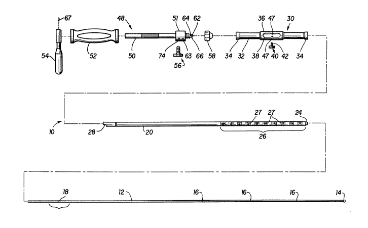
FIG. 1 is an exploded view of a tool for
reducing a fracture of a bone according to the present
invention illustrating separately a shaft, measuring
sleeve with thumb screw, collar, handle body, cam
knob, handle and handle arm, together with a guide
wire.
FIG. 2 is a side view of the fracture
reducing tool of FIG. 1 in assembled form together
with a guide wire positioned through the shaft and
handle body.
~(
1 32 1 520
FIG 3 is a partial cross-sectional side view o~
the tool as shown in FIG 1 tak-n along the lines 3-3 o~
FIG 2
FIG 4 is a cross-sectional view of the tool as
shown in FIG 1 taken along th- lines 4-4 o~ FIG 2
FIG S i~ a cros--sectional view of the handle
body and guid- wir tak n along th- lines 5-S Or FIG 2 and
10 an xpo~-d vi-w ot th- ca~ knob in the unlocked
configuration
FIG 6 i- a cro~ ctional view taken along Sh- lin-s 6-6
of FIG 5 illu~trating rotation of the cam~knob to lock the
guid- wir- wlthin th- ha~t
PIC 7 i- a cros~-s-ctional vi-w taken along th-
lin-s 7-7 o~ FIC S
FIG 8 i~ a cro-~-s-ctional view taken along the
lin-s 8-8 o~ FIG 2
.
PIC 9 i- a cros--s-ctional view taken along the
lin-~ 9-9 o~ FIC 8
qe
FIC 10 i~ a cros--s-ctiona} ViQW taken along
th- lin-- 10-10 ot FIC 2 illuAtrating the thumb scr~w
locking tho m ~-uring ~l--v- on the shatt
~ .
FIC 11 i~ a cro~s-sectional view of the shaft
~; ; 30 and moa-uring ~ v- tak n along lines 11-11 of FIG 2 and
an xpos-d vi-w o~ th- guid- wira within the sha~t
;
; FIC 12 is a partially cro~-sectional view of a
human ~emur with th- tool o~ th- pr-sent invention and a
1 32 1 520
guide wiro positioned within the medullary canal o~ the
femur
FIG 13 illustrate~ a patient with a ~ractured
5 femur resting in position on a fracture table
FIG. 14 illuBtrates expo8ure o~ the greater
trochant~r
FIG 15 illustrates enlargement of the entry
hole for the tool of the present invention
FIG 16 is an ~nd view of the proximal end of
the femur with th- enlarged entry hole
- 15
FIG 17 illustrate~ the positioning of the tool
together wit~ a guide wir- within the medullary canal of
the fractur-d femur
FIG 18 illustrates the positioning o~ the
20 mea uring sl-ev~ 80 a3 to abut the tip o~ the greater
trochant-r
FIG 19 illu~trates the window and indicator
arrow of th- measuring sleeve in position over the
25 graduation~ of th- ~haft 80 a~ to permit measurement of the
rod or nail to b- in~erted into tho medullary canal
FIC 20 lllu-trat-s a rod driver mounted onto a
rod
FIG 21 illu~trat-s th- secure fastening of the
s-lf-locking thr-ading bolt and the ~-handle wrench onto
th- rod
1 32 1 520
PIG 22 illustrates driving th- nail with the
sliding hammer as~embly
- FIG 23 illu~trat-~ ins-rtion of the rod cap
5 screw
FIG 24 illu~trate~ a cross-locking drill guid-
FIG 25 illu~trateo th- proximal end portion of
10 the cross-locking drill guid- of FIG 24
PIG 26 illu~trat-~ positioning of the
serrated-end guid- sleev through th- guid- and down
through to th- lateral cort-x
FIG 27 illu~trate~ marking a ~tarting point for
th- drill
FIG 28 illustrat-- drilling through both
cortic-o
FIG 29 illu8trat-8 both drills in position at
th- di~tal portion of th- f-mur
: ~ .
FIG 30 illu~trat-- ~-a~ur-ment Or thR top of
25 th- out-r drill ~ v-
FIC 31 illu~trat-s placing a transverse scr-w
through th- drill l--v-
30 FIC 32 illu~trate~ drilling through the
proximal nd of th- f-mur
FIG 33 illu~trat-~ placem-nt of the proximal
cros--scr-w~
" ~
::
.
,, .
, ~ - .
:
1 32 1 520
FIG. 34 illustrates placement of the rod cap
screw in the top of the rod.
FIG. 35 illustrates the closed wounds.
DETAILED DESCRIPTION OF THE INVENTION
In the description which follows, any
reference to either direction or orientation is
intended primarily and solely for purposes of
illustration and is not intended in any way as a
limitation of the scope of the present invention.
Also, the particular embodiments described herein,
although being preferred, are not to be considered as
limiting of the present invention. Furthermore, like
parts or elements in the various drawings hereto are
identified by like numerals for ease of reference.
A fracture reduction tool 10 according to
the present invention is shown in FIG. 1 in exploded
form to illustrate the various components involved in
its assembly. Also shown is a guide wire 12 which has
an overall length greater than the length of the tool
10 when assembled. Guide wire 12 is of a typical
straight construction and configuration and has a
uniform diameter with an enlarged or beaded distal end
14 which is inserted into the medullary canal of the
fractured bone such as a femur 15 as shown in FIG. 12.
The guide wire 12 is inscribed at the distal portion
with length marks 16 which typically are ten
centimeters apart and at the proximal portion with an
overall length marking 18.
As shown in FIG. 1, the tool 10 is formed of
an elongated hollow shaft 20 which has a bore 22 shown
more clearly in FIG. 11 for passage of guide wire 12.
-12- 1321520
Accordingly bore 22 has a uniform diameter which is at
least slightly larger than the diameter of guide wire 12
unrestricted pass~ge therethrough The distal end 24 of
shaft 20 is beveled which aids in the penetration of the
5 shaft 20 through the marrow within the medullary canal of
the f~mur 15 a~ shown in FIG 12 The distal portion 26 of
shaft 20 al~o has in~cribed thereon a plurality o~ length
markings or graduations 27 Preferably these graduations
27 are shown in one centimeter increments In the
10 pref-rred embodim nt, th- range of these increments extends
from about thirty centimeters to forty-eight centimeters
which provides for use of this tool 10 with a variety of
different sized bones and, $n particular, the femur of the
human body At its proximal end, the shaft 20 has a notch
15 28 as shown in FIG 1 which is helpful in the assembly of
tool 10 a~ xplain-d more fully below
i
Although as doscribed herein, the fracture
reducing tool 10 of th- pr-sent invention is preferred for
us- in reducing fracture~ of the femur, it i9 contemplat-d
20 that such tool 10 can also b- used with other fractured
bone~ with prop-r ~izing of tho shaft 20 so as to permit
it~ entry and pa~ag- within th- medullary canal of the
resp-ctiv- ~ractur-d bone Moreover, although the shaft 20
a~ shown in th- pr-f-rred embodiments herein in the FIGS
25 is genorally o~ a lin-ar configuration, the shaft 20 can
al~o be curv-d lf d-sir-d in order to accommodate or
corre~ond to th- g-n-ral curvature of the respective
medullary canal involved
The tool 10 also includes a measurement member
30 which i~ a ~orm-d o~ tubular sleeve 32 having flange
end~ 34 and a body portion 36 with an oval window 38 The
measurement sleev- 30 is sized in diameter so as to be
capable of moving along and over the shaft 20 in the
-13-
1 32 1 520
configuration a~ illustrated in FIG 11 Sleeve 30 also
includes a thumb set screw 40 which has a screw rod 42 as
shown in FIG 10 that is sized for threaded passage through
passageway 44 in body portion 36 Screw rod 42 thus can be
5 threaded against the outer surface of shaft 20 In this
fashion, the measurement sleeve 30 can be selectlvely
locked into position at a pr~detQrmined location of shaft
20 to allow for ease of measurement The thumb set screw
40 include~ a knurled disc 46 attached to the free end of
10 screw rod 42 to allow for easy rotation of the thumb set
screw 40 In operation, as the tubular sleeve 32 moves
over the distal portion 26 of shaft 20, the graduations 27
ar- viewable through wlndow 38 as shown in FIG 19
Pre~erably the body portion 36 has opposed arrow indicators
47 as shown in FIGS 1 and 19 which are positioned midway
of the window 38 so as to aid in determining or measuring
the length of th- nail or rod to be inserted into the
medullary canal for fixation
The tool 10, a8 furth-r shown in FIGS 1 and 2,
also include~ a handle 48 which is formed of a cylindrical
handle body 50 having an enlarged butt portion 51, handle
grip 52 and handl- rod 54 A cam rod 56 coupled to butt
; portion 51 allow- for loeking of the guide wire 12
sel-ctiv-ly in po-ition r-lative to the shaft 20 and handle
25 48 As ~hown in FIG 3, the handle body 50 has a bore 60
along it~ l-ngth and through which the guide wire 12 can
fre-ly pa~- A eollar 58 by mean~ of threads within its
bor- at a proximal portion can be threaded onto
corr-~ponding thr ad~ po~itioned as shown in FIG 3 on the
~30 di~tal end of handl- body 50 The distal end of handle
body 50 also $neIud-- an op-n end 62 into which the shaft
20 ean be in~-rt-d for eoupling with the handle 48 This
open end 62 is eonfigutred to reeeive the notch 28 which is
; tightly fitt-d th-rQin by means of wedge pin 63 as shown in
. ,,
~: ~
~, . . .
1 32 1 520
FIGS 1, 2 and 9 Wedg- pin 63 i~ tlush aount-d in butt
portion 51, i e , Lt is shaved or ground o~ after
insertion so as to be tlush with the outer surface of butt
portion 51 Similarly, the handle rod 54 is secured onto
5 handle body 50 by means ot wedge pin 67 This pin 67 as
shown in FIG 3, is flush mounted with the outer surface of
handle rod 54 and i8 pr-ferably press-fitted through
suitabl- holes in handle rod 54 and handle ~ody 50
The open end 62 also includes a series of cut-
away portion~ 64 which leav- fingers 66 as shown in FIG
which togeth-r with collar 58 function as a chuck for
secur~ng or loc~ing eh- shaft 20 together with the handle
48 As th- collar 58 is screwed onto the open end 62, the
15~ cut- way portions 64 allow th- fingers 66 to press into
engaging contact with th- out-r surface of shaft 20 as
~hown in PTG 3 ln this fashion the collar 58 tightens
down~th- ting-r~ 66 so a8 to s-curely retain the shaft 20
withln the op-n nd 62 ot handle 48 Th- bore 60 within
th- body handl- 50 upon a~embly is aligned coaxially with
tho bor- ~2 Or ~hatt 20;as shown in FIG ~3 This permits
guid~ wir- 12 to b- fr-ely and selectively passed
throughout th longth ot tool lO until such time as it is
look d in po~ition~by ~ an~ Or cam knob S6
~ R t-rring to FIG S, th~ cam knob 56 includes a
ca~ rod 68 which i- po-ition-d in a passageway in the butt
portion 51 and xt-ndJ ther trom to a free end A four
prong knob 70 i~ attach-d to th- cam rod 68 free end The
c--~rod 68 ha~ a groov- 72 which is suitably sized to
r-c-iv a w dgo pin~74 a~ shown in FIGS 1, 2 and 7 which
tlu~h ~ount-d -lmilar to wodgo pin 63 and press-fitted
through a hol- in butt portlon 51 Thus, the cam rod 68
can b- rotat-d whil- k-pt in position within the respective
pa~agow y Or butt portion 51 The cam rod 68 also has a
, ,: ", ,
;-,; ~
~ ~ ,
.
-15-
- 1 32 1 520
cam groove 76 whose outer surface acts as a cam as shown in
FIG 6 ~o that as the cam rotatQs, for example in the
direction o~ the arrow, a greater portion-of tho cam enter~
the bore 60 during at least a portion of one complete
5 rotation the cam rod 68 In this ~ashion, the cam groove
76 will contact the guide wire 20 and push it against and
into contact with th- opposing surface of bore 60 so as to
selectively lock the guide wire 12 within the bore 60 in
position relative to the butt portion 51 and shaft 20
10 This operation is demonstrated in FIG 6 wherein the guide
wire 12 shown in solid form is advance toward its position
shown in phantom line~
As noted, the guide wlre 12 with its beaded end
15 14 is assembled with tool 10 by insertion through bores 22
and 60 in the manner shown in FIG 2 Thereafter, the tool
10 and guide wire 12 can be inserted through a suitably
sized entry hol- 78 shown in FIG 16, and into the
medullary canal Or femur 15 as shown in FIG 12 The tool
10 can then b- positioned as desired within the medullary
canal and the guide wire 12 extended through the canal
portions~so as to manipulate th- various fragments of
fractur-d t-mur lS Onc- reduction is achieved, the guide
wir- 12 is ~ully ins-rted so that the beaded end 14 abuts
th- di-tal nd~o~ th-~medullary canal The tool 10 can be
25~ po~ition-d~so~that the proximal end of handle arm 54 is
flu~h with th- proximal end of guide wire 12 In this
configur~tion, th-~m-a~urement sleeve 30 is positioned so
a~ to abut th- gr ~t-r~trochanter of femur 15 as shown in
FIG 19 $h- ~rrow ind~cator~ 47 then provide a
30 measur-~-nt o~ th- pin or nail to be ins-rted into the
m dullary canal ror fixation Accordingly, the graduations
27 ar- calibr~t-d to provid- th- nail or rod measurement
wh-n th- tool 10 and guid- wire 12 are positioned as
d-scribed abov- Although the tool 10 is preferably
.,, :
-16-
1 32 1 520
utilized with the guide wire 12 for reduction,
alternatively the shaft 20 can be inserted as far into the
medullary canal as desired so that reduction of the
fragment portions can be achieved with shaft 20 itself
s
With respect to the tool 10, its use in
intramedullary ~i m ) rod surgical technique for a
positioning and fracture reduction will now be discussed
Place the patient 80 on the fracture table in
the lateral decubitus position as shown in FIG 13 The
supine position may also be used Reduce the fracture in
your usual manner With this technique, anatomical
reduction of the fracture prior to insertion of the
15 reduction tool 10 is not necessary However, the fracture
must be reducible Confirm this with the C-arm fluoroscope
82, positioned to permit good anteroposterior and lateral
views from th- hip to th- knee
Perform standard surgical preparation and
draping of the skin If cross-locking i to be done, the
surgeon must allow adequate clearance at the knee, as well
as the hip Mak a 10-centimeter incision proximally from
th- tip of th- greater trochanter in line with the fibers
of the gluteu~ maximus Expose the entry site as shown in
FIG 14
The prop-r entry site for the nail is very
important and must b- located directly over the medullary
canal The entry point is the fossa on the underside of
30 th- greater trochanter at its junction with the femoral
neck in the midline of thQ anteroposterior plane Drill a
3 2mm hip bolt guide pin 84 through the fossa into the
medullary canal Verify critical position of this pin in
th- canal with biplanar view8 on the fluoroscope 82
-
- 17 -
1 32 1 520
Enlarge the entry hole 78 using the 14mm hip
bolt reamer 86 over the guide pin as shown in FIG. 15.
Once this entry 78 illustrated in FIG 16 has been
made, remove the reamer 86 and guide pin 84.
The fracture reduction tool 10 is used to:
reduce the fracture, help pass the reaming guide wire
and measure the length of nail to be used. Load the
3.Omm x 1000mm bead-tip guide wire 12 into the
reduction tool 10 by passing it retrograde. Place the
locking T-handle 88 on the proximal end of the wire
12.
Insert the guide wire 12 and reduction tool
10 into the proximal fragment. Reduce the fracture
using the fracture reduction tool 10 as a lever to
manipulate the proximal fragment. Reduction is
usually easy, except in supracondylar fractures, where
flexion of the distal fragment may require an
additional anteriorly directed push on the proximal
end of the distal fragment by an assistant. Advance
the guide wire using the T-handle 88 to manipulate the
wire 12. If necessary, the reduction tool can be
locked onto the wire 12 to drive it into the
cancellous bone of the distal fragment. Verify proper
position of the guide wire 12 with the fluoroscope 82
as shown in FIG. 17.
Remove the locking T-handle 88. Slide the
reduction tool 10 until it is flush with the proximal
end of the guide wire 12, and lock it in place with
cam knob 56. Slide the measuring sleeve 30 down the
tool 10 until it abuts the tip of the greater
trochanter, and lock it in place with the thumbscrew
40 a~ shown in FIG. 18. Read the rod length from the
reduction tool 10 scale 26 as illustrated in FIG. 19.
Unlock and remove the reduction tool 10.
Alternatively, the scale 26 can be read after the tool
10 is removed.
,X
-18-
1 32 1 520
Ream th- medullary canal progressively u~ing
flexible reamers Begin with the smallest end-cuttlng
reamer In most cases, ream 1 5~m largor than the rod to
be used When cross-locking, ream 2mm larger if
5 nondisplaced shaft fractures are suspected or when fixing
distal third fractures The sup-rior strength of the Alta~
nail permits use of smaller diameter rods than previously
recommended with other sy~tems As reamer sizes vary
somewhat by ~anufacturer, the surgQon must check the size
10 f reamer~ used
Placement of the i m rod in the i m rod
surgical techniqu- will now b- de~cribed
Exchang- th- bead-tip 14 reaming guide wire 12
for a 3 2mm x lOOOmm smooth-end guide wire, using the
medullary tub- to maintain fracture reduction
Mount the rod driver 90 onto the proper size
nail or rod 92 u~ing th- self-locking threaded bolt and the
20 T-handl- wrench a- shown in FIGS 20, 21 Be certain that
th- bolt i- ~-cur ly tightened
Th- driving handl- may be attached to control
rotation a~ the nail or rod 92 i~ being driven
Plac- th- nail or rod driver 90 assembly over
the guid- wir- and into th- femoral entry site
Occa~ionally, om manipulation i8 required to pa~s the
rod-driv-r ~unctlon locking ov-r the end of the guide pin
With pilot point directed into the driver,
attach th- liding hamm-r a~--mbly 94 to the driving handle
Conflrm prop-r rotation orientation of the rod The
guid- wire mu~t b- deflected out the side of the driver
,
--19--
1 321 520
assembly and be monitored to be certain that it does not
advance with the nail Drive the nail 92 with gentle
blowJ The nail 92 may al~o be driven with a mallet
directly on the driving handle as shown in FIG 22 Drive
5 the nail 92 to th- dQsired position The rod 92 must
progress smoothly without excessive force If too much
resistance i9 encountered, verify proper rod size and
po~ition Additional reaming may be required Drive the
rod 92 until the proximal end is just above the level of
10 the superior femoral neck
Remove the guide wire and the sliding hammer
assembly 94 Then insert the rod cap screw 96, which
facilitates removal o~ the rod by preventing bone ingrowth,
15 using th- T-40 Torx bit (FIG 23) Note that the cap
scr-w will app-ar on X-ray~ to b- proud becauae of the
radiolucent washer
Th- cross-locking ~crew procedure for the i m
rod ~urgical't-chniqu- will now b- presented
Prior to driving the rod 92, assemble the
cross-locking drill guide 98 and set the cross-screw
, alignment Mount th- cross-locking drill guide ~8 onto the
; driver assembly 90 a~ shown in FIG 24 Note that the
25 ~hort-r 100 o~ th- two mounting pins 100 and 102 is placed
proximally Slid th- guide 98 over the mounting pins 100,
102 E~timat- su~icient clearance to allow for the soft
ti~su-~ o~ th- thiqh Lock the guide 98 into place by
;~ hand-t1ghtening Xnob A as shown in FIG 25 Place the two
; ~ pilot pin~ in th- distal guide holes on either side of the
numb-r,r~pr---nting the proper rod length
Unlock Xnob B, and ad~ust the guide 98 using the
knurled ad~ustment wheel C until the pilot pins drop
.' .
-20-
1 32 1 520
through the guide 98 into the center of the distal screw
holes in the rod 92 During this procedure it is important
to hold the rod 92 and cross-locking drill guide 98 only by
the driving handle 90 as shown in FIGS 24 and 25
Lock the guide 98 into position by tightening
Knob B with the T-wrench, th-n verify that alignment has
been maintained Remove the guide 98 by loosening the Knob
A and sliding the guide 98 over the mounting pins 100, 102
10 Place th- guide 98 qently on the sterile back table, being
careful not to loosen Xnob B Drive the rod 92 as
de~cribed hereinabove with respect to placement of the i m
rod
R-mount th- cross-locking drill guide 98 on the
driving handle 90 as previously described Be certain the
guide 98 do-- not imping- on th- patient 80 or any
quipm nt
Distal eross-locking is performed first, except
when only proximal ero~s-loeking is required This
maximizes aeeuraey ~or the distal screws Locate the
ineision sit- by plaeing two alignment pins through the
appropriat- guid- holes and marking the skin Make a
ingl- longitudinal ineision 104 and expose enough lateral
25 eort-x ot th- femur 15 80 that soft tissue impingement on
th- drill ~ v-- 106 doe- not oecur as shown in PIG 26
"
,'
Plae- th- outer, serrated-end guide sleeve,
drllI 8I--v- 106 and troear through the guide 98 and down
30 to th- lat-ral eortex Lightly tap a sharp trocar to
~; ereat- a pilot noteh a- shown in FIG 27 Be certain to
k--p th- troear~ harp Remove the trocar Mount the
long, ultra sharp Alta~ 4 0mm drill point on your power
soure- Insert th- drill into the drill sleeve Be
35 ab olutely eertain
-21-
1321520
that proper alignment i~ maintained. Avoid side pressure,
bending, etc. Extra caution is required with heavy or
cumbe some power systems. Do not use hand powered drills.
Drill through both cortices as shown in FIG. 28. Varify
5 with C-arm that the drill point is through the rod holes.
Leave the drill in place and repeat the sequence
with the second distal cross-screw as shown in FIG. 29.
Leaving the outer screw sleeve in place, remove
the inner drill sleove and th~ first drill bit. Measure
for the length of the transverse screw using the cross-
locking ~crew depth gauge. Read the measurement directly
- off the top o~ the outer drill sleeve as in FIG. 30.
Overdrill through the lateral cortex using the $mm drill
bit. Place the proper transverse screw through the screw
sleeve using the long T-25 Torx bit as in FIG. 31. Repeat
the process for the second distal cross-screw.
~ he proximal targeting sequence is identical to
20 distal targeting. Again, after placing the drills through
the rod 92 and both cortices, the surgeon may check
radiographically to make certain that they are through the
hole~ in the rod 92 as in FIG. 32.
Aftor placing th- proximal cross-screws as in
FIG. 33, Femov- the cros~-locking guide 98 and then the
driver a88-mbly 90. Place the rod cap screw 96 in the top
of th- rod 92 u~ing tho T-40 Torx bit as shown in FIG. 34.
The tip ot the screw cap should be at or slightly below the
30 tip of tho greater trochanter.
, ~
Close and dress all the wounds as in FIG. 35 and
take the final radiographs.
-22-
1 32 1 520
Variation~ of the above-described tool lO for
reducing the fracture of a bone which involve minor changes
are clearly contemplattd to be within the scope of the
present invention. In addition, minor variations in the
5 design, angles or materials of the various components of
the tool lO are also contemplated to be within the scope of
the present invention. These modifications and variations
may be made without departing from the spirit and scope of
the present invention, as will become apparent to those
10 skilled in the art. The specific embodiments described
herein are offered by way of example only, and the
invention i3 limited only by the terms of the appended
claims.