Note: Descriptions are shown in the official language in which they were submitted.
1 32~06~
BACKGROUND OF THE INVENTION
This invention relate~ to a hcad positioner
for use in an X-ray or ultra-short wave cephalo-
tomograph, computer tomograph, dental pantomograph,
temporomandibular ~oint radiograph, and the like.
Hitherto, X-ray cephalotomograph~ ha~ been
practiced mainly in neurosurgical diagnoses, while
in dental diagnoses such photography ha~ not been
in general practice. Today, however, needs for
radiotomography are increasing in dental f1elds
such as oral aurgery, temporomRndibular joints,
implantation, and orthodontic treatment, and it is
desired that accurate and quantitative information
be obtained repetitively in same condition~ by
radiotomography and otherwise on matters such as
buccolingual denture inclination and antagonistic
tipping angle; sectional configuration of jaw
bones and, more particularly, cortical-bone con-
figuration, ~pongy-bone dlstribution, and kinematic
position of inferior alveolar veins; and spacial
position of impacted third molar.
In this conJunction, various improvements
have been made with respect to tomo~raphic appa-
ratuees per ~e, and tomograph~ which can exhibit
good performance accuracy have been propo~ed.
1 322060
However, insofar a~ head po~itioner~ for u~e in
radiotomography, no such positioner has been pro-
posed which can exhibit good practical performance
accuracy.
In Japane~e Utility Model Laid-Open Publica-
tion No. 61-~4209, for example, there i~ described
a technique for fixing the head of a ~ub~ect in
position by means of an anchor band. In Japane~e
Patent ~aid-Open Publication NOB. 61-94639, 60-
58127, and 61-203948, there are disclosed techniques
such that a fixing member is applied to the ears,
cranial fossa, or the like portion of the ~ubject
to fix the head in position. However, these tech-
niques are designed to fix the head of the ~ubject
through the intermediary of a skin portion or the
hair o~ the sub~ect over which the fixing member
is applied, and therefore no satisfactory po~ition-
ing can be achieved because of the resiliency of
the skin or other portions; furthermore, the head
cannot be tightly bound by the anchor band or fix-
ing member, which fact aleo prevents the head ~rom
being completely fixed in position. As such, with
these head positioners, the head of the sub~ect
cannot be repeatedly fixed in ~ame conditions and
it i8 virtually impo~sible to obtain laminograms
1 322060
of the subject under same conditions during the
pre-treatment, in-treatment, and post-treatment
stages.
Japanese Utility Model Laid-Open Publication
No. 61-14006 discloses a head positioner which can
solve these problems to some extent. According to
this document, the head positioner comprises a
fixing device having a spherical element adapted
to be inserted in the palate of a subject laid
down on a bed and to be subjected to a bite by the
subject, the fixing device heing pivotally sup-
ported by a supporting member of an image inten-
sifier or the bed, and a mechanism for fixing the
fixing device to a desired rotation angle posi-
tion. However, since this positioner is such that
the spherical element of the fixing device is
inserted in the palate of the subject and sub-
jected to a bite, the positional relation between
the spherical element and the palate of the sub-
ject is unstable; and therefore the positioner
cannot be put in use unless the subject is laid
down on the bed. Another problem is that because
of such instability, the relative position of the
subject and the image intensifier cannot be con-
stantly defined, which fact means poor
duplicability.
a ~,
t 322060
Furthermore, the insertion of the spherical
element is a possible cause of pain to the sub-
ject, and the positioner cannot be employed for
purposes of jaw bone photographing.
BRIEF SUMMARY
The object of the invention is to provide a
head positioner which permits repetitive reproduc-
tion of initially set head positioning conditions
in the practice of cephalotomography and the like.
In accordance with the invention a jaw bone
anchored type head positioner is provided which
comprises:
(a) a die duplicated from an impression taken
with respect to the whole or a part of the
dentition, dentulous or edentulous, of one or both
of the upper and lower jaws of a subject,
(b) a supporting member formed integrally with or
separately from said die for supporting said die,
and
(c) fixing means for controlling said die con-
tinuously or stepwise to a desired position
through said supporting member and for removably
fixing said supporting member in position, whereby
when the subject is caused to have a bite of said
die, the jaw bones of the subject can be
- 4
1 322060
anchored to the die and accordin~ly the head of
the subject can be fixed in po~ition.
Ear rods and a nasion support adjusted to the
nose of the subject are supplementarily u~ed in
combination with the supporting member.
According to ~uch arrangement of the inven-
tion, a die is duplicated from the dentitlon of
one or both of the upper and lower ~aw~ of the ~ub-
~ect. When ths jaw bone or bones are dentulous,
the die i8 prepared with respect to the dentulous
dentition, or if the ~aw i9 toothless, in a~dition
to a model obtained on the baais of an impre~sion
taken from the alveolar ridge, a base plate is pre-
pared which, together with the model, i9 held in
position by mean~ of an edentulou~ ~aw skater. An
impression for die making may be taken with respect
to the whole or a part of the dentition. The die
duplicated on the baeis of the impression taken
~rom a subJect ha~ a ~upport member formed inte-
grally therewith or securely fi~ed thereto, by which
the die is ~upported in position. The supporting
member is ~itted in a fixing device mounted in a
tomo~raph or the like, in such a way th~t the die
fixed to the ~upporting member i~ adjusted to be
finally positioned and oriented properly in relation
1 322Q6~
to the ~ection to be tomographed.
The die fixed through the supporting member
to the fixin~ device i9 subjected to biting by the
~ub~ect, whereby the dentition and the die come
into enga~ement with each other ~o that the ~aws
and head of the subject are fixed to the fixing
device through the die.
BRIEF DESCRIPTION OF THE DRAWING8
Fig. 1 i~ a per~pective view ~howing one form
of ~aw bonc anchored type head positioner embody-
ing the invention;
Fig. 2 is a side view showlng a plaster model
of teeth for the preparation of a ~ie for the head
positioner in Fig. l;
Figs. ~, 4 and 5 are views showin~ other
embodiment~ of the invention, Fig~. ~ and 4 being
perspectlve views, Fig. 5 being a front vlew;
Fig. 6 is a p~r~pective view, partly in sec-
tion, showing a structure for the preparation of a
die repre8enting another embodiment o-f the inven-
tion; and
Fig. 7 i8 a side view showing, by way of
example, a conventional head positioner.
DETAILE~ DESCRIPTION OF THE INVENTION
One embodiment of the inventlon will now be
1 322060
de~cribed in detail with reference to the accom-
panying drawings.
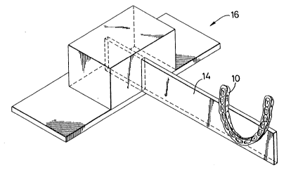
In ~ig. 1, the reference numeral 10 desig-
nates a die which, in the present embodiment, i~
patterned on an dentition impres~ion taken from
one of the upper and lower jaw~ of a sub~ect. To
produce die 10, a plaster model 12 a~ 8hown in Fig.
2 i9 fir~t made by a certain techniqlle convention-
ally employed in dental practice, and then a re~in
splint 10 removably attachable to the plaster model
10 i~ prepared. The die 10 is formed of a reaction
setting resin or the lika resin material which i~
less liable to volume ~hrinkage or expansion.
The site and direction of a particular section
to be tomographed using the die 10 and plaster model
12 thus prepared are ~elected, and the die 10 i8
fixed to a resin-made supporting member 14 in a
direction convenient for tomographing purpo~es.
The direction in which the die 10 i~ fixed to the
supporting member 14 may vary accordin~ to the type
of the tomograph to be employed, and is Yuitably
selected accordingly.
The supporting member 14 i~ mounted to a fi~-
ing device 16 placed on a table of the tomograph
not ~hown and is clamped to position by a clamp
1 322060
not shown. The fixing device 16 i9 held constant
in po~ition relative to the tomogr~ph 80 that the
supporting member 14 fixed to the fixing device
16 is liable to no variation in position relative
to the tomograph. Therefore, the die 10 which ha~
been fixed to the supporting member 1.4 after having
been suitably positioned and oriented in relation
thereto is fixed in po~ition a~ ~et relative to
the tomograph.
When the die 10 thu~ fixed to the tomograph
i~ placed between the jaws of the ~ub~ect for being
lightly bitten, the d.ie 10 goes into close engage-
ment with the dentition of the ~ubject and accord-
ingly the ~aw bone~ of the ~ubject are held ~table,
the head of the subJect being thu~ steadily fixed
to the tomograph through the fixi.ng devi.ce 16.
The supporting member 14 to which the die 10 is
fi~ed may be repeatedly mounted to and dismounted
from the fixing 16 without being liable to vari-
ation~ in setting position and direction with
respect to the die 10. The dentition Or the subject
is unlikely to change over a considerably long
period of ti~e, if the eub~ect is an adult, or
if the subject is an infant, the dentltion is sub-
ject to no or little change for a period of several
1 322060
to ten~ of months; therefore an initial die 10 may
be repetitively used. In the ca~e where ~ome change
ha~ been caused to the dentition of the ~ub~ect
due to the application of an implant or otherwise,
the die 10 may easily be re~tored to a usable con-
dition by eliminating a part of the ~ie 10 which
corre~pond~ to the site of such change in the denti-
tion.
The die permits proper occlusion throu~h clo~e
contact with the teeth of the sub~ect can produce
no backlash relative to the teeth. ~xcept where
unusual conditions are present, the die permits no
joltin~ with re~pect to the dentition and ,jaw bones.
By causing the subject to bite lightly the die,
the jaw bone~ are positionally stabilized and thus
the head of the ~ub~ect can be steadily fixed to
the fi~ing device.
In thi8 way the jaw bones and joints can be
accurately fixed in position simply by the subject
bein~ cau~ed to bite the die to bring it into good
occlusion relation with the teeth. Furthermore,
both the Jaw bone~ and the head can always be
~ixed in poeition under predetermined conditions
during all relevant stage~ includin~ pre-treatment,
under-treatment, and post-treatment ~ta~e~, and
_ g _
.
1 322060
thus more accurate diagno~is and tre~tment can be
given through comparative studies of laminograms
obtained under predetermined conditions.
In the ca~e where the die iB fixed to the
eupporting member after being positionally adju~ted
to the latter ~o that the site and orientation of
a particular section to be tomographe~ may readily
be located, same conditions can be repetitively
obtained simply by fixing the supporting member to
the fixing device. In the case where the die i~
fixed to the ~upporting member constant]y as preset
with respect to the positional relstionship between
the former and latter, the supporting member i~
fixed to the fixing device through suit~ble ad~ust-
ment made 80 as to provide the site and orientation
of the particular ~ection to be tomographed, and
thua, any time thereafter, the supporting member
can be fixed to the fi2ing device according to the
adjustment data, whereby ~ame conditions can be
repetitively obtQined for positional relation~hip
between the die and the fixing device.
For the purpose of jaw joint radiographing at
a desired ~ite o~ the lower ~aw, a fixation source
iB set on a molar tooth buccal surface of the upper
~aw. For die mounting and di~mountin~, a movabl~
-- 10 --
1 322060
clutch hinged at a median portion is prepared which
i8 to be accurately fixed to the molar tooth buccal
surface of the upper jaw. The hinge is graduated
to provide reference for purpo~es of duplicativity
acknowledgement.
One embodiment of the invention h~s been
described above; however, the invention may be
practiced in other forms.
A~ Fig. ~ shows, for example, a fixing device
18 compri~es a guide rod 20 provided in a tomograph,
a slid~ble-rotatable member 22 which i9 slidable
along and rotatable about the ~uide rod 20, and a
rotatable member 24 rotatably provided in the slid-
able-rotatable member 22, the guide rod 20, slidable-
rotatable member 22, and rotatable member 24 being
clamped by a clamp to position as set according to
the scale on each of them. The rotatable member
24 of the fixing device 18 has an insertion hole
bored therein ~or releasably receiving a supporting
member 26 to which the die 10 is fixed. The support-
ing member 26 is inserted at one end into the in-
~ertion hole and fixed in position.
According to this embodiment, the die 10 can
be fixed to the supporting member 26 a~ deslred,
and the orientation of the die 10, that is, the
1 3~206~)
site ~ld orientation of a particular section to
be tomographed, i9 suitably determin~d by the
fixing device 18. In thi~ embodiment, by record-
ing the graduation ~et on the fixing device it i8
possible to accurately duplicate the mounting
position for the d~e 10. As a modifi~d form of
this embodiment, the guide rod 20 of the fixing
device 18 may be adapted to be movable relative to
the tomograph.
In another embodiment, a~ Fig. 4 ~how~, guide
rod 20 of a fixing device 28 may be dispo~ed in
front of the ~ub~ect. In this embodiment, as well
as in Fig. 3 embodiment, various forms of fixing
device may be employed according to the site and
orientation of the section to be tomographed. It
i8 de~ired that the arrangement of the ~ixine device
be modified corre~pondingly to the type of the
tomograph to be used, as well as various other
kinds of apparatuse~ to be used, such a~ X-ray
television ~luoro~copic photo~raph and dental
pantomograph; and the present invention can immedi-
ately meet such requirement.
In another embodiment, as Fig. 5 shows, a
~ixing device 70 has a pair of guide rods ~2, and
a supporting member 34 extending between the guide
- 12 -
1 322060
rods 32 and obliquely fixed thereto 80 a9 to deter-
mine the 8ite and orientation of the s0ction to be
tomographed.
In the embodiments shown in Fig~. 3 to 5, the
guide rod or rods of the fixing device may be di~-
posed either vertically or hori7.0nt~11y ~o a9 to
meet varying conditions such that photographing
may be made with the sub~ect as postured in sitting
position or in lying position accordin~ to the type
of photographing apparatus used.
In the above described embodiments o~ the
invention, the die lO i~ ba~ed on a plaster model
made according to the conventional dent~l technique.
Such plaster model i~ made with respect to one of
the upper and lower jaw~ of the 8ub~ect that
requires diagnosis and treatment in particular.
A die can not only be made on the basis of a
plaeter model, but also it may be constructed in
such a way that, as Fig. 6 shows, for example, the
die compriees a structure 36 having a core 38
centrally formed therein, the core ~8 being covered
all over with a resin material 40. In thie embodi-
ment, the resin material 40 is a thermosetting
resin, for e~ample, and the eubject is caused to
take a bite of the structure ~6 formed of the
1 322060
thermosetting re~in 40 ~o that a mark of upper and
lower teeth i~ tran~ferred onto the surface of the
structure 36; thereafter, the ætructure 36 having
the teeth mark~ is heat treated so as to allow the
re~in 40 to be hardened into a die. The resin 40
may be a thermos~tting resin; and of course it i8
possible to construct a die of such re~ln.
In thi~ embodiment, the core 38 of the
structure 36 i~ formed integrally with a support-
ing member 42, whereby the trouble of fi~ing the
supporting member 42 to the die 36 is saved. The
core ~8 prevents the ~tructure 36 from being bitten
off when the sub~ect i8 cau~ed to have a bite of
the structure 36 to produce an impression of teeth
thereon, and it is al30 effective as a reinforce-
ment for the die 35 prepared. The core 38 may be
provided only for a part of the structure 36.
Several embodiments of the invention have
now been de~cribed. Not only can the apparatus
of the in~ention be employed for purposee of
dental hiagno~is and treatment, but al~o it can
be advantageously employed for cephalotomographic
purpoee~ in connection with neurosurgical diagnosie
and therapy and otherwi~e, and more particularly
in the case where laminograms in same conditions
1 322060
are repeatedly required at stages prior to during
and after treatment. It i9 not intended that the
invention iB limited to the art of tomography;
needles~ to say, the invention may be advantage-
ously employed in other photographic areas.
When using the jaw bone anchored type head
posit-ioner according to the invention, an ear rod
may be used in combination, or in place of or in
conjunction with the ear rod, a nasion support
ad~usted to particular condition~ of the ~ubJect
may be supplementarily used in combination.
The die may be formed with respect to only a
portion of the dentition. The fixing device may
be adapted for fine adjustment by a toothed wheel
or the like. Further, it is possible to arrange
for enabling the supporting member to be not only
continuously but also stepwise ad~usted by the fix-
ing device. It will be obvious to tho~e skilled
in the art, therefore, that many changes, modifi-
cation~, and variations of the invention may be
made without departing from the spirit and scope
of the invention.
(Advantages of the Invention)
As above de~cribed, according to the inven-
tion, a die con~tructed 80 as to meet particular
- 15 -
1 32206~
conditions of each individual sub~ect is applied
for fixing the jaw bone~ and ~oints, and head of
the sub~ect by cau~ing the sub~ect to have a bite
of the die, 80 that those parts of the sub~ect can
be properly fixed in position without the possi-
bility of becoming un~table.
The die is fixed to the fixing device through
the ~upporting member; therefore, the ~aw bones
and joints, and the head can be repetitively accu-
rately fi~ed in position under same conditions.
Therefore, the positioner of the invention provides
good duplicability, and yet it is comparatively
simple in con~truction and economical.
~ urthermore, the die i~ patterned on an
impression of the dentition of the sub~ect; and
therefore, when the sub~ect is caused to have a
bite of the die for occlusion, no feeling of physi-
cal disorder can be cau~ed to the sub~ect, nor can
any pain be caused.
- 16 -