Note: Descriptions are shown in the official language in which they were submitted.
(UIA-4)
1325590
--1--
,
CONC~NTRATED STABILIZED MICROBUBBLE-TYPE
ULTRASONIC IMAGING AGENT
This invention relates to ultrasonic imaging of the
human body for diagnostic purposes; and, more particularly, to
ultrasonic imaging agents.
- It has been known since 1968-70 that contrast echo-
cardiography can be used to delineate intracardiac structures,
assess valvular competence, demonstrate intracardiac shunts, and
identify pericardial effusion. (Gramiak and Shah, 1968; and Fei-
genbaum, et al., 1970.) Ultrasonic imaging of the heart poten-
tially has important advantages of convenience, safety, and re-
duced cost over present diagnostic procedures, such as angiogra-
phy, which requires the use of radio-opaque dyes for X-ray imag-
ing, or the use of radio-nuclide imaging agents for radio-imag-
ing. However, progress in practical applications of ultrasonic
imaging has been delayed by the lack of effective clinically-
usable imaging agents.
Ultrasonic imaging utilizes an ultrasonic scanner to
generate and receive sound waves. The scanner is placed on a
body surface overlying the area to be imaged, and sound waves
are directed toward that area. The scanner de~ects reflected
sound waves and translates that data into images. When ultra-
sonic energy is transmitted through a substance, the acoustic
properties of the substance depend upon the velocity of the
transmission and the density of the substance. Changes in the
substance's acoustic properties (e.g., variations in acoustic
impedence) are most prominent at the interfaces of different
substances, such as a liquid-solid or liquid-gas interface.
Consequently, when ultrasonic energy is directed through media,
changes in acoustic properties will result in more intense sound
reflection signals for detection by the ultrasonic scanner.
Ultrasonic imaging agents can consist of small solid
or gaseous particles which, when injected in the circulatory
system, provide improved sound reflection and image clarity.
Microbubble-type imaging agents consist of minute bubbles of a
gas (usually air) which are dispersed in a carrier liquid for
~ '.~ ' "' ' .
-~`` 132~90
--2--
parenteral injection. The "microbubbles" are carried by the
circulatory system to the organ being imaged.
It has been proposed to form a dispersion of air micro-
bubbles in a warm aqueous gelatin solution, and cooling the solu-
tion to a solidification temperature to trap the microbubbles.
For administration, the ~elled d~spersion is to be warmed until
it liquifies, and parenterally administered with the microbuh- ~ ---
bles dispersed in the liauified gelatin. (Tickner, et al. U.S.
Patent 4,276,885; and Tickner, et al., National Technical Infor-
mation Ser~ice Repor~ ~R-62917-lA, April, 1977).
Gelatin-trapped microbubbles on introduction into the
bloodstream have a short life-time. They rapidly dissapate.
Another disadvantage is that the microbubbles are too large to
pass through capillàry beds, and are therefore not suitable for
heart imaging by peripheral intravenous administration.
~he discovery by Dr. Steven B. Feinstein of sonication- -
produced microbubble imaging agents represented an important ad-
vance in this art. Using viscous aqueous solutions, such as 70
sorbitol or dextrose, Dr. Feinstein produced a dispersion of
microbubbles by high energy sonication of the solutions. The ``
resulting microbubbles had sizes less than 10 microns, and were ~;
capable of passing through capillary beds. The persistence of -
the microbubbles, although of the order of a few minu~es, per-
mitted the imaging agent to be pre~ared and administered intra-
venously for heart imaging. (Feinstein, et al., 1984; and
Feinstein U.S. Pate.nt 4,572,203.)
Subsequen~ly, Dr. Feinstein sought to improve the per-
sistence of the microbubbles. He found that hy sonication of a
heat-sensitive protein, such as albumin, microbubbles of
improved stability were obtained. (See Feinstein,
published PCT Application W0 84/02838, corresponding to
U.S. Patent No. 4,718,433 ~iled December 5, 1985).
Concentrations of microbubbles of 10 to 14 x 106
microbubbles per milliliter were obtained with bubble sizes
from 2 to 9 microns (Keller, Feinstein, and Watson, 1987).
The microbubbles persisted for 24 to 48 hours.
., .
',:
" ~
1325~9~
--3--
However, the sonication-produced albumin microbubble
imaging agent of Feinstein was not sufficiently stable for com-
mercial manufacture. Stabilities of the order of weeks or
months (rather than hours or days) are required to permit an
imaging agent to be manufactured at a central location and dis-
tributed to hospitals in the United States and other countries.
For commercially feasible manufacture, shipment and hospital
storage prior to use, a stability time of at least four weeks is
needed and pre~erably at least eight weeks or longer.
Further, for the most effec~ive imaging, it is desir-
able to have the highest obtainable concentration of microbub-
bles in the imaging agent. But the population of microbubbles
of the desired small sizes tends to decrease with holding of the
sonicated albumin solutions. The small bubble size attrition
can occur either by collapse of the microbubbles, or by coales-
ence to oversize microbubbles. Consequently a further important
objective has been to find means for increasing concentrations
of microbubbles in the imaging agent. An imaging agent of very
high microbubble concentration is inherently better, and a safe-
ty factor is provided. With a concentration of microbubbles
higher than the minimum required for effective imaging, some -
loss of the microbubbles of the desired size can be accepted.
The present invention provides an ultra-concentrated,
room-temperature stable microbubble-type imaging agent. This
improved imaging agent comprises a sterile aqueous medium con-
taining a ~ispersion of microspheres predominately of diameters
less than 10 microns, and is thereby suitable for parenteral
intravenous administration. The microspheres consist of gas
microbubbles encapsulated in a water-insolubilized biocompatible
material, such as albumin. Representing a substantial advance
in the art, the imaging agent of this invention has a homogene=
ously dispersed concentration of greater than 100 x 106 (e.g.,
10 ) microspheres per milliliter. ~his high concentration can
be maintained at ordinar~ room temperatures (20 to 25C) ~or ex-
tended periods of time (4 to a weeks or longer). In optimized
-
l32~a
--4--
embodiments, microspAere concentrations of the order of 300 to
500 x 106 microspheres per milliliter are achieved. Surprisin~-
ly, these ultra-high concentrations can be maintained for over
eight weeks. The imaging agents of this in~ention are therefore --
adapted for manufacture and distribution on a commercial basis.
Following shipment, they may be maintained in inventory by hos-
pitals for many ~eeks, being available for diagnostic use as re-
quired.
The imagin~ agents of this invention are preferably
produced from a heat-denaturable biocompatible protein by a step- ~-
wise sonication procedure. As with the Feinstein method, an :
aqueous solution of protein is subjected to sonication to form
gas microbubbles while concurrently heating the solution to in-
solubilize small portions of the protein. However, the improved -
sonication procedure, which results in the increased concentra-
tion of highly stable microbubbles utilizes a novel sequential
sonication. In the initial sonication phase, the sonicator horn
is directly contacted with the solution (viz. by immersion just
below the upper suface of the solution). This initial sonica-
tion is carried out without appreciable foaming of the solution.
In the next phase of the sonication, foaming is promoted. The
sonicator horn is withdrawn to a position in the ambient atmo-
sphere above but proximate to the surface of the solution. In-
tense foaming and aerosolating occurs. The population of micro-
bubbles is thereby greatly increased and the microbubbles are
encapsulated with denatured protein to obtain a dispersion of
highly stable microspheres. Moreover, the stability of the
microspheres permits them to be concentrated and/or fractionated.
By such manipulations, bubble concentration can be doubled or
tripled and oversize bubbles eliminated.
For example, the concentration of the microspheres as
initially produced can be from 50 to 150 x 106. By a float
separation concentration procedure, the microsphere concentra-
tion can be increased 200 to 600 x 106 microspheres per milli-
liter. Also, by another float-type separation, most of the mic-
robubbles of larger size than 10 microns can be removed, result- ~
:' '
~: :
.. .. . .
.:
1325~
ing in an imaging agent composed predominately of microspheres
of diameters substantially less than iO microns. For example,
at least 80~i of the microspheres can have diameters in the range
from 1 to 9 microns.
The accompanying drawings illustrate a preferred meth-
od of preparing the ultrasound imaging agent of this invention.
FIGS. lA to lD illustrate the steps in the sequential
sonication procedure.
FIG. 2 is a cross-sectional view taken on line 2-2 of
FIG. lB, illustrating the relation of the sonicator horn to the
inside of the syringe which contains the albumin solution being
sonicated.
FIG. 3 illustrates a separator vessel in which incre-
ments of the microsphere dispersions are pooled for float separa-
tion concentration.
FIGS. 4, 4A, and 4B illustrate a method of fractiona-
tion of microsphere dispersions to remove oversize microspheres.
FIG. 5 is a graph of experimental data showing the
concentration of the microspheres in the imaging agent as pro-
duced, and their storage stability.
The starting material for practicing this invention is
an aqueous solution of a suitable biocompatible material. The
encapsulating material should be heat-sensitive so that it can
be partially insolubilized by heating during sonication. More
specifically, coincident with the sonication, a small portion of
the dissolved biocompatible material is heated or otherwise
treated so that its solubility is reduced. This results in a
small volume of solid phase material, which forms the encapsu-
lating layers around the microspheres. Preferably a heat-
sensitive protein is selected such as albumin, hemoglobin, col-
lagen, etc. For administration to humans, human protein is pre-
ferred. Human serum albumin (HSA) is especially suitable. HSA
is available commercially as a sterile 5% aqueous solution, which
can be used directly as the starting material for preparing the ~ -
microspheres. However, other concentrations of albumin or other
heat-denaturable proteins can be used. HSA concentration can be
varied, for example, within the range from 1 to 25% by weight.
1325593
--6--
Co~mercially-available sonicator equipment may be used - -
in practicing this invention. Theoretically, sonicator vibra-
tion frequencies can vary over a considerable range, such as
from 5 to 30 kilo~erz (kHz), but most commercially-available
sonicators operate at 2~ kHz or 10 kHz. The 20 kHz sonicators
perform well for purpose of this in~ention. Such sonicator e-
quipment can be obtained from Heat Systems-Ultrasonics, Inc.,
Farmingdale, New York, and other companies. Ultrasonics~Model
W-380 or 5tmilar model can be used with a flat tip, high gain
sonicator horn. The power applied to the sonicator horn can be
varied over power settings scaled from 1 to 10 by the manufac-
turer, as with Ultrasonics Model W-380. An intermediate power
setting can be used (viz. from 4 to 8). The vibrational fre- -
quency and the power applied must be sufficient to produce cavi- -
tation in the liquid being sonicated.
The solution to be sonicated can be treated in small
increments. For example, 8 ml. quantities of the solution can
be individually sonicated. Initial sonication can be carried
out with the flat-ended sonicator horn in contact with the solu-
tion, preferably immersed in the upper portion of the solution.
Immersion is desirable in order to carry out the initial sonica-
tion without appreciable foaming. With a power setting of 4 to
6, the initial sonication can be performed in less than a minute
(viz. 15 to 45 seconds).
Immediately following the initial phase of the sonica-
tion, the sonicator horn is withdrawn to a position above the
solution but proximate to the upper surface of the solution. In
the second phase, the sonication is deliberately carried out in
such manner as to produce intense foaming of the solution, con-
trary to con~entional sonlcations, where it is desirable to
avoid foaming. Etor the purpose of the present invention, foam-
ing and aerosolating are important for obtaining the imaging
agent of enhanced concentration and stability.
To promote foaming, the power input to the sonicator
horn may be increased in the second stage. For example, the
power setting may be moved from an ~nitial setting of 4 to a
setting of 6. The second phase of the sonication can be carried
A
`, !. ~ t t
132~
out in less than a minute, (viz. from 15 to 45 seconds). The
total time for the sonication for both the first and second
phases can be o~ the order of one minute. ~or example, a 25 to
35 second sonic~ion can be used for each phase. The foaming
produced in the second phase of the sonication is immediately
detectable by the cloudy appearance of the solution, and ~y the
foam produced.
By means of the sequential sonication, comprising the
cavitation phase followed by a foaming phase, the concentration
of the encapsulated microbubbles, referred to herein as "micro-
spheres", can be greatly increased. Concentrations in excess of
25 x 106 microspheres per milliliter are easily obtainable, such ~ -
as from 50 to 150 x 10 concentrations. Moreover, the resulting
microspheres will be predominantly of diameters less than 10
microns. For example, 80% or more of the microspheres can have
diameters in the range from 1 to 9 microns with a mean diameter
of 4 to 6 microns.
When the son;cation is carried out in contact with air
as the ambient atmosphere, the microspheres will have air cen-
ters. Air is believed to be the most convenient ambient atmo-
~phere, but, if desired, sonication could be carried out under
other gas atmospheres (viz. nitrogen, oxygen, carbon dioxide,
etc.).
Following initial production, the microsphere disper-
sions can be further processed to increase the concentration
and/or to remove oversize microspheres. Since the microspheres
are buoyant they tend to rise to the surface of ~he dispersion.
By holding the dispersion without agitation for a number of
hours, (viz. for 4 to 12 hours), most of the microspheres will
rise to the surface and concen~rate in an upper layer above the
clarified solution~ By this "float-separation" of the micro-
spheres into an upper layer, portions of the clarified solution
can be removed from below the microspheres, thereby obtaining a
dispersion of greater microsphere concentration. For example,
from 50 to 75% of the solution volume may be removed in this -
concentration process.
:'
132559~
--8--
~ ither before or after the above-described concentra-
tion, float-separation of oversized microspheres ca~ be obtained.
Large size microsphexes such as one having diameters greater
than 10 microns have relatively greater buoyancy. They will
therefore rise ~ore rapidly to the surface of the solution. By
utilizing a short holding time, such as from 15 to 45 minutes,
the largest siæe microspheres can be selectively collected in a
small upper layer above a dispersion which will still contain
substantially all of the microspheres of small size. By removing
this microsphere dispersion from beneath the layer of oversize
microspheres, a fractionation may be achieved in which the
larger microspheres will remain in the vessel in which the frac-
tionation is carried out.
The imaging agent produced by this combination of two-
stage sonication and the float-separation concentration can have
a homogeneously-dispersed concentration of greater than 300 x
106, such as from 300 to 900 x 106 (3 to 9 x 10⁸) microspheres
per milliliter. High concentrations can be maintained for long
periods of holding at ambient room temperatures (20-25C). Con-
centrations above 200 and typically above 300 x lQ6 microspheres
per milliliter can be maintained for periods of at least four
and usually eight weeks or longer.
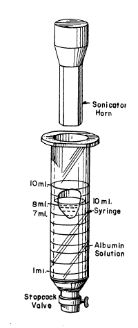
In FIG. lA, there is shown a 10 ml syringe having an
open top and a stopcock-type valve at its lower discharge end.
The syringe is filled to the 8 ml level with the 5% albumin (HSA)
solution. The sonicator horn is inserted in the syringe to the
7 ml level, indicated as the Tl position in FIG. lB. In this
position, the sonicator horn is immersed in the upper portion of
the solution, the solution level being as indicated in FIG. lB.
Initial sonication is carried out essentially without foaming of
the solution.
Immediately following initial sonication and without
turning of~ the sonicator, the horn is withdrawn to the 10 ml
level, indicated as the T2 position in FIG. lC. The power input
to the sonicator horn c~n also ~e ~ncreased as it is withdrawn
.'
~325~
g
to the T2 position. Immediately following the withdrawal, foam-
ing of the albumin solution commences and the solution becomes
milky in appearance. The solution will foam upwardly around the
sonicator horn during the second phase. The appearance of the
foamed solution is illustrated in FIG. lD, the microbubbles be-
ing indicated in greatly enlarged di~meter over their actual
micron ranye sizes.
The solution being sonicated contains both dissolved
and entrained air. The solution is in contact with the ambient
atmostphere around the sonicator horn. (The clearance between
the horn and the inside of the syringe can be seen in the cross-
sectional view of FIG. 2.) The air contact facilitates the
foaming and aerosolating of the solution in the second stage of
the sonication.
The dispersions from a plurality of sonication batches
can be pooled for concentration. For example,
a plurality of
the dispersion increments can be introduced into a separator
vessel, which may be a large syringe or separator funnel equipped
at its bottom with an outlet controlled by a drainage valve.
Such a separate vessel in the form of a large syringe is shown -
in FIG. 3. By holding the pooled dispersions for several hours
without agitation, such as overnight holding, the microspheres
will rise to the top of the solution and form a layer of float-
separated microspheres. Beneath the collected layer, the clari-
fied albumin solution will be substantially free of microspheres.
It is therefore possible to drain off a major portion of the
solution through the bottom outlet. For example, one-half to
three-fourths of the solution can be removed. However, it is
desirable to retain a sufficient solution volume to permit full
redispersion of the concentrated microspheres.
In PIG. 4 illustrates the microsphere concentrate with
the microspheres redispersed. The microspheres are sufficiently
stable that they do not adhere permanently to each other in a
concentrated layer, remaining as separate ~ntact microspheres.
They can readily be redispersed ~y mild agitation.
.,' ,:::
,
~32~59~
-10-
Aftex redispersion to an essentially homogeneous con-
dition, fxactionation may be carried out to remove oversize mi-
crospheres. ~y holding the redispersion ~or a short time, such
as around 30 minutes, the largest diamete~ microspheres will
pre~erentially rise to the top and colIect in a layer, as indi-
cated in FIG. 4A. When that has occurred, the microsphere dis- -~
persion beneath the oversize microspheres can be removed
through the drainage valve. When the collected oversize micro-
spheres approach the valve, the valve is closed so that the
oversize layer remains in the separator vessel, as indicated in
FIG. 4B. The product obtained is a concentrated fractionated
albumin m~crosphere product in which at least 80% of the micro-
spheres have diameters in the range from 1 to 9 microns. The
preferred product has at least 90% of the microspheres with di
ameters of from 2 to 8 microns. -
Further directional details of the presently preferred -
procedures are set out below under the appropriate headings.
Sonication:
Fill a 10 ml syringe of oval cross-section fitted at
its lower outlet end with a stopcock to the 8 ml mark with
sterile 5% human serum albumin. Position a sonicator probe of
smaller cross-section in the syringe so that the bottom of the
probe is at the 7 ml mark. Sonicate at energy setting 6 for 30
seconds then (with the sonicator still on) move the probe tip
to the 10 ml mark, while moving the energy setting to 8. Soni-
cate for an additional 25 seconds. Turn off sonicator, remove
probe and drain content~ of the syringe into a 60 ml syringe or
separatory funnel with a stopcock controlled bottom outlet.
From 5 to 6 syringe volumes are pooled.
Concentration:
Allow the pooled increments to stand overnight (8-12
hours) ~tho~t agitation in the separator vessel. When substan-
tially all the microspheres have ~ormed a layer on the top,
drain two-thirds of the volume from the ~ottom.
1325~9~
Fractionatlon:
Resuspend the microspheres and fill a 60 ml syringe
with them. Let sit 30 minutes, then drain all but about the last
3-4 ml into a collection vessel. The oversize microspheres are
left. Count a sample and calculate the concentration, mean di-
ameter, and percentage less than 10 ~. If less than 99.5% are
less than 10~, re-fractionate. If required for redispersion,
concentration may be ad~usted with 5% HS~.
.
RESULTS
Concentration measurements are set out below in Table
A for three representative runs using the procedures described
above. The initial concentration of the disperions after soni-
cation was of the order of 130 to 1~0 x 106/ml. This was in-
creased by the float-separation concentration to 340 to 450 x
106/ml.
For product control, the microspheres may be counted
by a Coulter Counter, obtainable from Coulter Electronics, Inc.,
Highleah, Florida (viz. Coulter Counter Model TAII). Micro-
sphere counts set out above were determined in this way.
The stability of a representative product was examined
in a study lasting for 20 weeks. The initial concentration was
approximately 4.31 x 10⁸ (431 x 106) microspheres per milliliter.
Concentration measurements were made at about weekly intervals.
The results are summarized in Table B. The measurements, which
were made by means of a Coulter Counter, are presented graphi-
cally in FIG. 5. The samples were held at ambient room tempera-
ture (20-25C). The concentration of about 400 X106 microspheres
per milliliter was maintained for 20 weeks. This evidences a
high degree o f room temperature stability.
The stability of the microspheres can be affected by
unusually hot or cold temperatures. However, even at tempera-
tures as low as 4C or as high as 37C, microsphere concentra-
tions in excess o 200 x 106/ml can be maintained for periods of
eight weeks or longer. Nevertheless, for commercial distribution
or long-term holding very high or low temperatures should be
avoided. Room temperature holding is preferred. Temperature i~
::
~ 32~5~
-12-
protection~of the microspheres during C?hipment can be used.
TABLE A
Concentration Measurements
Microspheres/ml Microspheres/ml
RunsAfter Sonication After Concentration
A135 x 106 386 x 106 : :
~141 x 106 4~3 x 10⁶
C 133 x 106 440 x 1o6 .: :
132~59~ -
-13-
TABLE B
Microsphere
WeekConcentration x 108
0 4 31
1 4.49
2 4.20
4 3-91 -
3.86
4.25
6 4.06
7 4.12
8 3.92
9 ' 3.94
3.97
11 3.48
12 3.48
13 4.09
14 3 70 ; ;
4.92
17 4.15
18 3.99
19 4.14 ~;
; .,' . ''
~ i~:';"
; .
: ",''. '
,-',"',':
1325~90
-14-
-
REFERENCES
Feigenbaum, et al. (1970), Circulation 41:615-621
Feinstein, U.S. Patent 4,572,203.
Feinstein PCT Application WO 84/02838.
Feinstein, et al. (1984), J Am. Coll. Cardiol. 3:14-20.
Gramiak and Shah (1968), Invest. Radiol. 3:356-358.
Keller, Feinstein and Watson (1987), Amer. Heart J., 114:570-575.
Tickner et al. U.S. Patent 4,276,885.
Tickner et al., National Technical Information Service Report
HR 62917-lA, April, 1977, pages 34-40.