Note: Descriptions are shown in the official language in which they were submitted.
1 33 1 ~9
.
A LIF~ING MONITORING AND EXERCISE
TRAINING SYSTEM
sAcKGRouND OF THE INVENTION
1. Field of the Invention:
The inYention is directed to a system for monito~-
ing the lifing motion and/or the exercise training of an
individual. Both systems comprise a preprogrammed m.icropro-
cessor that is operatively coupled to an electromyographic
sensor that is used to measure muscle force for a prede-
termined muscle group. However an alternate embodiment of -
the lift training system, does not employ electromyographic
sensors instead relying solely on a goniometer to monitor ~ - -
lifting angle
2. Description of the Prior Art~
Annularly millions of workers suffer from work `~
related low back pain, most of which is attributed to
improper lifting techniques. Such injuries result in work
time lost and disability claims costing employers large
amounts of money each year.
A number of devices have been proposed to monit~r
and provide feedback as to a person's correct posture. Such ~i
devices may comprise longitudinal belts that are wrapped
from a person's waist over his or her shoulder, these ;~
aevices monitor belt tension insuring that the user's back
is being held upright, See U.S. Patents 3,608,541,
4,007 7 733, and 4,055,168. Other devices include convention-
al belts that are fittea with sensors for monitoring stomach ~ ;
sag, which indicates improper posture because of relaxation ~ ;;
of the stomach muscles, See U.S. Patents 3,582,935, and ;~ `
3,670,320. U.S. Patent 3,644,919, discloses a signaling
device indicating the improper position of a skier's legs
during skiing.
-1- ~;~ "'''
,. ~ ~,,, '
1 331 4~q
In addition to monitoring lifting technique and
motion it is also important t~ monitor a person's exe~-cist
program during physical therapy to insure that the physical
therapy is being done properly, for the correct intensity
and duration. Devices for measuring overall physical loads
have been proposed, See U.S. Patent 4,394,865; but these
devices do not tend to be directed to a specific muscle
group for measuring the muscle force used in an exercise or
the duration of that exercise.
SVMMARY
The amount of force exerted by a muscle is direct-
ly related to its enervation by virture of the amplitude and
frequency of constituent action potentials. Therefore it is
possible to measure muscle force with electromyographic
(EMG) techniques. In integrated electromyography (IEMG) the
myoelectric signal is rectified and time averaged to prod~lce
an accurate representation of the EMG signal energy which ;
can be related to muscle force.
: :~
In the lift monitoring mode of the present in~
vent~on, an electromyographic sensor is secured to a belt
that is wrapped around a user's waist so that electrodes of ; ~ ;
the sensor are positioned adjacent to the lower back muscles
of the user's back. In this way the amount of muscle force
exerted by the lower back muscles during a lifting operation
can be monitored. It is also important to measure lumbar
angle during a lifting operation to insure that heavy
weights are lifted correctly, as such the belt is also
provided with a goniometer for measuring lumbar angle during
a lifting operation. Both the muscle force signal and the
goniometer output are applied to a microprocessor which
compares these signals with preprogrammed lifting parame~
ters. If these signals exceed the preproqrammed li~ting
-2-
i.i ~,~.' ~' " ~'.''.
~ ~ 1 331'48~
parameters an indicating means is activated to indicate to
the user he has exceeded these parameters. An electronic
memory is coupled to the microprocessor recording these
e~ents. ~he microprocessor can be coupled to a compliance
computer which reads the memory and tabula~es the lifting
operations for evaluating various lifting operations and
compliance with the preprogrammed parameters. The
microprocessor and EMG sensor together with a signal source
are used to measure interelectrode impedance to establish
that the device is actually worn and used.
An alternate embodiment of this system comprises
using a goniometer to measure lifting angle and logging into
the memory of the microprocessor any time a user exceeds the
lifting or lumbar angle parameters. The belt can also be
fitted with temperature and/or motion sensors to monitor if
the belt is being worn by a user.
A similar system is used in physical therapy
wherein the therapist prescribes that a muscle or muscle
group be isometrically exercised for a period of time during
a specified time interval, such as a day. An
electromyographic sensor is used to monitor IEMG and is
coupled to a microprocessor which displays the IEMG
intensity on a bar graph. The microprocessor is also
provided with a clock which first indicates when an exercise
!, 25 program is to begin; second when to contract the muscle or
muscle group; and third when to relax the muscle or muscle
group. The microprocessor is also provided with an `
electronic memory for recording the actual time, duration of
the tension:ing, and the muscle force exerted. The
microprocessor can be coupled to a compliance computer which
reads the electronic memory and tabulates the exercise
-3-
~ 1331~89
results, indicating compliance with predetermined exercise
program.
The electrodes for the electromyographic sensor
can be mounted in cotton gauze webbing that is the inner
layer of a cast. In this way ,arm and leg muscles can be
exercised and monitored while being encased in a cast.
Additionally the electrodes can be mounted on cylindrical
objects that can be fitted into natural body orifices for
measuring muscle force exerted by the muscles attempting to
close these orifices.
BRIEF DESCRIPTION OF THE DRAWINGS
Figures 1 and 2 are perspective views of the lift
training belt secured to a user.
Figure 3 is a top view of the belt.
Figure 4 is an electrical block diagram of the
lift training system.
Figures 5a-5d are graphs of muscles force and
lifting angle versus time for various lifting scenarios.
Figure 6 is a block diagram o~ the lift training ~ `
operating system.
Figure 7 is a front view of the exercise training
device.
Figure 8 is an electrical block diagram of the
exercise training system.
Figure 9 is a flow chart of the auto ranging
technique for the bar graph display of this exercise ~ -
training system.
Figure 10 is a cross sectional view of a cast
using the exercise training electrodes.
Figure 11 is a side view of a cylindrical mounting
assembly for the sensing electrodes that is adapted to be
inserted into a female's vagina. ;~
-4-
i, ' . " ' '~, ~
;~'"' "''`" :~.
, ' . .. ' ""
.: . . .
~ ~ I 1 331 489
Figure 12 is a side view of cylindrical mounting
assembly for the sensing electrode that is adapted to be
inserted into a user's anus.
Figure 13 is an electrical block diagram of an
alternate embodiment of the lift training system.
DETAILED D]3SCRIPTION
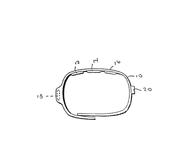
Figures 1-3 illustrate the belt mounted lift
training system. Belt 10 is slecured to just above the waist
of a user in a conventional manner. The belt is provided
with three electrodes 12, 14 and 16 which are electr;~all~
coupled to monitoring device 18 through wires (not sho~Jn in
these figures). The electrodes are secured to the belt so
that as the belt is worn the electrodes are located adjacent
to a patient's lower backO The training and monitoring
device is located in a pocket on the belt. Goniometer 20 is
also mounted on the belt and is located so that it is
positioned adjacent to a patient's side so that as a
patient bends the goniometer can monitor the bending angle.
It should be noted that by mounting the tranining and
monitoring device so that it too is located on the patent's `~
side, the goniometer can be located in the device rather
than having a' separate mounting location on the belt.
The belt can be fabricated from a light weight
elastomeric fabric and is designed to be worn just above the -
~ waist. The belt fastener or securing member can be made
from hook and pile fasteners located at the adjoining ends
of the belt. The electrodes themselves are silver element
pads that serve as surface electrodes of an
electromyographic sensor. The ~oniometer and the electrodes
are connected to the monitoring device via wires located in
the fabric that terminate in metallic snaps that can be
; '
' 1 331 489
coupled to mating snaps located in the training and
monitoring device.
Figure 4 is an electxical block diagram of the
training and monitoring device. The monitoring device
comprises electromyographic sensor 22 which is operatively ~ -
connected to control means ~4 through an analog to digital
converter 26. Goniometer 20 is also coupled to the control
means through converter 26. The control means comprises a
microprocessor unit acting also as an internal clock and is
interfaced to an electronic memory 25 that forms a recording ~ ;
.. means. The microprocessor is coupled to a indicator means ;;
27, which ~an be auditory and/or vibrational for indicating
to the user a lifting condition which exceeds preset
parameters programmed into the microprocessor.
In operation the myoelectric signals from the
three electrodes are amplified by high gain differential ~;
amplifier 28, filtered by bandpass filter 30 and directed to
envelope detector 32 which converts the raw EMG waveform of 1
the myoelectric sinals into an approximation of the total
myoelectric energy which essentially comprises a muscle
force signal. As the resulting muscle force signal is a~
analog signal it is converted into a digital format ~ccept- `
able to the microprocessor. Similarly the goniometer formc;
a horizontal angle signal that comprises a lifting angle
signal that is also converted from an analog to a digital
format before being directed to the microprocessor. It ~^
should be noted that goniometer measures the lumbar angle
including anterior and/or left/right lateral angles. ~ -
Figure 5 reflects the idealized behavior of lumbar ; ~ -
angle and l~MG measurement under several lifting conditions.
THe EMG curves shown do not not include components of ,,-
intertia and body weight.
-6
1,' ' ' .' ''" ','''','~ '
1 3~1 4~9
Figure 5a and 5c are graphical presentations of
lifting no loads in a back straight position and back bent
position. As can be seen in the back straight position t}le
horizontal angle changes only slightly whereas i]. the bac)~
bent position the horizontal angle changes from nearl~ zer~)
degrees to ninety degrees. However since no additional load
i5 involved in either lifting sequence the arnount of muscle
force iEMG) re~uired is mini~a:L. In Figures 5b and 5d a
load is lifted and although the lifting angle is identical
to the no load sequence, the amount of muscle force required
in each sequence vaxies considerably because of the lifting
methodology. In the back bent position the amount of muscle
force required from the lower back tends to mirror the
change in lifting angle where as in the back straight
position during the initial lifting motion the amount of
lower back muscle force is considerably reduced because the
legs are doing the lifting.
In training a user of the system, a teacher
programs the microprocessor ~ia the compliance computer
38with a 5et of lifting parameters which include limits as
to muscle load and horizontal angle. As there is interplay
between these parameters the teacher can set up a system
wherein a combination of the parameters triggers a feedback
warning signal. For example in Figures 5c the user has
taken an incorrect lifting angle but since the user i5 not ~
lifting any load the indicator is not triggered. However in ~;
Figure 5d the user has taken an incorrect lifting position ~ ~
and is lifting a load, therefore the indicator is triggered. ~ ;
As such the present system gives the teacher the ability to
program triqgering parameters that are a combination of the
lifting angle and muscle force required.
-7-
- 1331~89
The monitoring sy~tem is battery operated ~nd
located in a lockable housing so that after the teacher has
programmed the microprocessor, the housing is locked and t}-e
battery cannot be tampered with by the user. The
microprocessor is provided with interface 36 comprising a
plug for coupling the microprocessor to compliance computer
38. The compliance computer can be an IBM PC compatible
unit and is used to interrograte the memory so that a
training session can be tabulated for evaluation by the
teacher. In addition this interface can be used for
programming the microprocessor with the programmed lifting -~
parameters. As can be seen in Figure 6, the compliance
computer is provided with monitor 42, input keyboard 44, and
printer 46.
~o insure that the monitoring system is operating
correctly the microprocessor periodically activates inter-
electrode impedance test 48 to check if electrode contact is
sufficient. The test applies a bipolar sinusoidal signal
across the EMG inputs, the impedance is then measured by the
microprocessor. In addition the microprocessor can be ~
provided with a testing system for testing battery voltage ;
to insure proper voltage to the monitoring system. In the
event that the contacts fail the impedance test or the
battery has insufficient voltage the microprocessor signals
the user through the indicator means and turns off the
system. -
Figures 7-12 are directed to an exercise training
system which is similar to the lift training monitoring
system. As can be seen in Figure 8 the circuitry is similar
except that the exercise training system is provided with
visual feedback display means 50 comprising a bar graph, and
alerting means 5~ comprising three light emitting diodes.
. ,' ~
'.' ''' :" ''
1 331 489
The auditory eedback element 27, which in the lift;n~
training system is an indicating means~ in this embodiment
ig used in conjunction with the visible display means and
the alerting means to inform the patient audibly that these
displays have been triggered.
Bar graph 50 is a liquid crystal or light emitting
diodedisplay that is used for clisplaying muscle force used
during use. The exercise training system is auto ranging
with respect to the bar graph, the alogorithm for auto
ranging the bar graph is disclosed in Figure 9. During an
exercise period light emitting diode 54 lights up indicating
to the u~er to contract the muscle group that is equipped
with the electromyographic electrodes. The user keeps that
muscle contracted until light emitting diode 54 is turned
lS off, and light emitting diode 56 lights up indicating to the
user to relax the muæcle group. Contract/relax cycles are -
repeated as determined by the preprogramed microprocessor.
The intensity of the muscle contractions is fed back to the
user by viewing bar graph 50 which indicates muscle force
used.
A physical therapist first applies the
electromyographic electrodes to a patient adjacent to the
muscle group to be exercised. Then the therapist programs
the microprocessor via the compliance computer of the
training system, by programming a time interval in which the
exercise routine is to begin, the`timed interval for
contracting a muscle group and relaxing a muscle group, and
the ~umber oiE repetitions. The therapist then couples the
unit to the electrode leads and the patient can then conduct
his own physical therapy by using isometric exercises for
contracting the desired muscle group for the required
_g~
,,. . .,, , ., ,, ~ ~ ~,
~ 1 331 489
duration and repetitions and monitoring the intensity of the
exercise on the bax graph.
As with the lift training and monitoring system
the exercise training system can be coupled to compliance
computer 3~ through interface 36, which can comprise a
simple jack. The compliance colmputer is used to program the
microporcessor and to tabulate the patient's performance
with the exercise program by interrogating the electronic
memory which recorded the exercise session. The therapist
can then program into the microprocessor a new training ~ ;
routine ba~ed upon the patient's actual performance in the
last training session. As with the lift training system the ;;
compliance computer is also used to program the '
microprocessor. ;
Figure 7 is a front view of the training and ~-
monitoring device which iS relatively compact. The
circuitry including the microprocessor, the electronic ~ ,
memory, and the electromyographic processing circuitry are
contained in housing 60. The device is provided with a -~
start/stop switch 72 for overriding the exercise routine
programmed into the microprocessor, and a third light
emitting diode 73 indicates the device is not functioning
correctly based upon its self testing, which is identical to
the self testing of the lift training device. ~;
Figure 9 discloses a flow chart illustrating the
method of auto ranging the bar graph display. At the start
of an exercLse session the exercise parameters programed
into electronic memory via the compliance computer are read
by the microprocessor and are used to initialize relevant
variables. The auto ranging method then through subsequent
EMG ~muscle force~ readings sets a continually updated top
value and bottom value for the bar graph scale. The method
-10- , ...... ..
., .
. -",
~,.,
~ 1 331 489
then calculates a new EMG reading located between the top
and bottom value as a ratio of the E~G range and as such
displays this ratio by lighting up the correct number of bar
graph display elements.
Figure 10-12 disclose different devices for
securing the electrodes of the electromyographic sensor to
selected body location. In the embodiment illustrated in
Figure lO, the electrodes are secured to cotton gauze 74
that forms the inner liner of a cast for a limb. The
monitoring housing and related circuitry because of its
compact nature, can then be embedded in casting material 75
of the outer cast layer. The bar graph display is located
at an angle to the housing to facilitate viewing by the
patient.
Figures 11 and 12 are directed to electrode
mounting assemblies that were designed to be inserted into a
naturally occurring body orfices. These assemblies are
cylindrical and have three stainless steel electrode bands -
located about their circumference. The embodiment
illustrated in Figure 11, comprises cylindrical member 80
which is inserted into a female vagina~ so that the female
patient can monitor the exercise of associated vaginal
muscles. The embodiment illustrated in Figure 12 comprises
cylindrical member 81 and is inserted into a patient's anus
for monitoring a patient's exercise of the anal sphincter
muscles. ~oth units are made from injected molded plastic,
and are provided with depth ga~ges 82 which can be
adjustabl~positioned and fixed on the cylindrical members
by the therapist.
An alternate embodiment of the lift training
system is illustrated in Figure 13 and comprises a lift
training system that is not provided with an -~
- 11 1331489
electromyographic sensor. Instead only goniometer 20 is
used to measure lumbar angle. ~emperature sensor 90 and/or
motion sensor 92 are also mounted on the belt and indicate
the belt is being worn by a user. In this way, the actual
usage of the lift training system is logged together with a
log of incorrect lifting angle.
As with the previously discussed lift train;ng
system the microprocessor is programmed with liftin~
parameters ~ia the compliance computer that when exce~(led ::
,," ,
trigger indicator means 27 to alert the user. The
compllance computer is used to interrogate the electronic
memory for evaluating and tabulating the results of the lift ~ ;
monitoring session.
It should be noted that the ouput signals of usage ~-
sensors 90 and 92 do not have to be applied to converter 26 -
if the signals are already in digital form. In addition the
usage sensors can be used on the belt disclosed in Figures
1-3,
The invention should not be limited to the ;~
above-described embodiments but should be limited solely to -
~the claims that follow.
-12-
:"."','
. . ' ~
. ',
I; . , ~':
': ~