Note: Descriptions are shown in the official language in which they were submitted.
~ ~ 3 ~ r~
OPTICAL VIEWING DEVICE
Description
Technical Field
, ............................................................... .
lV This invention relates generally to optical
viewing devices and more particularly concerns a dispos-
able endoscope for use in viewing a region within a body
cavity or the like.
Backqround
Instruments which permit visualization of
typically inaccessible areas and organs within a pa-
tient's body are known. Such optical viewing instru-
ments, or "endoscopes," can often obviate the need for
20 excising specimens from an internal organ of the living `~
body for examination with a conventional microscope.
Also, as disclosed in, for example, U.S. Patent Nos.
3,677,262 to Zukowqki and 4,392,485 to Hiltebrandt,
endoscopes may further be provided with a means for
supporting and guiding surgical instruments within a
patient's body.
Structurally, endoscopes ~ypically include a
light pipe for illuminating the region to be viewed, at
least one lens assembly for focusing and relaying the
image of the illuminated object, and a housing for the
entire assembly which is structured so as to minimize
tissue damage upon examination. Examples of such endo-
scopes may be found in U.S. Patent Nos. 3,089,484 to
.
'
.,...~
. .. A
~. ~
--2--
Hett, 3,257,902 to Hopkins, 3,556,085 to Takahashi,
4,267,828 to Matsuo and 4,273,110 to Groux.
There are several drawbacks in the endoscopes
of the prior art to which the present invention is
addressed, including the expense and complexity of the
known optical viewing devices and the corresponding
difficulty in volume production. The devices of the
prior art incorporate expensive and carefully fabricated
ground glass lenses in structures that are complicated
and difficult to manufacture. Typically, as in, for
example, V.S. Patent No. 3,257,902 to Hopkins, endo-
scopes include a rather complicated design so as to
correct for axial color aberration. Because of this, it
has not been possible to make such endoscopes dispos-
able, i.e. out of plastic materials, or easily producedin quantity.
,-.~.
Disclosure_of the Invention
Accordingly, it is an object of the present
invention to overcome the aforementioned disadvantages
of the prior art.
It is another object of the invention to pro-
vide a disposable optical device of simple and inexpen-
sive construction.
It is still another object of the invention to
provide a disposable endoscope in which the objective,
relay and viewing lens assemblies are fabricated of
polymeric material. ~-~
It is yet another object of the invention to
3~ provide such a disposable endoscope which is configured
for direct, visual observation of an area.
It is a further object of the invention to
provide such a disposable endoscope which is configured
for preparation of a photographic or electronic record
'
:!.
~33~ 5~
--3--
or display, i.e. by coupling the endoscope to a display means or to a camera or other recording
means.
It is still a further object of the invention to provide a disposable endoscope as above
in which the light pipe also serves as the support means for the entire op~ical assembly, thereby
reducing the number and complexity of parts within the device.
It is still another object of the invention to provide a disposable endoscope which is
sealed so as to be both airtight and watertight.
Further objects, advantages and novel features of the invention will be set forth in part
in the description which follows, and in part will become apparent to those skilled in the art on
examination ofthe following, or may be learned by practice ofthe invention.
In one aspect of the invention an apparatus for viewing a region within a body cavity
of the like is provided which enables direct examination of that region. The apparatus includes
a light pipe having a distal end, a proximal end, and an elongate axis, designed to direct light
from the proximal to the distal end to illuminate a region to be viewed near the pipe's distal
end; carried at the distal end of ht light pipe, and objective lens system cornprising a plurality
of polymeric objective lens elements and constructed to form an image of the region
illuminated by the light pipe; a polymeric relay lens assembly mounted within the light pipe for
relaying the image formed by the imaging system to a proximal region of the light pipe,
wherein the relay lens assembly comprises symmetrical pairs of polymeric rod lenses arranged
end-to-end within the light pipe, the light pipe structured to support and align the objective
lens elements and the rod lenses; and carried adjacent such proximal region of the light pipe, a
viewing lens system including a plurality of polymeric viewing lens elements constructed and
arranged to allow viewing of the image relayed by the relay assembly.
In another aspect of the invention there is provid,od an optic train useful in an
endoscope, the optic train defined along a generally elongate axis and having a distal end and a
proximal end, comprising: at the distal end of the optic train, an objective lens system
including a plurality of polymeric objective lens elements; at the proximal end of the optic
train, a viewing lens system comprising al least two polymeric viewing lens elements; and a
series of polymeric relay lenses disposed end-to-end along the elongate axis and between the
objective and viewing lens systems, wherein the relay lens assembly comprises symmetrical
pairs of polymeric rod lenses placed end-to-end.
1 The field of view provided by these embodiments is generally on the order of 60 to
70 although the objective lens assembly may be constructed so as to provide a narrower or
wider field of view. A Fresnel lens or other optic may be incorporated within the device at the
~1
i~ ~
I !
1~3~
distal end of the light pipe. Such an assembly refracts the light from the light pipe into a larger .
cone and thus gives a larger illuminated region. This assembly may also be used to deviate the .
centroid of the illumination pattern.
In order to ensure economy of manufacture, the light pipe and the objective, relay an
viewing lens assemblies are preferably all fabricated of a polymeric material which lends itself
to injection molding. Suitable materials include acrylics, polystyrenes, -.. .
.' '' '','.''''',
c :- - ~ . . .. ~ .
, ..
4 ' :~
,.:::-. : ~ -:: .: -: '.:. :
s- ~ ~ 3 ~
polycarbonates and styrene-acrylonitrile (SAN) copoly-
mers.
Optionally, a means for adapting the device to
be coupled to a recording or display means may be in-
cluded at ~he proximal end of the device. With a re-
cording means, for example, a photographic or electronic
record may be made of an endoscopic examination.
Brief Descri~tion of the Drawin~s
FIG. 1 is a cross-sectional side view of a
preferred embodiment of an endoscope embodied by the
: ~ -
invention
FIG. 2 is an exploded, perspective view of a
preferred embodiment of the endoscope showing the series
of rod lenses placed end-to-end within the molded light
pipe; and
FIG. 3 is a cross-sectional view taken along
the 3-3 lines of FIG. 1, and specifically illustrates
the placement of one of the rod lenses within the light
pipe.
FIG. 4 is an optical layout of a preferred
embodiment of the invention and illustrates chief rays -
and image orientation.
Modes for Carrvinq out the Invention
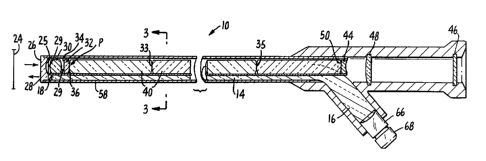
Turning now to the drawings in detail, the
optical viewing apparatus is shown generally at 10 and
is positioned in Figures 1 and 2 so as to enable a
viewer 12 to examine a region 24 within a body cavity or
the like.
An elongate light pipe 14 disposed within the
apparatus 10 is provided with a distal end 18 positioned
near the region 24 to be viewed and with a proximal end
16. The light pipe directs light from a suitable light
source 20 located at proximal end 16 along its length to
,~
-6- ~ ~3
distal end 18. Carried at the distal end of the light
pipe is an objective lens system collectively designated
22 which includes a plurality of polymeric lens ele-
ments. It is preferred that the polymeric lens elements
include at least one aspheric surface to improve the
clarity of the image over an extended field of view.
After suitable placement of the apparatus, light di~
rected by light pipe 14 onto the region to be examined -~
is reflected therefrom and imaged by objective lens
system 22.
In the preferred embodiment illustrated in the
drawings, objective lens system 22 includes the follow-
ing lenses, which preferably provide a focused, real ~-
image of the illuminated region at point P, i.e. prior -
to relay. Surface 26 of distal negative lens 28 di-
rectly receives light reflected from the illuminated
region, and is preferably planar in order to avoid
change of optical power when the endoscope is immersed
in a liquid. Lens 28 is preferably plano-concave as
shown and aspheric on concave surface 25. The image is
then processed by primary positive lens 30 which is
preferably a double-convex objective lens with two
aspheric surfaces. Lenses 28 and 30 together comprise a
reversed telephoto lens that has a relatively short
focal length and which covers a field of view on the
order of 60 to 70. Field lens 32 is located proximal
to the image formed by the objective lens 30 and is
preferably also a double-convex lens as shown. Like
distal negative lens 28, the surfaces 34 and 36 of field
lens 32 are preferably aspheric to ensure elimination of
spherical and other aberration, i.e. to provide a better
degree of correction and to reduce the number of lens
elements needed. The primary purpose of field lens 32
is to reduce or eliminate the vignetting at the edge of
, ~ . .
~7~ ~ ~ 3 ~
the field of view. The lens is placed in the vicinity
of the plane of the image. Lenses 28, 30 and 32, which
together comprise the objective lens assembly herein,
are made of a polymeric material such as acrylic, poly-
styrene, polycarbonate or SAN, preferably of a lowdispersion material such as acrylic. Fabrication into
suitable structures such as those illustrated may be
effected by means of injection molding, conventional
grinding and polishing, or diamond turning, although
injection molding is the preferred method.
This placement of lenses 28, 30 and 32 at the
distal end of the viewing device obviates the need for a
focusing assembly, as the device is optimized to provide
a focused image for areas viewed within the range of
distances generally associated with therapeutic use.
Outside of the typical therapeutic range, it is prefer-
red that the device be provided with a focusing means.
Relay lens assembly 38 includes a plurality of
rod lenses 40 arranged end-to-end so as to transmit the
image provided by objective 22 through the elongate
section of the apparatus to its proximal end. Like the
imaging lenses, the relay lenses are fabricated from a
polymeric material which lends itself to injection
molding, e.g. styrene, polycarbonate, acrylic, SAN and
the like. As above, low dispersion materials, acrylic
in particular, are preferred. The number of relay
lenses is selected so as to reduce the number of surface
refractions which degrade the image while still allowing
for transmission of sufficient light. For direct,
visual observation of an area, the number of relay
lens~s is preferably an odd multiple of`2, i.e. 2(2n+1)
where n is zero or an integer. A particularly preferred
number for such an embodiment which optimizes the afore-
mentioned considerations is 6. As illustrated in Figure
' .. ~
-8- ~ 3~ $~
4, proper image orientation thus typically requires an
odd number of symmetrically placed pairs of rod relay
lenses. Alternatively, the use of an inverted prism
such as a dove prism will allow for the use of an even
number of relay lens pairs.
The image formed at P is collimated and refo-
cused several times during relay, e.g., where 6 rod
lenses are incorporated within the device, the image
will be collimated and refocused three times. Because
10 the placement of rod lenses is symmetrical, correction ;~
for lateral chromatic aberration is automatic, that is,
inherent in the structure of the relay assembly. The
device does not incorporate significant means of cor-
recting for axial color, as the eye is not particularly
sensitive to axial chromatic aberration; this allows for
a relatively simple and inexpensive construction. The
symmetry of the relay system also eliminates distortion
and coma.
The individual rod lenses are difficult to
make if not molded, since the radius of curvature is
about half the length of the rod (thus, it would be
difficult to fit several on a block for grinding). In a
preferred embodiment, the rod lenses are fabricated by
injection molding on standard equipment. The polymeric
material is emplaced in a suitable mold and heated to at
least about 350C. A suitable mold clamping force is
applied, followed by a cooling hold. Generally, a mold
runner diameter about equal to the diameter of the rod ~-
lens optimizes the results obtained.
The rod lenses are preferably identical, dou-
ble-convex lenses having entrant and exit refracting
surfaces 33 and 35 of the same focal léngth. The longer
the rod lenses, the darker the system appears because of
a reduction in overall aperture (f-number) of the optic
.'' ' ~
9 ~ ~ 3 ~
train. The length of the rod lenses is thus optimized
to allow for transmission of sufficient light while at
the same time providing for an endoscope of sufficient
physical length. In a preferred embodiment, the length
of each of the rod lenses is designed to be approxi-
mately equal to the focal length of the refracting
surfaces. That is, for a rod lens having an index of
refraction n and one surface with a focal length f, the
lens will focus a distance nf away from the surface; the
overall f-number of the system is thus f/d where d is
the diameter of the lens. The overall f-number of the
relay system is preferably optimized at between about 4
and 6. The diameter of the rod lenses is preferably
between about 5 mm and about 7 mm, and the index of
refraction for the materials used, e.g. acrylic or
styrene, is on the order of about 1.48-1.49.
A viewing lens system 42 is housed adjacent
the proximal end of the light pipe, and processes the
transmitted image from the relay lens assembly 38. In
one embodiment, a reverse telephoto lens assembly is
used to increase the overall length of the device and
the illumination of the image viewed. In such a case,
viewing lens system 42 includes only two lenses,
post-rod lens 44 and positive lens 48, with a window at
46. In a second embodiment, viewing lens assembly 42
includes three lenses, negative post-rod lens 44,
proximal negative lens 46 (which replaces the window in
the reverse telephoto assembly) and a strong positive
lens 48 disposed therebetween. The post-rod lens 44 is
3~ preferably plano-concave, with the planar surface 50
facing the relay lens assembly and dire~tly receiving
the image transmitted therethrough. Like the objective
and relay lenses, viewing lenses 44, 46 and 48 are
I
~ ~ .
~"-. .-
.~ ,. . ~
t '''~
' ~' . ~ . ' . ~ . ' , .
o- ~ ~ 3 ~
fabricated from a suitable low dispersion polymeric
material which lends itself to injection molding.
It should be noted at this point that appli-
cants' endoscope -and in particular the polymeric
5 aspheric and relay lenses -is thus completely fabricated
from inexpensive materials which easily lend themselves
to volume production. In a preferred embodiment, the
light pipe itself is fabricated from a polymeric
material such as styrene, acrylic or polycarbonate,
10 preferably from a polymeric material with a relatively
high refractive index such as polycarbonate (n*.*1.58).
In the embodiment described above, the field
of view provided by the imaging lens assembly 22 is
about 60 to about 70. If desired, a Fresnel lens such
15 as that shown at 52 may be provided so as to disperse
light and thereby increase the uniformity of
illumination within the field of view. The Fresnel lens
is incorporated within the structure by placement at the
distal end 18 of the light pipe, thereby refracting the
20 light directed onto the region to be examined and
providing a wider region of illumination.
Light pipe 14 is provided with an elongate
cradlelike cavity 54 along its elongate distal section
56. This cradlelike cavity provides a support means for
25 the objective lens assembly as well as the relay lens
assembly. The relay lenses 40 extend along the pipe's
distal section and are arranged end-to-end as described
¦ above. A housing such as an elongate, substantially
I rigid tube 58 encases the light pipe 14 as well as the
1 30 various lens assemblies. The tube is preferably con-
structed of a relatively strong, lightweight material
3 such as aluminum, stainless steel, plastic and the like.~ As illustrated by Figure 3, the relay lenses are
¦ securely wedged between cavity 54 and tube 58 so that
~ :
; ~
'
.
3 3 ~
the lenses are held in axial alignment along the length
of the tube. In a preferred embodiment, the lenses of
imaging lens system 22 are wedged between light pipe 14
and tube 58 in the same manner. Tube 58 is preferably
sealed with adhesive, filler, or the like so as to
provide an airtight, watertight seal.
The light pipe thus doubles as a mechanical
support for the optic train and provides a means for
easily aligning and centering the individual lens ele-
ments. The lens systems can thus be assembled withoutthe need for complicated aligning fixtures. Although
the light pipe is preferably comprised of a polymeric
material which can be injection molded, it can also be
fabricated from either glass or plastic fibers.
Optionally, a shielding means 60 such as dark
paper, ~yla-r~~or other opaque material may be disposed
between light pipe 14 and the relay lenses 40 so as to
eliminate degradation of the transmitted image by light
scattered from the light pipe. Shielding means 60 also
helps baffle nonimaged light, i.e. light from the light
pipe is prevented from entering the rod lenses directly.
Spacers 62 and 64 may also be included to provide
physical separation of the light pipe 14 from the relay
lenses.
Aæ may be seen in Figures l and 2, the light
pipe is angled at "A" within handle 62 and becomes
j completely annular proximal to angle "A" where it is
coupled to light source 20 by suitable means, e.g. by
means of adapters 66 and 68. Although necessary to
3Q eliminate any structural interference of the proximal
end of the light pipe with the viewing lens assembly,
angle A is preferably minimized at about 30 or less so
es to prevent loss of trensmitted light.
~ '
~ .
!`~
..
--12-- ~ ~ 3 ~ ~? ~ ~ .
Assembly of the endoscope is a relatively uneomplicated procedure.
All lens elements except for distal negative lens 28 are ini~ally placed in
shielding paper as described and then inseIted into ~he light pipe. Spacers
29 are provided between ~e objective system elements so as to ensure axial
5 separation. Distal negative lens 28 caps the distal end of ~e endoscope as
illustrated in Figure 1, and the remainder of the optic train is slid toward thedistal end to set axial spacing. Centering occurs upon sliding of the
elements of the optic train into tube 58. Light pipe 14 and tube 58 are both,
as noted above, fabricated from a strong, rigid material so as to prevent
10 buclding during insertion and to ensure sufficient support and centering for
the optic train. The pipe is aUowed~ however, some compression flexibility.
The invention also comprises an optic ~ain of polymeric lens
elements, preferably fabricated from a low dispersion, optical quali~r plastic
such as acrylic. The optic train includes: (1) an objective lens assembly for
15 forming a real image of an illuminated region ~hat is substantially
uncorrected for axial color; and (2) a relay lens assembly, similarly
substantially uncorrected, which comprises an odd number of symmetrical
pairs of polymeric rod lenses (or an even number used in conjunction with
an inverted prism), which rod lenses are designed to relay the image along
20 the leng~ of the endoscope to form an image that can be observed and
optionally magnified. In a preferred embodiment, the objective and relay
! lens assemblies are as described above and illustrated in Figure 1.
In still another embodiment of the invention~? eyepiece section 70 is
provided with a means for coupling ~e viewing device to a display, came~?
25 or other
~ 13- ~ ~3~ p~
recording means so that a display or a photographic or
other record may be made of an endoscopic examination.
Thus, as may be deduced from the above, the
optical viewing device of the present invention is
relatively inexpensive to fabricate; in contrast to
known analogous devices, which are comprised of a number
of ground glass lenses and mirrors, the present inven-
tion incorporates a large number of inexpensive poly-
meric components, including the light pipe as well as
the relay, objective and viewing lenses. Finally,
because in the apparatus of the present invention, the
light pipe doubles as the support means for the system
of relay lenses, the diameter and overall complexity of
the device are substantially reduced.
While the invention has been described in
conjunction with the preferred specific embodiments
thereof, it will be understood that this description is
intended to illustrate and not limit the scope of the
invention, which is defined by the appended claims. It
should in particular be noted that while the optical
viewing device of the present invention has been de-
scribed in conjunction with its use as an endoscope,
other uses of the device -in viewing poorly lit and
remote areas, generally -are clearly within the purview
of the invention.
. .. . ~ , ~ ., . , - , .
. :~
. ~ ~ .; , . , . , ~ . . . . .