Note: Descriptions are shown in the official language in which they were submitted.
61211-918D
1332~4~ `
Background of the Invention
The present invention relates in general to the treat-
ment of disease, tumors, etc., by the use of ultrasound. More
particularly, the present invention relates to a combined -~
visualization and treatment device using ultrasound for both
functions. Treatment is achieved by ablation of tissue
representing the disease entity.
~: -. . ;. .
A large number of diseases manifest themselves in
whole or in part in a focal manner. These include, for example,
diseases of or in the brain, breast, liver and prostate. While
: ,~ ~ .,, - .....
surgical procedures have traditionally been employed when
medicinal approaches were not suitable or effective, surgery
still represents a significant risk to the patient and a chance ;~
that the entirety of the disease entity will not be completely
. . . .
removed.
There is no disputeas to the value of noninvasive
treatment as such as producing volume lesions with focused
ultrasound. One difficulty though with ultrasound treatment
.,. .. :
procedures is the need to visualize the disease entity and `
thereby determine the size, shape and location. While this
concern does not normally exist with invasive techniques such ;
as surgery, it is of critical concern in noninvasive procedures.
In our pending Canadian Application Serial No. 592,604, -~
entitled ULTRASOUND BRAIN LESIONING SYSTEM, filed March 2, 1989,
a visualization technique is described for volume lesioning
treatment of a brain tumor. The technique involves a use of
ultrasound or CT or MRI scan transparencies whose data is
.
,: "
', ~,, :,:
" ;
61211-918D
~332~4~
digitized into a computer and the landmark references from a
skull fixation apparatus are used to preprogram the drive system
for the transducer. By computer control, the brain tumors are
located and the transducer automatically programmed for position-
ing such that the focused ultrasound beam is directed at each
tumor and the dosage set to produce volume lesions.
An alternative to this position translation technique
for brain tumors is to use ultrasound to visualize the disease
entity. Since brain lesioning is somewhat unique due to the CT
or MRI scans and the skull fixation apparatus, the visualization
technique of our co-pending application may not be the most
appropriate technique for ablation of other focal disease sites.
Since some of these other disease sites may be most
effectively treated by the use of ultrasound in either a
transcutaneous or intraoperative mode, there is a need to insure
that the transducer components which are designed and the
materials selected be such so as to be suitable for steam auto- ;~
claving.
The present invention provides an ultrasound localiza-
tion and therapy system which is designed with both avisualization transducer and a therapy transducer. Those
portions of the structure which must be sterilized are
constructed from selected materials which are steam autoclavable.
Another concern with the treatment of disease in a
transcutaneous mode by ultrasound is the physical size and shape
of the probe. Since the transducer design of the co-pending
application is used external to the patient, size and packaging
61211-918D
1332~41 ~
considerations are not substantial. However, with the modes of
examination and treatment such as transrectal, transesophogeal,
etc., the probe design is critical. While the specifics of
our co-pending transducer design may be used in some embodiments ~ ;
of the present invention, it will require some scaling down in .:~
size. Further, if the transducer assembly is going to be steam .:~
autoclavable, certain material changes are advisable in order :
to provide a finished product which will withstand the high
autoclaving temperatures. .;;:~:
In a related embodiment the concept of utilizing a
visualization transducer in combination with a treatment trans- .:
ducer is disclosed for treatment of the prostate. This .... : .
particular configuration is adaptable for use in other body
cavities. The therapy treatment from within such body cavities ; .
by ultrasound, where ultrasound is also used for imaging of the
area to be treated, has not heretofore been done. ~
Summary of the Invention . .
A visualization.and treatment transducer for producing : :
lesions in diseased tissue sites according to one embodiment of
the present invention comprises.a transducer housing having a .
main section and a detachable.enclosure, movable visualization .
transducer means disposed within the detachable enclosure, ~ ::
movable treatment transducer means disposed within the detachable
enclosure, first drive means providing rotary motion to the
visualization transducer means in two degrees of freedom, second .
drive means providing rotary motion to the treatment transducer ~ .
means in two degrees of freedom, the visualization transducer
: 61211-918D
1332~41
means and treatment transducer means having generally coaxial
focal axes and the first and second drive means being operable
independently of each other.
. A transrectal or other body cavity visualization and
treatment transducer assembly for ultrasonic visualization and - .
treatment by producing lesions in diseased tissue sites accord-
ing to another embodiment of the present invention comprises a ~ ;
fluid-filled, flexible-walled enclosure, a movable visualization ; .
transducer disposed within the enclosure, a movable treatment
transducer disposed within the enclosure, a reflective scanner .
disposed within the enclosure and aligned with the treatment
transducer for changing the direction of the focused ultrasound
beam from the treatment transducer, first drive means providing ~: :
rotary motion to the treatment transducer, second drive means
providing linear motion to the visualization transducer, the
first and second drive means being operable independently of
each other, and third drive means providing rotary motion to
the visualization transducer and the treatment transducer .`
concurrently.
In accordance with the present invention, there is
provided an ultrasound treatment transducer assembly for
directing a focused beam at an anatomical site, said transducer
assembly comprising: an acoustic focusing lens having a concave -
front surface and a substantially flat back surface; a
substantially flat piezoelectric transducer plate disposed in
spaced relation to said focusing lens and having a rear surface :~
and a front surface which is disposed at a fixed distance of
separation with respect to the back surface of said acoustic :
4 :
.',',.',"''
; 61211-918D
1332441
focusing lens; an acoustic coupling medium disposed between the
back surface of said focusing lens and the front surface of
said transducer plate; first pressurizing means cooperatively ;~
arranged with said acoustic coupling medium for maintaining
said acoustic coupling medium between said transducer plate ;
and said focusing lens at a desired pressure; and air pressure ~
means cooperatively arranged relative to the rear surface of ;
said transducer plate and adapted to apply air pressure against
said rear surface, wherein the pressure applied against said
rear surface by said air pressure means is higher than the
pressure on the front surface of said transducer plate due to ` -
said acoustic coupling medium.
One object of the present invention is to provide an
improved transducer assembly including both a visualization
transducer and a cooperating treatment transducer. -
Related objects and advantages of the present
invention will be apparent from the following description. ~
Brief Description of the Drawings - ;
FIG. 1 is a diagrammatic illustration of an ultrasound
treatment apparatus according to a typical embodiment of the `
present invention.
FIG. 2 is a side elevation, diagrammatic illustration
in full section of a transducer assembly which is suitable for
use in the FIG. 1 apparatus. `
FIG. 3 is a front elevation, diagrammatic illustration
in full section of a transducer design suitable for use in the ;~
FIG. 2 transducer assembly.
~; 61211-918D
1332~1
FIG. 4 is a perspective, diagrammatic illustration of
an ultrasonic probe for prostate visualization and treatment.
FIG. 5 is a side elevation, diagrammatic illustration
of the FIG. 4 ultrasonic probe.
FIG. 6 is a lateral section view of the FIG. 4 ultra-
sonic probe detailing the configuration and support of a
reflective scanner.
FIG. 7 is a lateral section view of the ultrasonic
probe detailing the arrangement and support of the treatment
transducer.
FIG. 8 is a side elevation, diagrammatic illustration
in full section of a control unit which is coupled to the FIG. 4
probe for imaging and treatment control.
Description of the Preferred Embodiment
For the purposes of promoting an understanding of the
principles of the invention, reference will now be made to the
embodiment illustrated in the drawings and specific language
will be used to describe the same. It will nevertheless be
understood that no limitation of the scope of the invention is ~
thereby intended, such alterations and further modifications in ~-
the illustrated device, and such further applications of the
principles of the invention as illustrated therein being
contemplated as would normally occur to one skilled in the art
to which the invention relates.
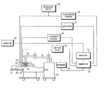
Referring to FIG. 1, there is illustrated an ultrasound
treatment system generally in block diagram form with the ~--
: ;:: ~ .
patient 20 lying on an appropriate table 21 with the transducer
. - .
- . -':. ';' '
6 ~
-~ 61211-918D ;
1 3 3 2 ~
housing 22 diaphragm 23 in contact with the patient. A suitable ~ ~ ;
coupling medium is used between the diaphragm and patient and - -
the therapy transducer 24 is disposed in a volume of degassed
water 25. In an intraoperative mode, sterile housing 22 with
its diaphragm 23 is brought into contact with sterile fluid ;~ `
overlying on the internal organ or tissue directly. Guidance
to the tissue or organ site is provided by ultrasound visualiza-
tion element 2,8 located inside housing 22. The relative sizes
and positional relationships of therapy transducer 24 and
visualization element 28 which is an imaging transducer is best
illustrated in FIG. 2. ;
Housing 22 is manually placed in position by the
operator while ~eing guided by transducer 28 with the ultrasound -
image displayed on monitor 29. Housing 22 is supported by
articulating arms 30 and 31 with rotation axes as shown by the
rotary arrows. Vertical motion is shown emanating from base
support 32. Once the system is appropriately located for
treatment, the articulating arms and rotation axes are locked
in place. From the scanning of visualization transducer 28,
the treatment v~lume is defined and stored in computer 35. The
spatial position of the treatment volume is also defined with
respect to depth and orientation to surrounding tissues. By ;
interacting with the tissue and organs displayed on monitor 36,
the treatment spatial regimen is computed. Dosage parameters
of sound intensity and time-on period are entered into computer
35.
Once the treatment regimen is established, the system
automatically progresses through the treatment volume by placing
. :
-~ 61211-918D
1 3 3 2 ~ ~ 1
individual focal ablative lesions. Power amplifier 37 provides
the drive energy to therapy transducer 24 for each focal site
under control of computer 35. Degassed water system 38
provides degassed water to the interior of transducer housing
22 and temperature control system 39 keeps this degassed water ;~
at a constant temperature during the therapy procedure. The -
procedure can be interrupted at any time by the operator and
restarted at the last stopped position, if that is desired.
In the event the operating and control electronics
are remote from the patient, which would be the typical case,
local keyboard control 42 1s provided for at-site interfacing
with the computer 35. Also interfacing with computer 35 are
the ultrasound guidance and site placement system 43 and the
motion drive and control apparatus 44.
Referring to FIG. 2, transducer assembly 27 is
illustrated. Assembly 27 includes visualization transducer 28
which is a spherical ceramic piezoelectric element mounted in
a metal ring. Hollow metal rod 47 attaches to this metal ring
and runs through O-ring seal 48 in metal housing 49. Housing
49 is attached to metal housing 50 which runs through plate 51
and is sealed by O-ring 52. Transducer 28 is mechanically
rotated (as shown by arrow) in a sector motion by rotation of
f ` rod 47 which is driven through bevel gears 55 and 56. Gear 55
is attached to rod (shaft) 47 and gear 56 is attached to drive ;
shaft 57. Shaft 57 is driven in a rotary fashion by motor 58 ~;
which incorporates an encoder so that the angular position of
transducer 28 is known. Knowing the angular position of trans~
ducer 28 provides angular information for the sector format ;~
61211-918D
1332~41
(visualization) display. Electrical driving pulses and receiving
pulses to transducer 28 go through wire lead 59 which attaches to
the piezoelectric element in transducer 28 through the center
hollow portion of rod 47. Transducer 28 is rotated in a plane
normal to the plane of the paper from beneath transducer 22 by
rotating tubular housing 50 using attached gear 62 which meshes
with gear 63 driven by stepping motor 64 which has an encoder
to establish the position of transducer 28 in this particular
plane of rotary motion.
Transducer 24 is rotated on axis elements 67. This -
rotation is accomplished through sprocket gear 68 driven by
belt 69 which in turn is driven by sprocket 70. Sprocket 70 is
driven by shaft 71 which in turn is driven in a rotary manner
by meshed bevel gears 72 and 73. Bevel gear 73 is attached to
and driven by shaft 74. Shaft 74 is rotatable through O-ring -
seal 75 in top plate 76 which is attached to tubular housing 77.
Transducers 28 and 24 are positioned so that their respective
ultrasound beam focal axes are substantially coaxial to each -
other.
Tubular housing 77 is movable up and down relative to
plate 51 through O-ring seal 80. Plate 51 is rotatable in ring
81 through ring gear 82 mounted to plate 51 and running entirely
around the apparatus (360 circle). The parts including and
below plate 51 and ring 81 are detachable from ring 83 for
autoclaving. Ring 83a is illustrated as a separate piece but
is in fact rigidly attached to ring 83. These components remain -~
with the support (articulating) arms during the autoclaving ~;
-- 61211-918D
133244~ `
procedure for the parts which are detached. Similarly, gear 85
is not removed for autoclaving. For autoclaving top plate 76
is removed with housing 77 and plate 51.
After autoclaving plate 51 and ring gear 82 are
inserted in ring 83 and attached by a plurality of pins 84
positioned around the periphery of ring 83a. Rotation of plate
51 is accomplished through circular ring gear 82 driven by gear
85 attached to stepping motor 86 which includes an encoder. ~ ~;
When plate 51 rotates all attached members including transducer
24 rotate concurrently. Plate 76 meshes with tube 89 on
insertion of plate 51 and ring 81. When plate 76 meshes, drive
shaft 74 meshes with shaft 90 which is attached to stepper~;~
motor 91 which includes an encoder.
Electrical drive power to transducer 24 is also coupled
as is pressure system 92 when plate 51 and ring 81 are inserted.
Vertical motion of transducer 24 is accomplished through ring 95
attached to tube 89 which links with plate 76. Ring 95 can-~
rotate freely in element 96 which is driven up and down by gear ;
rack 97 attached to element 96. Element 96 is constrained by
slide system 98. Gear rack 97 is driven by gear 99 which is -
attached to stepper motor 100 and includes an encoder which is
supported off the top surface 101 of ring 83 by member 102.
Filling of chamber 103 with degassed water 25 is accomplished ~;
through tubing member 104 which is coupled through O-rings 105
to ring 81. Bath temperature in 103 is maintained by coils~ ;
which circulate controlled-temperature fluid introduced through ~ ~
tubing 104. , ;
''', .
' '~
~ ,
61211-918D ~
1 3 3 2 ~
Therapy transducer 24 is provided with three degreès
of freedom. The unit can be rotated about axis 106, it can be
moved up and down as shown by arrow 107, and it can be rotated
about axis 108. Use of these motions permits volume lesions
to be made after unit 27 is locked in position.
Referring to FIG. 3, internal details of therapy
transducer 24 are illustrated in greater detail. It should be
noted that this illustration does not include axis elements 67
and the power cable which is diagrammatically shown in FIG. 2
as a coiled wire connecting to the transducer is, in the FIG. 3
illustration, a coaxial cable. While FIG. 2 discloses an air
pressure system 92 for some of the interior spaces, FIG. 3
further includes a similar air pressure system 188 and an air
pressure system 196 for controlling the silicone oil pressure
for other interior spaces within transducer 24. ;
Referring to FIG. 3, transducer 24 is configured with
several unique features which are provided in order for a -~
stable acoustic output to be obtained at all preselected driving
levels. These driving levels are required in order to produce
controlled focal lesions. In order to achieve this necessary
objective, it is necessary to have a stable sound-producing
source such as generally circular (disc) quartz plate 161 which
is used in this particular embodiment. The quartz plate 161 is
able to-be maintained flat and parallel to generally circular,
plano-concave lens 162 by the structure which will be described
hereinafter. Lens 162 is a hard anodized aluminum lens with an
elliptic concave surface for minimizing the half-intensity
13 3 2 4 4 ~ 61211--918D
length of the beam at the focus. In order to maintain flatness
and parallelism of plate 161 and lens 162 with a fixed spacing
distance therebetween, the aluminum flat side of the lens is
precisely machine flat with at least one l/8-inch diameter rod
163 machined on the surface to extend a distance above the lens
surface equal to a 1/4 wave length in the silicone oil which is
disposed in space 165.
In order to maintain this 1/4 wave length spacing to
within plus or minus 0.0001 inches, it is required that the
outer peripheral lip 162a of aluminum lens 162 provide
unanodized surfaces (flat top and bottom surfaces and outer edge
surface) which rest directly in contact with the flat machined
surface of housing 164 and end plate 164a. Housing 164 includes
an inwardly and upwardly directed lip 164b, of an annular ring ~;
configuration, whose underside abuts against the top surface of
lip 162a and whose top surface supports plate 161. The height ;
of this lip is precisely machined since it is the means to fix
the 1/4 wave length separation between the plate 161 and lens -
162. Rod 163 provides center stabilizing for the plate due to
its span between peripheral edge supports and the pressure
differential between the top and bottom surfaces of the quartz ;
plate. The space 165 between the plate 161 and lens 162 (the ;~
1/4 wave length spacing) is filled with silicone oil 166 which ;- -
is freely exchanged through radially open channels in lip 164b. ;~
A suitable silicone oil for this application is *Dow Corning
710 fluid. Gasket 164c seals the oil in space 165.
* ~ .
Trade-mark ~-
12
^ 61211-918D
1332~41
One gold-plated and polished electrode, electrically
connected to quartz plate 161, rests in direct contact with the
top machined surface of lip 164b and provides the electrical
ground contact for the quartz plate.
In order to keep plate 161 in pressure contact with : ~.
housing 164, a flat, flexible gasket 171 is firmly pressed ~ :
against plate 161 through metal member 172. In order to provide .
electrical contact for power to plate 161 an electrode 173
fabricated of an approximate .001 thick soft metal foil (gold, .:
brass, silver) extends part-way under compression gasket 171,
while the remainder of gasket 171 acts as a seal for the silicone ~.
oil. The power and ground electrodes on plate 161 do not extend ~
to the edge of plate 161 and the silicone oil provides insulation .:: :
around the edge. The foil electrode 173 is attached to metal
member 172 with a series of metal screws 174. ~:
To provide RF power to drive quartz plate 161 a
coaxial.cable 179, with metal sheath 180 drawn back and clamped
under plate 181 to metal plate 182, is provided. The coaxial
cable has an end plug 184 which side pressure contacts plate
(metal member) 172 through a central hole. Space 185 is an
air space so that the quartz plate 161 is not back acoustically .
: loaded thereby directing all its acoustic output through the
' ~ interspace 165 and lens 162 into the fluid which is in front of :~.
lenS 162. To insure flatness of-quartz plate 161 and
parallelism with the flat surface of lens 162, the air space 185
and all other air spaces in the transducer housing 164 are
pressurized through tube 86 into element 187. This air
13
61211-918D
1~3244i
, .: .-'
pressure holds quartz plate 161 against machined rod 163 to
maintain the necessary parallelism. Pressure is applied from
source 188.
In order to maintain a positive differential pressure
in space 185 relative to the pressure in interspace 165, flow
communication is provided from interspace 165 via flow access
channels 189 into column 190 and well 191. These areas are all
silicone oil filled and in pressure equilibrium is a thin
flexible diaphragm 192 which covers well 191. Above diaphragm
192, the air space 193 is exhausted through flexible tubing 194 ;~
and rigid tube 195 to the outside atmosphere. ~ ; ,
A further feature to suppress cavitation in the oil in
space 165 between the quartz plate 161 and lens 162 when the
system is run at the highest acoustic output power is provided
by pressure system 96 providing greater-than-atmospheric
pressure to space 193. Typically this pressure will be that ~ :
which prevents any cavitation in space 165 (of the order of
40-50 pounds per square inch). This pressure in space 193 is ;
readily transmitted through diaphragm 192 to the silicone oil
in well 191 and hence through column 190 into space 165. The
pressure provided by source 188 is in the order of 2 pounds per ~`
square inch higher than the pressure in system 196 in order to -
k~eep plate 161 flat and held against lens 162 through rod 163. ~ ~-
Element 199 in the transducer assembly is an insulating
member to which element 172 is bolted by screw(s) 200. Gasket
201 keeps the silicone oil contained in column 190 from reaching
the coaxial cable 179. Metal plate 182 is bolted to housing 164
around the outer periphery of plate 182. Oil is kept in column
.`., ' ',
14
-- 61211-918D
1332~41
190 and well 191 by the use of O-ring seal 203 positioned
between housing 164 and plate 182 and by gasket 205. Member 206 .
is bolted and sealed to plate 182. Top metal plate 207 is
bolted by screws 203 to housing 164 and sealed thereto through
O-rings 209. Metal tube 195 is sealed to element 187 through
seal 210. The coaxial cable 179 is water-tight and sealed to ~:
top plate 207 through member 211 and O-ring 212.
In ~rder to accomplish the task of producing lesions
of any complex size or shape with intense focused ultrasound it .
is necessary to provide for ultrasound dosage conditions which
produce individual focal lesions (from which the complex volume
can be generated), which do not compromise tissue outside the
intended focal lesions side and permit subsequent individual
focal lesions in a contiguous manner. When transducer 24 is
used for the treatment of brain tumors by creating lesions in
deep brain sites in both gray and white matter and abnormal
brain tissue, it is necessary to inhibit the production of micro-
bubble formation at the primary focal site so that there can be
no vascular dispersion of such microbubbles away from the primary
-, ....
focal site which microbubbles could initiate off primary site .
lesion production and hemorrhage due to ultrasound passage through : -~
microbubble comprised tissue.
! ' In order to accomplish this task while being able to
accomplish primary site lesions, it is necessary to derive -
these sound intensities as a function of frequency which will
not produce microbubbles at the primary lesion site. This
requires that for a 1 MHz sound frequency (a frequency necessary
~:
61211-918D ~
1 ~ 3 2 4 ~ ~
to achieve deep penetration into the human brain)l the primary
site sound intensity must not exceed 300 watts per square
centimeter. At this intensity and for lower intensities, gray
and white matter lesions on a multiplicity of individual
contiguous sites can be produced without undesirable side effects
(microbubbles). As the frequency is increased above 1 MHz, the
primary site sound intensity can be increased and produce no
microbubbles but the penetration capability in brain tissue
returns as the sound frequency is increased. At 4 MHz frequency ;
which is the upper frequency which can be considered for more
superficial brain lesion production, the intensity which will
not lead to microbubble formation is at least 2,100 watts per
square centimeter. At these intensity limits, the time-on
period of sound irradiation at each individual site can be
extended to as many seconds as is needed to ablate the tissue at
the focal site without microbubble formation.
In order to constrict the individual lesion sites so ~ ~-
that the boundaries of desired volume lesions can be constrained, ~ ~ ;
the transducer configuration used will give a half intensity
length at the lesion focal region in the order of 15 mm at 1 MHz ~ -~
operating frequency. This length of half intensity is ;~
consistent with the necessity of constraining lesions in the
` human brain so that the extending of individual lesions into
white matter (white matter is more sensitive than gray matter)
can also be constrained. `
Still referring to FIG. 3, in order to make the trans-
ducer assembly 27 capable of being steam autoclaved, gasket 171
needs to be made from fluorosilicone in order to take the high ~;
~.:, .~
16 ~
: 13 3 2 4 41 61211-918D
autoclave temperature and resist the uptake of the silicone oil
which is used within the assembly. A suitable silicone oil for
this application is Dow Corning 710 fluid which has the
necessary high temperature resistance. All gaskets in contact
with the Dow Corning 710 fluid must be made of fluorosilicone.
All other O-rings and gaskets not in contact with the Dow
Corning 710 fluid can be made of silicone. Insulator 199 must
be a high temperature plastic, such as, for example, General
Electric's Ultem. Coaxial cable 179 must also include high
temperature materials such as Teflon insulation. The volume
expansion chamber (well) 191 requires a fluorosilicone membrane
192 which must be capable of taking the volumetric expansion of
the silicone oil during the autoclaving procedure. The system
design requires that all differential expansions be accounted
for when the steam autoclaving is performed.
As previously pointed out, one of the primary concerns
with transcutaneous and intraoperative modes of ultrasound
treatment is the need to design the transducer assembly so that
those portions that need to be autoclaved can be steam
autoclaved. Recognizing that the entirety of the assembly will
not be contaminated by use in the prior treatment procedure,
only selected components need to be autoclaved and these are
detachable as previously described. Another concern is the
ability to visualize the area for treatment. In order to guide
and maneuver the therapy (treatment) transducer to the ~
appropriate ablation sites within the body, some visualization -
Trade-mark
17
61211-918D ~ ~ ~
1 3 3 2 ~ 4 1
means must be employed. In the disclosed embodiment of FIGS. l-
3, the visualization means is the visualization transducer 28.
Yet another concern with a transcutaneous mode of treatment is ;
the size and shape of the probe (transducer assembly and housing).
Transcutaneous modes may include transrectal or transesophogeal,
for example.
Localization and treatment (tissue destruction) of the ;~
prostate by way of a transrectal route requires both the ability
to localize the treatment volume and then to apply the treatment ~ ;
regimen in that identified volume. One configuration to
accompLish this particular task is described in FIGS. 4-8. -
In the FIG. 4 embodiment, ultrasound probe 240 is
illustrated as inserted into the rectum and positioned for
visualization and treatment of the prostate 241. Also
illustrated and positioned in FIG. 4 are the urinary bladder 242
and rectum 243. Diagrammatically illustrated is a cross-section
area of the tapered stem of probe 240 in order to show the entry
diameter 244. The probe is inserted by way of the rectal entry
region 245.
Referring to FIG 5, the internal features and components
of probe 240 are diagrammatically illustrated. In answer to ;
concerns previously mentioned, probe 240 includes a focused -
transducer 248 for delivering the therapy (abalation) which is
supported by and movable relative to arm elements 249 positioned j-
within flexible envelope 250. Envelope 250 is filled with water ~ -
so as to expand to contact the rectal wall, but by removal of -
some water and some rotation of transducer 248 and mirror 256,
,
., . ' ~, .
18 ;:;~
~"."',- :,
~ 61211-918D
1332~1
the size is reduced to make entry easier. Arm elements are
curved so that when the unit is in the rectum, the diameter at
the entry of the probe is smaller than the remainder.
Visualization element 253 includes in ltS interior
space transducer 254 which is operable to generate ultrasound
imaging beam 255 in the direction of the prostate. Transducer
248 is movable in a rotary manner relative to elements 249 and
has a focused beam directed at circular (disc) mirror 256
which is adapted to bend and redirect beam 257 toward the
desired region of the prostate. The movement of transducer 248
relative to mirror 256 is used to affect the depth of the beam -
(focused spot) into the prostate. Since the transducer beam
has a fixed focus, the less of the beam length used between the
transducer and mirror, the longer the beam length reflected from
the mirror. Transducer 248 is also movable linearly with mirror
256 along the longitudinal axis of probe 240. The entire probe ~ `
portion is rotatable by external means as illustrated in FIG. 8.
Referring to FIG. 6, the support of mirror 256 by arm
elements 249 and related components is illustrated in greater
detail. As previously described, elements 249 which support
transducer 248 and by means of rotary and sliding extensions 260
also support mirror 256. Arm elements 249 are thin-walled,
holiow, flexible tubes open at their proximal end for the
exiting of elements 262 and 263 (FIG. 7) and bands 273 and 274
(FIG. 7). Extensions 260 rigidly attach to mirror 256 and
extend through slot 267 so that linear movement of the mirror
relative to elements 249 can be affected. Extensions 260 fit
19
61211-918D
1 3 3 2 4 ~
within elements 262 for rotary motion and elements 262 traveI
in top and bottom tracks 268 formed as part of the interior wall
surface of element 249. Referring to FIG. 7, the extension of
elements 249 and their coupling to transducer 248 is illustrated.
Both FIGS. 6 and 7 should be regarded as lateral sections looking
along the longitudinal axis of the ultrasonic probe 240 with the
mirror and transducer oriented so as to reveal their full disc
(circular) configuration. The structure of FIG. 7 is virtually ~-
the same as FIG. 6 with one main difference. The rotational;~
and linear travel linkage made up of elements 263, 270, 272 and
274 for transducer 248 is outward, relative to element 249,
from elements 262, 269, 271 and 273 for the mirror. This allows
the linear travel of the mirror to be separately controlled as
well as the rotation relative to element 263, without inter-
ference between the transducer and mirror and their linkages.
Referring now additionally to FIG. 8, control unit
261 which attaches to the reduced diameter end of probe 240 is ;~
,.: ; ",: :.:
illustrated. The linear movements on both transducer 248 and
mirror 256 are accomplished by the linear translation of ;
elements 262 and 263 which are flexible strips or bands so that ;
they are able to accommodate the configurational bend in arm
elements 249. Elements 262 and 263 are coupled to linear ,,~-
actuators and encoders, all of which are represented by block
254 through couplers 265 and 266. This arrangement permits ~-
coordinate linear translation of the therapy transducer and;~
reflective mirror with respect to the visualization element 253 -
and the beam 255 generated by transducer 254. Rotation of
~` 61211-918D
1 332~41
focused transducer 248 and reflective mirror 256 is accomplished
by crank arms 269 and 270. The crank arms with pins 271 and 272
are in turn driven by bands 273 and 274. These bands are
connected to the linear actuators and encoders represented by
block 264 by way of couplers 275 and 276. This particular
arrangement permits the coordination of linear translation and/or
relative translations to rotate the focused transducer 248 and
reflective mirror 256.
On insertion of ultrasonic probe 240 into the rectal
area, the focused transducer and reflective mirror are rotated
so as to reduce as much as possible the overall outside contour
of the probe upon entry into the patient. The mirror may be
rotated on axis, i.e., relative to elements 249 in order to
gain additional space within the probe for movement of the
visualization transducer 254. Except for these two instances of
mirror rotation, it remains rotationally fixed. ~ ~ -
When visualizing the prostate elements, the focused
transducer and reflective mirror are translated as shown so ;
that free visualization of the prostate can be accomplished and
then the transducer 248 and reflective mirror 256 positioned in
order to place beam 257 at the positions delineated by beam
255. These position determinations are made through encoder ~ -
determinations arrived at by computer computations. Visualiza-
tion element 253 is linearly translated and encoded by rack 279,
pinion 280, shaft 281, and drive motor with encoder 282.
In order to provide rotary motion in the rectum, the
entire system including control unit 261 can be rotated through
21
-~ 61211-918D ~
1332441 ` ~
ring gear 285 driven by pinion or drive gear 286 and motor
encoder 287.
- .
Filling (and emptying) of the unit with degassed water ~
is done through tubes 288 and 289 so that the entire system is ~-
water-filled and means for removing trapped air bubbles
provided. This arrangement avoids sliding seals at the juncture
: -:
between the insertable elements and the exterior elements.
Flexible enve~ope 250 is attached to control unit 261 by slipping -~
band 290 over the outer surface of envelope 250. ;
All electrical leads, some of which are shown
diagrammatically, pass through water-tight seals in control
unit 261. Electrical power to the focused transducer 248 is -~
provided by an electrical lead which travels along arm element
249.
While the invention has been illustrated and described
in detail in the drawings and foregoing description, the same is
to be considered as illustrative and not restrictive in
character, it being understood that only the preferred embodi~
ment has been shown and described and that all changes and
modifications that come within the spirit of the invention are
,. : :
desired to be protected.
' .,~
.~ : :: :
22 ~
' ' ' '' :
~'i,`''~.',i",''.''.`,.'' .'',, .;:' :','`'`'